
Elderly patient presenting with gradually worsening vertigo , no sudden onset , no ear infection on examination, blind in right eye , extraocular movements normal , no evident nystagmus, rhombergs test positive
vertebrobasilar insufficiency posterior circulation stroke MRI
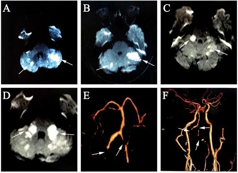
A multi-panel figure containing radiological studies for a case of posterior circulation stroke. (A-D) Axial diffusion-weighted imaging (DWI) MRI frames demonstrate progressive ischemic changes. Panels A and B (Day 3) show multifocal hyperintense areas within the bilateral cerebellar hemispheres, consistent with acute infarction. Panels C and D (Day 10) illustrate new, confluent hyperintensities extending into the bilateral middle cerebellar peduncles (MCP), indicating evolving infarcts. Arrows highlight the specific foci of restricted diffusion. (E, F) 3D reconstruction and coronal views from a computed tomography angiography (CTA) of the head and neck. These vascular images reveal significant pathology of the vertebrobasilar system, including total occlusion of the bilateral vertebral artery (VA) at the V3 and proximal V4 segments, as well as occlusion of both anterior inferior cerebellar arteries (AICA). Severe focal stenosis is also visible in the distal left VA V4 segment. This series provides a diagnostic correlation between multi-segment arterial occlusion and bilateral cerebellar territory infarction.
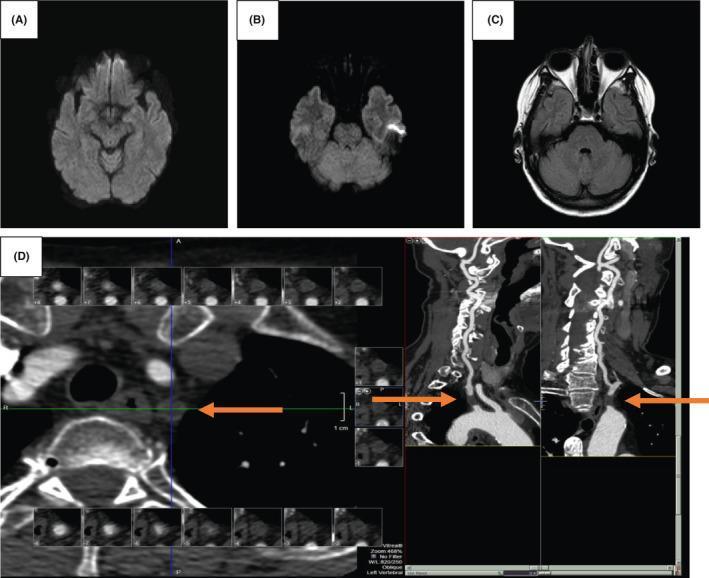
A multi-panel diagnostic imaging study comprising MRI of the brain and CTA of the neck. Panels (A) and (B) show axial Diffusion-Weighted Imaging (DWI) sequences of the brain parenchyma, demonstrating normal signal intensity without evidence of restricted diffusion or acute ischemic stroke in the posterior circulation. Panel (C) is an axial T2-FLAIR sequence showing no hyperintense lesions or edema in the cerebellum or brainstem. Panel (D) features a Computed Tomography Angiogram (CTA) of the neck in axial and sagittal reformats. Orange arrows highlight a complete occlusion of the left subclavian artery. The blockage is located distally to the vessel's aortic origin and proximally to the origin of the left vertebral artery. This clinical finding is significant for the evaluation of subclavian steal syndrome in patients presenting with vertebrobasilar insufficiency symptoms such as vertigo and ataxia.
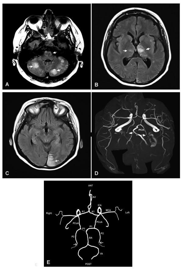
This composite figure illustrates an acute vertebrobasilar territory stroke using diagnostic imaging and anatomical diagrams. Panels A, B, and C are axial MRI (FLAIR/T2) images of the head demonstrating multiple areas of high signal intensity (hypersignal) representing ischemic infarcts. Specifically, panel A shows bilateral cerebellar and bulboprotuberancial junction involvement; panel B shows bilateral thalamic infarcts, more extensive on the left; and panel C shows an infarct in the left occipital lobe. Panel D is an MRI Time-of-Flight (TOF) angiography of the Circle of Willis, showing bilateral occlusion of the posterior cerebral arteries (PCA), marked by arrows. Panel E is a corresponding anatomical diagram mapping the patient's vascular configuration. It highlights a 'fetal type' right posterior communicating artery (PCoA) with a hypoplastic P1 segment of the PCA, where the right posterior territory is primarily supplied by the internal carotid artery (ICA). The diagram confirms the sites of bilateral distal occlusion (arrows) relative to the basilar artery (BA) and vertebral arteries (VA). This case demonstrates the clinical presentation of embolic stroke within the posterior circulation.
elderly gradual vertigo monocular blindness positive Romberg no nystagmus carotid vertebrobasilar diagnosis
Clinical Analysis: Elderly Patient with Gradual Vertigo + Right Eye Blindness + Positive Romberg
Putting the Clues Together
| Finding | Significance |
|---|---|
| Elderly patient | Atherosclerosis, vascular risk factors likely present |
| Gradually worsening vertigo | Favors central cause - peripheral causes (BPPV, vestibular neuritis) are usually sudden in onset |
| No sudden onset | Rules against acute labyrinthitis, vestibular neuritis, perilymph fistula |
| No ear infection on exam | Peripheral infectious cause excluded |
| Blind in right eye | Critical clue - suggests ipsilateral ophthalmic artery or internal carotid artery disease |
| Extraocular movements normal | Brainstem cranial nerve nuclei (III, IV, VI) are intact |
| No nystagmus | Central vertigo can present without nystagmus, unlike most peripheral causes |
| Romberg positive | Sensory ataxia - patient stable with eyes open, falls with eyes closed = proprioceptive or vestibular pathway dysfunction |
Most Likely Diagnosis: Ipsilateral Internal Carotid Artery (ICA) Disease with Carotid-Vertebrobasilar Insufficiency
- Monocular blindness (right eye) = ophthalmic artery is the first branch of the internal carotid artery (ICA). Permanent monocular blindness in elderly = likely ICA occlusion or high-grade stenosis on the right causing ophthalmic artery hypoperfusion/occlusion. This is distinct from amaurosis fugax (transient), which is the classic TIA presentation. The permanent nature suggests completed infarction of the retina via the central retinal artery.
- Gradual vertigo = reduced perfusion to the vestibular nuclei and labyrinth via the vertebrobasilar system (posterior inferior cerebellar artery and anterior inferior cerebellar artery territory). As noted in K.J. Lee's Essential Otolaryngology: "The symptoms of vertebrobasilar insufficiency include vertigo, hemiparesis, visual disturbances, dysarthria, headache, and vomiting. These symptoms are a result of a drop in blood flow to the vestibular nuclei and surrounding structures." - K.J. Lee's Essential Otolaryngology
- No nystagmus - This is actually more consistent with a central/vascular lesion than a peripheral cause. Peripheral vestibular disorders almost always produce nystagmus. As noted in Rosen's Emergency Medicine: "Nystagmus is the cardinal sign of inner ear disease and the principal objective evidence of abnormal vestibular function" - absence of it makes a peripheral cause unlikely. Central ischemia affecting vestibular pathways may produce vertigo without frank nystagmus.
- Positive Romberg = the patient can stand with eyes open (compensating via vision) but falls with eyes closed, indicating loss of proprioceptive/vestibular input. This reflects sensory ataxia from vestibular pathway involvement. It is NOT cerebellar ataxia (in cerebellar ataxia, the patient is unsteady even with eyes open).
- Normal EOMs = no involvement of cranial nerve nuclei III, IV, VI, ruling out complete brainstem stroke (Wallenberg syndrome or basilar artery occlusion).
Differential Diagnosis (Ranked by Probability)
1. Ipsilateral ICA Disease + Vertebrobasilar Insufficiency (Most Likely)
- Right ICA atherosclerotic occlusion/stenosis causing ophthalmic artery territory infarction (permanent right monocular blindness)
- Concurrent vertebrobasilar atherosclerosis causing gradual vestibular symptoms
- Common in elderly with cardiovascular risk factors (hypertension, DM, hyperlipidemia, smoking)
- May show ipsilateral carotid bruit on auscultation over the neck
2. Embolic Posterior Circulation TIA / Stroke (Consider)
- Cardiac embolism (atrial fibrillation) or aortic arch atheroma
- Could cause multi-territory ischemia (ophthalmic artery + vertebrobasilar territory)
- Gradual progression is less typical but possible with recurrent emboli
3. Subclavian Steal Syndrome (Consider in appropriate context)
- Subclavian artery stenosis causing retrograde vertebral artery flow
- Vertigo on arm exercise, supraclavicular bruit
- Can co-exist with ipsilateral arm symptoms
4. Lateral Medullary Syndrome (Wallenberg) (Less likely here)
- Would typically present with ipsilateral facial numbness, contralateral limb sensory loss, hoarseness, Horner syndrome - none reported here
- EOMs usually normal in pure PICA infarction which fits, but presentation is usually acute not gradual
5. Large Vessel Atherosclerotic Disease with Chronic Multifocal Ischemia (Possible)
- Small vessel disease or chronic hypoperfusion causing progressive central vestibular dysfunction
Why This is NOT Peripheral Vertigo
| Feature | Peripheral | This Patient |
|---|---|---|
| Onset | Sudden | Gradual ✓ = Central |
| Duration | Seconds to days (intermittent) | Weeks/months progressive ✓ = Central |
| Nystagmus | Almost always present, horizontal-torsional | Absent ✓ = Central |
| Neurologic deficits | None | Monocular blindness ✓ = Central |
| Ear findings | Often present | Normal ears ✓ |
Positive Romberg Interpretation
- Positive Romberg here = vestibular pathway failure (central vestibular nuclei ischemia) is preventing adequate proprioceptive-vestibular integration
- The patient compensates with vision when eyes are open (therefore stable) but decompensates with eyes closed
- This is distinct from cerebellar ataxia where the patient is unsteady even with eyes open
Recommended Workup
- Carotid Duplex Ultrasound - right ICA stenosis/occlusion (first-line, non-invasive)
- MRI Brain (DWI + FLAIR) - look for acute or subacute posterior circulation infarcts (cerebellum, brainstem, thalamus, occipital lobes) and white matter ischemic changes
- MRA Head and Neck - evaluate vertebrobasilar system and ICA anatomy
- CT Angiography if MRA not available
- ECG + 24h Holter - rule out atrial fibrillation as embolic source
- Echocardiogram - rule out cardiac thrombus or valvular disease
- Ophthalmic evaluation - right eye fundoscopy: look for pale disc (optic atrophy from chronic ischemia), Hollenhorst plaques (cholesterol emboli in retinal vessels), central retinal artery occlusion changes
- Lipid profile, HbA1c, BP monitoring - cardiovascular risk factor assessment
- Vestibular function tests (VNG/ENG, VEMP) - if diagnosis uncertain
Imaging Examples
Management Principles
- Antiplatelet therapy - aspirin or clopidogrel if not already on; dual antiplatelet for high-risk TIA/minor stroke (POINT/CHANCE trial criteria)
- Anticoagulation - if AF found, start oral anticoagulation (NOAC preferred in elderly)
- Carotid endarterectomy (CEA) or stenting - if right ICA stenosis >70% and the patient has had ipsilateral ischemic events; however, since the eye is already permanently blind, the benefit is now primarily for stroke prevention
- Aggressive vascular risk factor control - statins (target LDL <70 mg/dL), antihypertensives, DM management
- Vestibular rehabilitation - for ongoing balance dysfunction
- Fall prevention - positive Romberg indicates significant fall risk; occupational therapy referral
Summary
Sources: Rosen's Emergency Medicine; K.J. Lee's Essential Otolaryngology; Localization in Clinical Neurology 8e; Adams and Victor's Principles of Neurology 12e; Tintinalli's Emergency Medicine
The blindness in right eye is chronic for years and not associated with this vertigo
Revised Clinical Analysis: The Romberg Test is Now Unreliable / Falsely Positive
The Crucial Insight
- Proprioception (dorsal columns)
- Vestibular input (inner ear + CNS pathways)
- Vision (the compensating input that is removed when eyes are closed)
"Close the eyes → remove visual compensation → unmask deficits in proprioception/vestibular system"
- With eyes open, he/she is already functioning on monocular vision - reduced visual input for balance compensation
- Closing both eyes does not simply "remove visual compensation" - it removes the only remaining eye
- The positive Romberg in this patient may simply reflect the loss of monocular visual compensation rather than any true vestibular or proprioceptive deficit
- The Romberg test result is therefore unreliable and cannot be correctly interpreted as indicating a vestibular or dorsal column pathology in isolation
Re-Analysis Without the Romberg
| Finding | Significance |
|---|---|
| Elderly | Degenerative/vascular disease likely |
| Gradual onset vertigo | Central > peripheral |
| No sudden onset | Against BPPV, vestibular neuritis, labyrinthitis |
| No ear infection | Against acute labyrinthitis, otitis media-related vertigo |
| No nystagmus on exam | Against most peripheral causes; can be seen in central/chronic compensated |
| Normal EOMs | No brainstem CN nuclei (III, IV, VI) involvement |
| Chronic right eye blindness (pre-existing, unrelated) | Not a new finding; excluded from current diagnosis |
Revised Differential Diagnosis
The gradual onset + no nystagmus + elderly profile points strongly to:
1. Vertebrobasilar Insufficiency (VBI) - Most Likely
- Gradual reduction in posterior circulation blood flow to vestibular nuclei
- Common in elderly with atherosclerosis
- Vertigo can be the predominant or sole symptom
- No nystagmus between episodes (or suppressed by central compensation)
- Drop attacks and visual symptoms (not the chronic blindness) are associated features
- K.J. Lee's Essential Otolaryngology: "The symptoms of vertebrobasilar insufficiency include vertigo, hemiparesis, visual disturbances, dysarthria, headache, and vomiting. These symptoms are a result of a drop in blood flow to the vestibular nuclei and surrounding structures."
2. Chronic/Compensated Unilateral Vestibular Hypofunction - Consider
- Gradual loss of one labyrinth (e.g., from age-related degeneration, or an old undetected lesion)
- Central compensation takes months to develop - hence gradual course
- With compensation complete, nystagmus is suppressed - which explains absence of nystagmus
- Positive Romberg would be expected here too (but is confounded by monocular blindness)
3. Bilateral Vestibular Hypofunction (BVH) - Important to Consider
- Both labyrinths failing gradually - common in elderly
- Causes: aminoglycoside toxicity, meningitis, autoimmune, idiopathic
- Presents as chronic unsteadiness/oscillopsia rather than episodic true vertigo
- No nystagmus is characteristic (equal bilateral loss = no asymmetry = no nystagmus)
- Romberg would genuinely be positive here (not a confounder issue)
4. Posterior Fossa Space-Occupying Lesion - Must Exclude
- Cerebellar or brainstem tumor (primary or metastatic)
- Acoustic neuroma (vestibular schwannoma) - more common presentation with unilateral tinnitus + hearing loss, but can present with gradual vertigo
- Gradual onset is the key feature that raises this concern
- Normal EOMs and absent nystagmus fit an early/slowly growing lesion
5. Cervical Spondylotic Vertigo - Contributory
- Very common in the elderly
- Osteophytes compressing vertebral arteries causing intermittent VBI
- Vertigo worsened on neck movement
- K.J. Lee's: cervical vertigo in elderly can arise from vertebral artery compression, sympathetic plexus irritation, or proprioceptive loss from cervical joints
6. Presbyvestibulopathy (Age-related Vestibular Degeneration) - Contributory
- Gradual deterioration of vestibular hair cells and nerve with age
- Chronic imbalance and mild vertigo without dramatic episodes
- Increasingly recognized as a diagnosis in elderly patients
Why No Nystagmus?
- Central cause - central lesions affecting vestibular nuclei can produce vertigo without nystagmus (especially chronic, gradual onset)
- Compensated peripheral lesion - if the peripheral deficit developed slowly, the CNS compensates and suppresses nystagmus over time
- Visual fixation suppressing nystagmus - nystagmus is suppressed by visual fixation; the patient was likely tested in normal light; Frenzel glasses (removing fixation) may reveal nystagmus not seen on routine exam
Recommended Approach
Immediate Workup
- MRI Brain with gadolinium - posterior fossa (cerebellum, brainstem, CP angle) - rule out tumor, infarct, demyelination
- MRA of posterior circulation - vertebrobasilar system assessment
- Frenzel glasses examination - re-examine for nystagmus with fixation removed; may unmask subtle nystagmus invisible on standard exam
- Caloric testing / Video Head Impulse Test (vHIT) - objectively assess unilateral or bilateral vestibular hypofunction
- Audiometry - unilateral sensorineural hearing loss would suggest acoustic neuroma or Meniere's
- Cervical spine X-ray / MRI - if cervical spondylosis suspected
If VBI/Vascular Cause Suspected
- ECG, Holter monitor (for AF)
- Carotid + vertebral duplex ultrasound
- Vascular risk factor panel
Important Caveat on Romberg
Summary
| Key Point | Detail |
|---|---|
| Chronic right eye blindness | Pre-existing, unrelated - makes Romberg unreliable |
| Gradual onset | Strongly favors central or chronic peripheral etiology |
| No nystagmus | Consistent with central vertigo OR compensated peripheral deficit |
| No ear pathology | Against acute peripheral causes |
| Elderly | VBI, presbyvestibulopathy, cervical spondylosis, posterior fossa lesion all plausible |
| Priority diagnosis | VBI or posterior fossa lesion - needs MRI urgently |
| Key next step | MRI brain + MRA posterior circulation + vHIT/caloric testing |
Sources: Scott-Brown's Otorhinolaryngology Head & Neck Surgery, K.J. Lee's Essential Otolaryngology, Rosen's Emergency Medicine, Tintinalli's Emergency Medicine
Should I give betahistine or prochlorperazine to help with the symptoms?
Betahistine vs Prochlorperazine in This Patient
Prochlorperazine - Use With Caution / Preferably Avoid
What it does
Why it is problematic in this patient
Betahistine - The More Reasonable Choice
What it does
- Improving microvascular circulation in the stria vascularis (relevant to endolymphatic hydrops / Meniere's)
- Inhibiting activity of the vestibular nuclei - which may reduce vertigo perception - Scott-Brown's Otorhinolaryngology Vol 1
- Increasing cochlear blood flow and decreasing peripheral vestibular inputs - Tintinalli's Emergency Medicine
Why it is more suitable here
- Tintinalli's: "Betahistine... may facilitate vestibular compensation" - unlike prochlorperazine, it does not actively block compensation
- It has a favourable side effect profile in the elderly: mainly GI disturbance, headache, rashes - far less dangerous than phenothiazines
- If VBI or vascular vertigo is the cause, betahistine's vasodilatory effect on cochlear/vestibular microvasculature may be directly relevant
- Does not cause sedation, extrapyramidal effects, or worsen blurred vision - critical given monocular blindness and fall risk
Caveats
- Evidence base is primarily for Meniere's disease - evidence for VBI or non-Meniere's vertigo is less robust
- Contraindicated in phaeochromocytoma (exclude this)
- Use with caution in asthma and peptic ulcer disease
- Dose: 16 mg three times daily (maintenance 24-48 mg/day) - Scott-Brown's Otorhinolaryngology
The Bigger Picture - What Tintinalli's Says Explicitly
"Do not treat patients with nonvertiginous dizziness and disequilibrium of aging with antivertigo medications." - Tintinalli's Emergency Medicine
Summary Recommendation
| Drug | Use in This Patient | Reason |
|---|---|---|
| Prochlorperazine | Avoid long-term; caution short-term | Suppresses vestibular compensation; extrapyramidal effects in elderly; sedation + vision risk dangerous with monocular blindness |
| Betahistine | Reasonable adjunct | Does not suppress compensation; may facilitate it; safe side effect profile; relevant if vascular cause |
Sources: Scott-Brown's Otorhinolaryngology Head & Neck Surgery (Vols 1 & 2); Tintinalli's Emergency Medicine; Goldman-Cecil Medicine