Pathogenesis of sickle cell disease. Pathology 5-mark question
Reading File
Finding Sources
Finding Sources
Searching PubMed
"sickle cell disease"[MeSH Terms] AND pathogenesis
Reading File
Finding Sources
Finding Sources
Loading Image
Reading File
Reading File
Reading File
Loading Image
I now have everything needed for a comprehensive 5-mark pathology answer. Here is the complete response:
Pathogenesis of Sickle Cell Disease
Genetic Basis
Sickle cell disease is an autosomal recessive hemoglobinopathy caused by a point mutation in the β-globin gene on chromosome 11. A single nucleotide change (GAG → GTG) at codon 6 replaces glutamic acid (hydrophilic) with valine (hydrophobic) at the 6th position of the β-globin chain. This produces abnormal hemoglobin called HbS (α₂β^S₂).
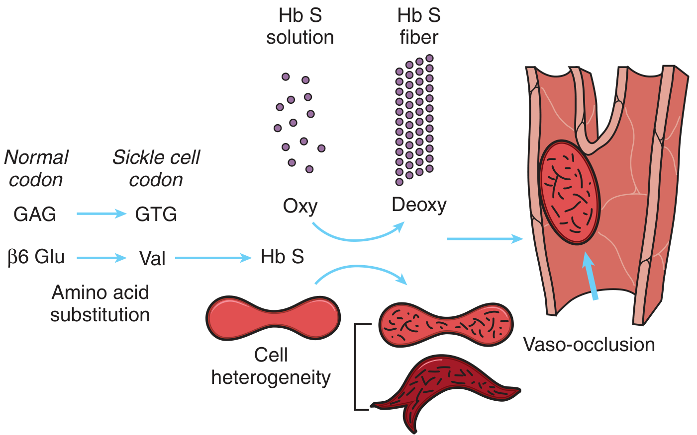
Figure: Pathogenesis of sickle cell disease (Thompson & Thompson Genetics in Medicine)
Step-by-Step Pathogenetic Cascade
1. HbS Polymerization Under Hypoxia
- In the oxygenated state, HbS functions almost normally (solubility is close to HbA).
- Under deoxygenated conditions, HbS becomes only one-fifth as soluble as HbA.
- The hydrophobic valine residue creates a complementary "sticky patch" that allows deoxygenated HbS molecules to undergo intermolecular contacts and aggregate into rod-shaped polymers/fibers.
- These polymers distort the red cell membrane, producing the elongated, crescentic sickle shape.
2. Reversible vs. Irreversible Sickling
- Early sickling episodes are reversible upon reoxygenation.
- However, repeated cycles of sickling cause calcium influx into the red cell, leading to loss of potassium and water (cellular dehydration) and progressive membrane skeleton damage.
- Over time, this cumulative injury creates irreversibly sickled cells (ISCs) - these cannot regain normal shape even upon reoxygenation, and are prone to intravascular hemolysis.
3. Factors Governing Clinical Severity of Sickling
Three key determinants control whether significant HbS polymerization occurs:
| Factor | Effect |
|---|---|
| Intracellular concentration of non-HbS hemoglobin | HbA and HbF both inhibit HbS polymerization. In heterozygotes (~40% HbS), sickling is rare. High HbF levels (as in neonates) protect until ~5-6 months of age. |
| Intracellular HbS concentration | Dehydration increases Hb concentration, promoting polymerization. Co-existing α-thalassemia reduces Hb concentration and is protective. |
| Microvascular transit time | Normal capillary transit is too brief for significant polymerization. Sluggish flow (spleen, bone marrow) or factors like infection/inflammation prolong transit, enabling sickling. |
4. Two Major Pathological Consequences
A. Hemolytic Anemia
- Membrane damage from repeated sickling reduces red cell lifespan to ~20 days (normal: ~120 days) - one-sixth of normal.
- Leads to moderate-to-severe normochromic, normocytic hemolytic anemia.
- Severity correlates with the fraction of irreversibly sickled cells in blood.
- Hemolysis releases free hemoglobin, which scavenges nitric oxide (NO), impairing vasodilation and worsening ischemia.
B. Vaso-occlusion and Ischemic Injury
- Sickle cells physically obstruct the microvasculature.
- Repeated sickling makes the red cell membrane abnormally "sticky" (increased adhesion to endothelium).
- Vaso-occlusion is not directly proportional to ISC numbers; it is triggered by precipitating factors: infection, inflammation, dehydration, acidosis, hypoxia, and cold.
- Inflammation slows blood flow further by increasing leukocyte-endothelial adhesion and leaky vessels - creating a vicious cycle.
Pathological Changes (Morphology)
| Organ/System | Change |
|---|---|
| Spleen | Initially congestive splenomegaly (up to 500 g in children) → repeated infarcts → autosplenectomy (fibrotic nubbin) by adulthood |
| Peripheral blood smear | Elongated, boat-shaped sickled cells, anisocytosis, poikilocytosis, target cells |
| Bone marrow | Compensatory erythroid hyperplasia → bone resorption + new bone formation → "crew-cut" pattern on skull X-ray, prominent cheekbones |
| Multiple organs | Vascular thrombosis and infarction in bones, kidneys, brain, retina, lungs, skin |
| Liver/Heart | Fatty changes from hypoxia-induced ischemia |
| Gallbladder | Pigment gallstones (from chronic hemolysis and bilirubin excess) |
| Kidney | Papillary necrosis, hematuria, hyposthenuria |
| Penis | Priapism → penile fibrosis, erectile dysfunction |
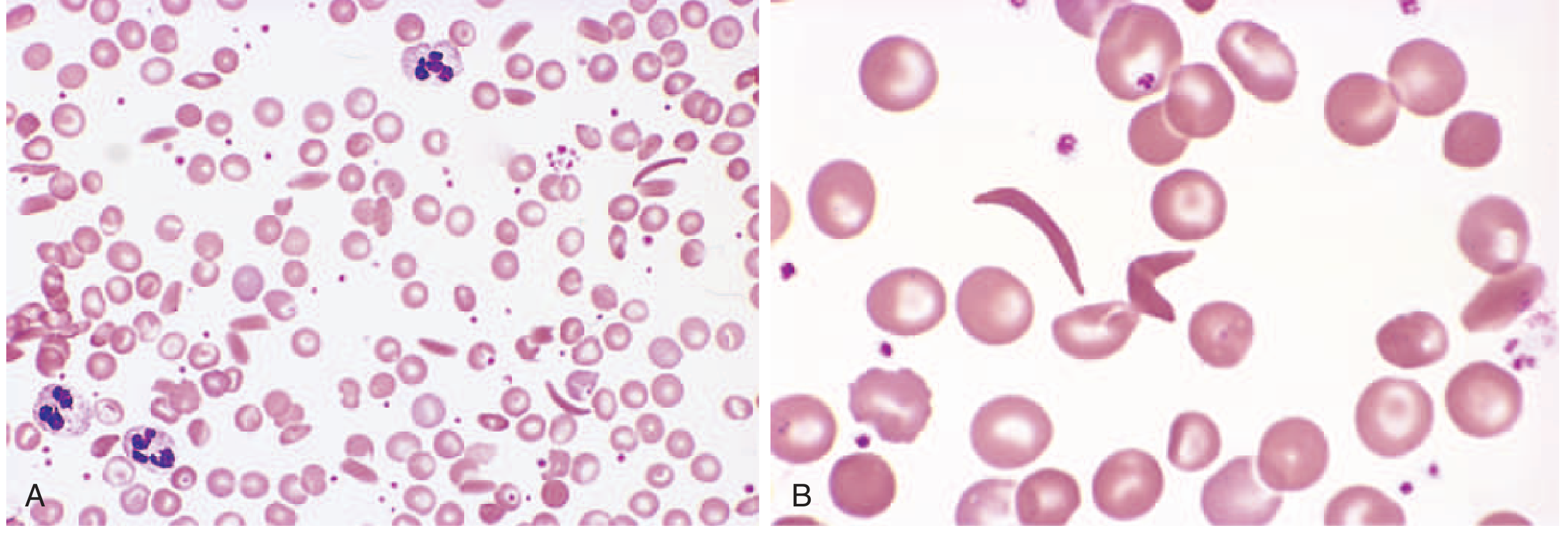
Fig: Sickle cell anemia - peripheral blood smear (Robbins Basic Pathology)
Role of Modifier Genes (HbF)
- HbF (α₂γ₂) inhibits HbS polymerization by failing to form intermolecular contacts with deoxygenated HbS.
- Higher HbF = less clinical severity (reduced pain crises, longer survival).
- BCL11A gene product is a transcription factor that silences γ-globin expression postnatally. Loss-of-function of BCL11A → hereditary persistence of fetal hemoglobin (HPFH), which ameliorates sickle cell disease.
- Hydroxyurea therapy works by increasing HbF levels (via a poorly understood mechanism), reducing sickling frequency.
Summary (5 Key Points for Exam)
- Molecular defect: GAG→GTG mutation → Glu→Val at β-globin codon 6 → HbS
- Polymerization: Deoxygenated HbS forms rigid polymers → red cell deforms into sickle shape
- Reversible → Irreversible: Repeated sickling causes Ca²⁺ influx, membrane damage, irreversibly sickled cells
- Hemolysis: RBC lifespan reduced to ~20 days; free Hb scavenges NO → worsens ischemia
- Vaso-occlusion: Sticky sickled cells + sluggish flow → microvascular obstruction → ischemia, infarction, pain crises (bones, spleen, brain, kidney)
Sources: Robbins & Kumar Basic Pathology (9780323790185); Thompson & Thompson Genetics and Genomics in Medicine (9780323547628); Robbins, Cotran & Kumar Pathologic Basis of Disease (9780443264528)
Pathogenesis of sickle cell disease. Pathology 5-mark question. Give in flow chats
Here is the pathogenesis of sickle cell disease presented as flow charts:
FLOW CHART 1: Molecular Basis (The Core Defect)
Point mutation in β-globin gene (chromosome 11)
                    ↓
        GAG → GTG (codon 6)
                    ↓
   Glutamic acid → Valine substitution
   (hydrophilic)    (hydrophobic)
                    ↓
        Abnormal hemoglobin = HbS (α₂β^S₂)
                    ↓
    In homozygotes (HbSS): HbA completely replaced by HbS
                    ↓
            SICKLE CELL DISEASE
FLOW CHART 2: Sickling Mechanism
                    HbS
                    ↓
        ┌───────────────────────┐
        ↓                       ↓
  OXYGENATED state         DEOXYGENATED state
  (normal solubility)      (1/5th solubility of HbA)
        ↓                       ↓
  No sickling           Hydrophobic valine creates
                        "sticky patches" on HbS
                                ↓
                  HbS molecules undergo conformational change
                                ↓
                  Intermolecular contacts → Rod-shaped
                  HbS polymers/fibers form
                                ↓
                  Distortion of red cell membrane
                                ↓
                  Elongated, crescentic SICKLE SHAPE
FLOW CHART 3: Reversible → Irreversible Sickling
    First few sickling episodes
                ↓
        REVERSIBLE sickling
     (re-oxygenation restores shape)
                ↓
    Repeated cycles of sickling
                ↓
    Ca²⁺ influx into red cell
                ↓
    Loss of K⁺ and water → Cell dehydration
                ↓
    Membrane skeleton damage (cumulative)
                ↓
    IRREVERSIBLY SICKLED CELLS (ISCs)
    (cannot regain normal shape even on re-oxygenation)
                ↓
    Prone to intravascular HEMOLYSIS
FLOW CHART 4: Two Major Pathological Arms
                    SICKLED RED CELLS
                          ↓
           ┌──────────────────────────────┐
           ↓                              ↓
    ARM 1: HEMOLYSIS               ARM 2: VASO-OCCLUSION
           ↓                              ↓
  RBC lifespan ↓ to ~20 days    Sticky sickled cells adhere
  (normal = 120 days)           to vascular endothelium
           ↓                              ↓
  Hemolytic anemia              Microvascular obstruction
  (moderate to severe)                    ↓
           ↓                    Ischemia → Infarction
  Free Hb released                        ↓
           ↓                    PAIN CRISES + organ damage
  Scavenges Nitric Oxide (NO)
           ↓
  Impaired vasodilation
  → Worsens ischemia
FLOW CHART 5: Vaso-occlusion in Detail
    Precipitating factors:
    Infection / Inflammation / Dehydration / Acidosis / Hypoxia / Cold
                    ↓
    Sluggish blood flow in microvasculature
    (especially spleen, bone marrow)
                    ↓
    Prolonged transit time of sickle RBCs
                    ↓
    More time for HbS polymerization to occur
                    ↓
    Increased RBC adhesion to endothelium
    (membrane damage → abnormally "sticky" cells)
                    ↓
    Leukocyte adhesion + plasma exudation
    (inflammation worsens flow)
                    ↓
    ┌─────────────────────────────────────────┐
    ↓               ↓               ↓         ↓
  Bones         Spleen           Brain      Kidney
  (infarcts)  (autosplenectomy) (stroke)  (papillary necrosis)
    ↓               ↓               ↓         ↓
  Lung           Retina           Liver     Penis
(acute chest)  (blindness)     (ischemia) (priapism)
FLOW CHART 6: Splenic Changes (Classic Sequence)
    Childhood
        ↓
    Sickled RBCs trapped in splenic red pulp
        ↓
    Congestive SPLENOMEGALY (up to 500 g)
        ↓
    Repeated hypoxic damage + infarcts
        ↓
    Progressive fibrosis and shrinkage
        ↓
    AUTOSPLENECTOMY (by adulthood)
    (small, fibrotic, non-functional nubbin)
        ↓
    Functional asplenia
        ↓
    Susceptibility to encapsulated organisms
    (Streptococcus pneumoniae, H. influenzae, Salmonella)
FLOW CHART 7: Role of HbF (Modifier Gene)
    BCL11A gene → silences γ-globin → ↓ HbF postnatally
                    ↓
    Low HbF → HbS polymerization proceeds freely
                    ↓
            Severe disease
    ─────────────────────────────────
    High HbF (e.g., HPFH, hydroxyurea therapy)
                    ↓
    HbF interacts weakly with deoxygenated HbS
                    ↓
    Inhibits HbS polymer formation
                    ↓
    ↓ Sickling → ↓ Pain crises → Milder disease
MASTER SUMMARY FLOW CHART
β-globin gene mutation (Glu6Val)
            ↓
           HbS
            ↓
   Deoxygenation in tissues
            ↓
   HbS polymerization → Fiber formation
            ↓
   Red cell sickling
        ↙           ↘
HEMOLYSIS         VASO-OCCLUSION
   ↓                    ↓
Anemia            Ischemia & Infarction
   ↓                    ↓
Jaundice         Pain crises, Organ damage
Pigment stones   Stroke, Autosplenectomy
                 Acute chest syndrome
Sources: Robbins Basic Pathology; Thompson & Thompson Genetics in Medicine; Goldman-Cecil Medicine
Create a visual infographic poster of sickle cell pathogenesis flowchart
Generating Image
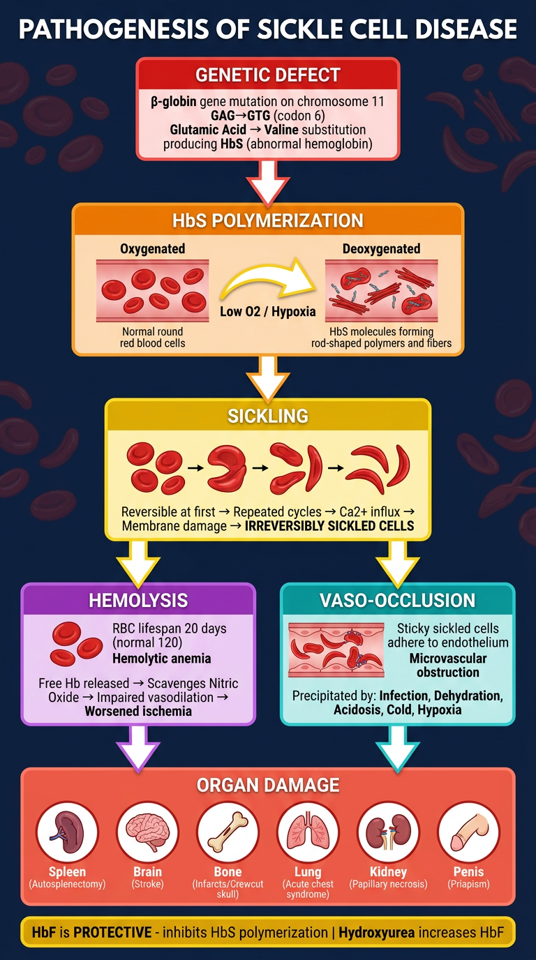
Here is your visual infographic poster covering the full pathogenesis cascade:
- Top - the genetic root cause (GAG→GTG mutation → Glu→Val → HbS)
- Middle - HbS polymerization under deoxygenation → sickling (reversible → irreversible)
- Split arms - the two major consequences: Hemolysis (left) and Vaso-occlusion (right)
- Bottom - end-organ damage across 6 systems (spleen, brain, bone, lung, kidney, penis)
- Footer banner - the protective role of HbF and hydroxyurea therapy
This is ideal for study notes, presentations, or classroom display. Let me know if you'd like any section emphasized differently or a version with more detail on any specific arm.
This is a shared conversation. Sign in to Orris to start your own chat.