
Papillary lesions of the breast 10 marks md pathology exam
papillary lesion breast histology microscopy
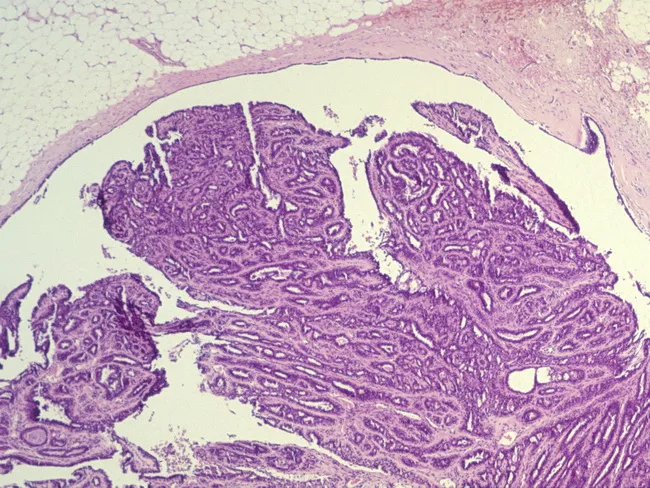
Imaging modality: Light microscopy of hematoxylin and eosin (H&E) stained breast tissue section illustrating a palpable intraductal papilloma. The lesion measures 2.1 cm in greatest dimension, consistent with a solitary ductal papilloma within a lactiferous duct. The histology reveals papillary architecture with fibrovascular cores projecting into ductal lumens. Each papillary frond is lined by a two-layered epithelium comprising an inner luminal epithelial cell layer and an outer myoepithelial cell layer, a characteristic feature distinguishing benign papillary lesions from invasive carcinoma. Ductal epithelium displays mild hyperplasia without overt cytologic atypia; no invasion of surrounding stroma is evident. The ducts appear distended with a mild fibrous stroma and occasional periductal inflammation. The presence of preserved myoepithelial mantle supports benignity. The overall pattern is nodular, with elongated papillae and a fibrous stroma backdrop. Pathologic significance: benign breast lesion that can explain a clinically palpable mass and possibly nipple discharge. Diagnostic considerations include solitary intraductal papilloma with or without epithelial hyperplasia, papillomatosis, and radiologically occult microcalcifications. Clinical relevance: complete excision is often curative; follow-up recommended to exclude atypia or malignant transformation in broader ductal pathology. This image is educational for pathology, breast disease differential diagnosis, and surgical planning. Useful for teaching, research, and reviews.
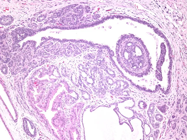
Histology: Light microscopy of a breast tissue section stained with Hematoxylin and Eosin (H&E). A dilated lactiferous duct is the central focus, showing a prominent intraductal papilloma with a delicate fibrovascular core. The papillary stalks project into the ductal lumen and are lined by two cell layers: inner luminal epithelium and an outer myoepithelial layer, consistent with a benign intraductal process. The epithelium exhibits uniform cuboidal to columnar cells without significant cytologic atypia or mitotic activity. Surrounding the dilated duct are fibrocystic changes, including stromal fibrosis and scattered cystic dilatations with mucinous or apocrine metaplasia. The overall architecture remains organized, with no invasion into the surrounding stroma. Basally oriented myoepithelial cells are preserved, supporting benignity. The features differentiate intraductal papilloma from papillary ductal carcinoma in situ, where myoepithelial absence and cellular atypia would be more evident. Clinically, such lesions can present with occasional nipple discharge and are typically managed with conservative surgical excision when symptomatic. The image provides a representative example of a recurring benign papillary duct lesion within a dilated duct and accompanying fibrocystic breast parenchyma, underscoring the importance of recognizing papillary architecture, mucinous luminal contents, and the protective myoepithelial layer in diagnostic practice. For educational use and clinical correlation.
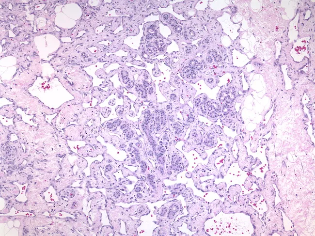
Breast histopathology using light microscopy of formalin-fixed, paraffin-embedded tissue demonstrates a low-grade, well-differentiated angiosarcoma. The architecture is dominated by proliferating, open, anastomosing vascular channels that diffusely infiltrate the breast parenchyma. Lumenal spaces contain erythrocytes and show irregular calibers, contributing to a ragged vascular network. The endothelial lining displays bland cytology with minimal atypia and a low mitotic rate, distinguishing this entity from higher-grade sarcomas. Papillary formations are absent or infrequent, helping to differentiate from certain papillary endothelial proliferations. The tumor margins may blend with surrounding adipose and fibrous stroma, reflecting an infiltrative growth pattern within a limited biopsy core. Overall, the lesion’s low-grade phenotype correlates with a relatively indolent histologic appearance compared with high-grade angiosarcoma, yet clinical behavior can be unpredictable and warrants thorough evaluation. The gold-standard imaging context is histology, not radiography, with staining by Hematoxylin and Eosin providing contrast between endothelial cells and luminal spaces. Immunohistochemical confirmation (CD31, CD34, and factor VIII–related antigen) supports endothelial origin. Clinically, recognizing this pattern in breast tissue is critical for accurate diagnosis, prognosis assessment, and treatment planning, including surgical excision and consideration of adjuvant therapy, radiotherapy, or systemic therapy as indicated. This image is essential for education, pattern recognition, and research datasets globally.
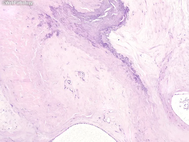
Imaging modality: Histopathology (Light Microscopy) after Hematoxylin and Eosin staining of breast tissue. Anatomical location: Breast tissue, mammary gland stroma; central region shows clusters of cysts separated by dense fibrous stroma. The collagenized stroma forms a stellate scar-like configuration with radiating fibrous septa extending into surrounding parenchyma. Dystrophic calcifications are present within the stromal matrix between cystic spaces and the scar, appearing as granular basophilic deposits. The epithelial component lining ducts within cysts is minimal and lacks cytologic atypia; occasional sclerosing adenosis-like features may be seen; overall architecture resembles complex sclerosing lesion or radial scar. Notable features: calcified stromal dystrophy, stellate radiating architecture, benign-appearing ductal epithelium; absence of significant pleomorphism or mitotic activity. Diagnostic significance: benign fibrocystic-related radial scar-like lesion; on imaging, such patterns may mimic carcinoma, necessitating complete excisional biopsy or careful margin assessment; differential diagnosis includes papillary lesions, fibroadenoma with sclerosis, and invasive carcinoma with desmoplasia. Clinical correlation: in asymptomatic patients, correlate with imaging findings; may be incidental; in patients with calcifications or a palpable mass, correlate with radiology to determine need for excision. Potential clinical use cases: teaching histology of breast lesions, correlating radiologic-pathologic findings, evaluating sclerosing breast lesions in diagnosis and surgical planning.
intraductal papilloma breast fibrovascular core myoepithelial
Histology: Light microscopy of a breast tissue section stained with Hematoxylin and Eosin (H&E). A dilated lactiferous duct is the central focus, showing a prominent intraductal papilloma with a delicate fibrovascular core. The papillary stalks project into the ductal lumen and are lined by two cell layers: inner luminal epithelium and an outer myoepithelial layer, consistent with a benign intraductal process. The epithelium exhibits uniform cuboidal to columnar cells without significant cytologic atypia or mitotic activity. Surrounding the dilated duct are fibrocystic changes, including stromal fibrosis and scattered cystic dilatations with mucinous or apocrine metaplasia. The overall architecture remains organized, with no invasion into the surrounding stroma. Basally oriented myoepithelial cells are preserved, supporting benignity. The features differentiate intraductal papilloma from papillary ductal carcinoma in situ, where myoepithelial absence and cellular atypia would be more evident. Clinically, such lesions can present with occasional nipple discharge and are typically managed with conservative surgical excision when symptomatic. The image provides a representative example of a recurring benign papillary duct lesion within a dilated duct and accompanying fibrocystic breast parenchyma, underscoring the importance of recognizing papillary architecture, mucinous luminal contents, and the protective myoepithelial layer in diagnostic practice. For educational use and clinical correlation.
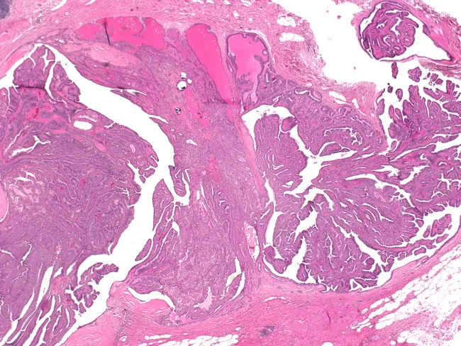
Imaging modality: light microscopy; specimen type: breast ductal tissue; technique: hematoxylin and eosin staining. The slide demonstrates a 1.5 cm intraductal papilloma arising within a central/subareolar duct with a palpable mass and associated bloody nipple discharge clinically. Histologically, the lesion consists of multiple papillary fronds protruding into a dilated duct lumen, each frond supported by a fibrovascular core and lined by a dual epithelial and myoepithelial layer. The epithelial component is typically columnar to cuboidal with uniform nuclei and minimal cytologic atypia; myoepithelial cells encircle the papillae, helping to distinguish benign intraductal papilloma from invasive carcinoma. The surrounding ductal epithelium shows mild ductal ectasia and focal hyperplasia but lacks invasion, high-grade atypia, or marked mitotic activity. The architecture is characteristic of benign papillary lesions of the breast, though larger papillomas can be solitary and clinically present as subareolar masses with Bloody discharge. The lesion may be associated with ductal dilation and mild stromal inflammation; no necrosis or stromal desmoplasia is evident. Clinically, this finding correlates with nipple discharge spectrum (bloody) and mass effect; management typically involves surgical excision to rule out atypia or malignant transformation and ensure complete removal of the papillary lesion.
papillary DCIS breast duct carcinoma in situ
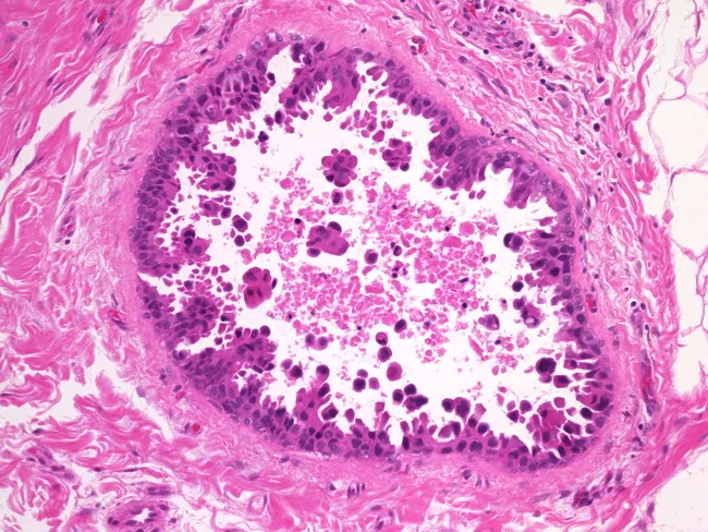
This image depicts a hematoxylin and eosin stained breast tissue section examined by bright-field light microscopy, highlighting micropapillary ductal carcinoma in situ (DCIS). The lesion shows micropapillary architecture with papillary fronds projecting into the duct lumen. Papillary elements range from small bumps or mounds to slender, elongated fronds; in several instances the fronds are transected, creating small detached irregular clusters of tumor cells. The intraductal space contains scattered cellular debris, a feature associated with higher-grade disease. The tumor cells exhibit high nuclear grade, characterized by enlarged, hyperchromatic nuclei and conspicuous nucleoli within a compact epithelial lining. The surrounding tissue shows a distinct ductal distribution without obvious stromal invasion in this field, consistent with an in situ process rather than invasive carcinoma. The micropapillary pattern may be linked to increased risk of local recurrence and challenges in complete excision, underscoring the need for meticulous histologic sampling to exclude invasion. Clinically, recognizing this variant informs prognosis and treatment planning in breast cancer, including surgical margins, adjuvant therapy considerations, and radiologic correlation for residual disease. This image serves educational and research purposes, illustrating the micropapillary DCIS subtype and its distinguishing morphologic features for pathologists, residents, and students.
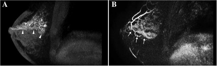
This diagnostic imaging composite displays breast Magnetic Resonance Imaging (MRI) Maximum Intensity Projection (MIP) views from a 53-year-old woman. Panel A shows a pre-contrast T1-weighted image highlighting significant duct dilation, indicated by arrowheads, appearing as hyperintense linear/branching structures against the dark fibroglandular background of the right breast. Panel B displays a post-contrast subtracted MIP image revealing an abnormal enhancing lesion with a distinct segmental distribution, marked by arrows. The enhancement pattern follows a ductal network originating from the nipple and extending deep into a breast segment, characterized by non-mass enhancement (NME). This combination of imaging findings—ductal dilation and segmental NME—is clinically significant for identifying high-risk or malignant lesions. In this specific case, the pathology confirmed an intraductal papilloma associated with ductal carcinoma in situ (DCIS), illustrating how segmental enhancement patterns on breast MRI can serve as markers for co-existing malignancy in papillary lesions.
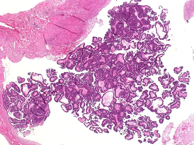
This histopathology image depicts a breast lesion consistent with encapsulated intracystic papillary carcinoma. Stained with Hematoxylin and Eosin and examined under light microscopy at multiple magnifications (4x–40x), the lesion is sharply circumscribed within a dilated duct, producing a mural nodule projecting into a cystic space. Papillary architecture is evident, with slender fibrovascular cores lined by neoplastic epithelium. The epithelial layer is typically bland, with low to intermediate cytologic grade and minimal mitotic activity; myoepithelial cells may be present around the papillary fronds in in-situ-appearing regions, while invasive foci, when present, disrupt the capsule and show irregular infiltration. The capsule is fibrous and distinct from surrounding ductal tissue. In some samples, accompanying DCIS or invasion is limited to the pericapsular region, correlating with clinical reports that encapsulated papillary carcinoma often follows an indolent course but may harbor an invasive component. Immunophenotype often shows variable estrogen and progesterone receptor positivity with variable HER2 status. Clinically, this entity occurs in older women and carries a favorable prognosis after complete excision. Differential considerations include intraductal papilloma, mucinous carcinoma, and invasive papillary carcinoma without an encapsulating border. The image highlights the need to assess invasion, capsule integrity, and ductal architecture for accurate diagnosis and treatment planning.
Papillary Lesions of the Breast
Classification
| Category | Entity |
|---|---|
| Benign | Intraductal papilloma (solitary / central), Peripheral papillomatosis |
| High-risk / Borderline | Atypical papilloma (with ADH/ALH), Intraductal papilloma with DCIS |
| Malignant In Situ | Papillary DCIS, Encapsulated papillary carcinoma (EPC), Solid papillary carcinoma (SPC) |
| Malignant Invasive | Invasive papillary carcinoma, Invasive EPC |
1. Intraductal Papilloma (Benign)
Subtypes
- Solitary (central) papilloma: arises in a major lactiferous duct near the nipple-areolar complex; the most common papillary lesion
- Peripheral papillomatosis: multiple small papillomas arising in terminal duct-lobular units (TDLUs), >5 cm from the nipple
Pathology (Gross)
- Small (~0.5 cm; rarely up to 5 cm), soft, tan, friable nodule resembling "miniature broccoli florets" attached to the duct wall by a stalk
- Resides within a dilated duct or cyst, often causing duct obstruction
Histology
- Multiple branching papillae with fibrovascular cores projecting into the ductal lumen
- Covered by a bilayered epithelium: inner luminal epithelial cells + outer myoepithelial cells
- Myoepithelial layer is intact and continuous - this is the key feature of benignity
- Variable fibrosis between epithelial components; apocrine metaplasia may be seen
- Occasionally shows ductal epithelial hyperplasia (usual type) without atypia
IHC
- CK5/6, p63, smooth muscle actin (SMA), calponin: positive in myoepithelial layer (outer layer) - confirms benignity
- ER: focally positive; low Ki-67

Clinical Features
- Solitary papilloma: presents with unilateral, spontaneous, serous or bloody nipple discharge from a single duct (most common cause of bloody nipple discharge in premenopausal women); may form a subareolar palpable mass
- Peripheral papillomatosis: may be bilateral; palpable or incidental on imaging; associated with higher malignant potential
- Ultrasound: intraductal nodule; MRI may show segmental ductal enhancement
Risk of Malignancy
-
Solitary central papilloma: slight increase in relative risk (~2.1x) of future breast cancer in either breast
-
Peripheral papillomatosis: markedly higher relative risk (3-3.5x); more likely to harbor atypia or undergo malignant transformation
-
Approximately 12% of adjacent tissue in excised papillomas contains mammary dysplasia
-
Fischer's Mastery of Surgery, 8th ed., p. 1382-1383
2. Atypical Papilloma
- Intraductal papilloma that contains foci of atypical ductal hyperplasia (ADH) or atypical lobular hyperplasia (ALH)
- Confers a significantly higher upgrading rate to carcinoma on excision (~5-15%)
- Surgical excision is always warranted
3. Papillary DCIS (Ductal Carcinoma In Situ - Papillary type)
Histology
- Ducts distended by papillary fronds with fibrovascular cores lined by a monotonous population of malignant epithelial cells
- No myoepithelial cells on the papillae (key differentiating feature from benign papilloma)
- Nuclear atypia, increased mitotic activity, may show necrosis
- Micropapillary variant: small club-shaped or Roman-arch projections without true fibrovascular cores
IHC
- CK5/6, p63, SMA: absent on papillary fronds (no myoepithelium)
- ER: usually strongly positive
- High Ki-67 in high-grade lesions
4. Encapsulated Papillary Carcinoma (EPC) / Intracystic Papillary Carcinoma
- Formerly called "intracystic papillary carcinoma"
- A circumscribed, cyst-like structure containing papillary carcinoma, surrounded by a fibrous capsule
- Now considered a low-grade in situ carcinoma (or at the border of in situ and minimally invasive)
Gross
- Well-circumscribed, encapsulated nodule; cut section shows a cystic space with papillary projections and hemorrhagic fluid
Histology
- Papillae with fibrovascular cores lined by atypical/malignant cells
- Fibrous capsule encloses the lesion
- Myoepithelial cells absent at the periphery of the capsule (unlike benign papilloma)
- Often low-to-intermediate nuclear grade; ER-positive in majority
IHC
- p63, SMA, calponin: absent at the lesional periphery (distinguishes from benign lesion)
- ER strongly positive (>90%)
Clinical Significance
- Generally indolent behavior; excellent prognosis after complete excision
- Occurs more often in older/postmenopausal women
- If invasive component is present beyond capsule: classified as invasive EPC
5. Solid Papillary Carcinoma (SPC)
- A variant where ducts are filled with solid proliferations of cells that still contain small fibrovascular cores visible on careful examination
- Cells often have neuroendocrine differentiation (chromogranin, synaptophysin positive); may contain mucinous material
- Typically low-grade, ER-positive, occurs in older women
- In situ form (without stromal invasion) has excellent prognosis
6. Invasive Papillary Carcinoma
- Accounts for ~2% of all invasive breast cancers
- Occurs predominantly in seventh decade; disproportionately in non-white women
- Typically small (rarely >3 cm)
Histology
- Invasive tumor with papillae bearing fibrovascular stalks and multilayered epithelium infiltrating the stroma
- Low grade; limited desmoplasia
- 87% are ER-positive (SEER database data)
- Low frequency of axillary lymph node metastases
Prognosis
-
Favorable - 5- and 10-year survival rates comparable to mucinous and tubular carcinoma
-
Similar prognosis to other special-type low-grade breast cancers
-
Schwartz's Principles of Surgery, 11th ed., p. 369; Fischer's Mastery of Surgery, 8th ed., p. 1754
Key Distinguishing Features: Benign vs Malignant Papillary Lesions
| Feature | Benign Papilloma | Papillary Carcinoma |
|---|---|---|
| Myoepithelial layer | Present (intact, bilayered) | Absent |
| Nuclear atypia | Absent / minimal | Present |
| Epithelial monotony | Polymorphic (two cell types) | Monomorphic |
| IHC: CK5/6, p63, SMA | Positive | Negative on fronds |
| ER expression | Focal, heterogeneous | Diffuse, strong |
| Necrosis | Absent | May be present |
| Architecture | Complex papillae | May be solid/micropapillary |
Diagnostic Approach on Core Needle Biopsy
- Benign papilloma on CNB may be observed (no excision) IF:
- Large-bore needle (≤14 gauge) used
- Lesion adequately sampled (>50% removed)
- No atypia present
- Lesion ≤10-15 mm
- Radiologic-pathologic concordance
- No concurrent ipsilateral breast cancer (if present, upstaging rate >20%)
- Peripheral papillomatosis: always excise (significant malignant upgrade rate)
- Atypical papilloma: always excise
- Any discordant lesion: excise
Summary Mnemonics
- "FAME" for benign papilloma = Fibrovascular cores + Apocrine change possible + Myoepithelial layer intact + Epithelium bilayered
- "MONA" for malignant papillary lesion = Myoepithelial absent + Only one cell type (monotonous) + Nuclear atypia + Absence of CK5/6
- Robbins, Cotran & Kumar - Pathologic Basis of Disease (Breast chapter)
- Fischer's Mastery of Surgery, 8th ed., pp. 1382-1384 (Proliferative Breast Disease; Papillary Carcinoma)
- Schwartz's Principles of Surgery, 11th ed. (Invasive Breast Carcinoma)
- Berek & Novak's Gynecology (Breast Papilloma section)