

What will be the treatment of it accrued 2 times already
keloid recurrence treatment earlobe
PMID: 38789810
earlobe keloid treatment
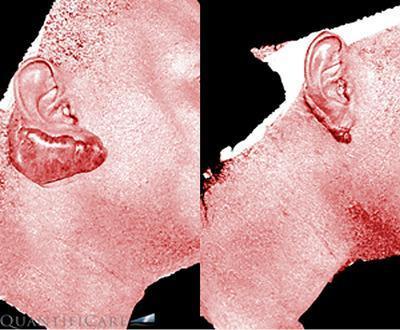
This clinical comparison shows a side-by-side progression of an earlobe keloid before and after CO2 laser treatment, visualized using the Vectra H2 digital vascular filter system. The images are presented in a reddish-tinted digital mode to highlight superficial vascularity. In the 'before' image (left), a large, pedunculated (Type I Chang-Park classification) keloid mass is visible, originating from the inferior aspect of the earlobe. The mass is prominently raised, with an irregular, bumpy texture and high vascular density indicated by darker red pigmentation compared to surrounding skin. The 'after' image (right), taken at a 6-month follow-up, demonstrates significant clinical improvement. There is a substantial reduction in the lesion's volume and height, returning the earlobe to a more anatomical shape. The vascularity appears decreased, evidenced by the lighter, more uniform color intensity across the treated site. This comparison illustrates the efficacy of laser therapy for hypertrophic scarring in dark-skinned patients (phototypes III-VI) and the utility of 3D vascular imaging for monitoring treatment response.
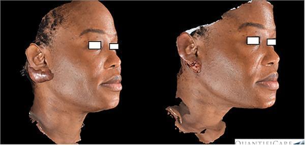
Side-by-side comparison of 3D digital clinical images (Quantificare system) showing a patient with a prominent earlobe keloid before and immediately after CO2 laser treatment. In the 'before' image (left), a large, smooth, pedunculated keloid mass is visible, significantly distorting the natural anatomy of the right earlobe. The patient has a high Fitzpatrick skin phototype. In the 'after' image (right), the bulky keloid has been ablated, resulting in a dramatic reduction in lesion volume. The treatment site shows immediate post-operative changes, including a raw, slightly uneven surface texture with pinpoint bleeding and removal of the primary mass, restoring the approximate contour of the earlobe. The images illustrate the efficacy of CO2 laser ablation in treating hypertrophic and keloid scarring in dermatological practice, particularly for complex anatomical areas like the ear helix and lobe.
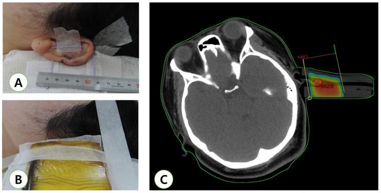
A multi-panel figure illustrating the workflow for adjuvant radiotherapy of an earlobe keloid following surgical excision. Image A is a clinical photograph showing the immediate post-operative state of the earlobe, featuring surgical sutures and medical tape for positioning, with a millimeter ruler included for precise dimensional measurement. Image B displays the simulation phase for radiotherapy, showing a bolus material applied over the surgical site to ensure optimal surface dose distribution. Image C is an axial Computed Tomography (CT) scan used for treatment planning. The CT shows transverse cross-sections of the cranium and orbits. Superimposed on the left earlobe region is a color-coded dose-volume histogram (DVH) representation, with red indicating the high-dose target area and yellow contours defining the radiation field boundaries, labeled 'Field 1'. This composite image demonstrates the integration of clinical surgery, simulation, and radiological planning in dermatology and radiation oncology.
Diagnosis: Recurrent Earlobe Keloid
Treatment for a Twice-Recurrent Earlobe Keloid
Why simple excision fails
Best Evidence-Based Options (Ranked by Evidence)
1. Surgical Excision + Postoperative Radiotherapy (STRONGEST evidence)
- Excision + radiotherapy significantly improved recurrence-free survival (p=0.003) vs. excision alone
- Electron beam radiotherapy showed advantages over X-ray treatment
- Radiotherapy should begin within 24-48 hours of excision
- Median recurrence-free interval with combination therapy: 10 months
2. Surgical Excision + Perioperative Intralesional Corticosteroid Injections
- Triamcinolone acetonide (TA): 10-40 mg/mL, injected intralesionally
- Important: use 10 mg/mL not 40 mg/mL to avoid fat atrophy and hypopigmentation
- Schedule: weekly or bimonthly early on, then monthly
- Excision + steroid combination improves success to 72-92% vs. steroid alone
- The 2024 meta-analysis confirmed this also significantly outperforms excision alone (p=0.003)
- Cummings Otolaryngology, p. 366
3. Triple Therapy: Excision + Steroid + 5-Fluorouracil (5-FU)
- Intralesional 5-FU inhibits fibroblast proliferation and collagen synthesis
- Excision + 5-FU gives a recurrence rate of 19% at 1 year - better than steroids alone
- TA + 5-FU combined outperforms either alone (RCT evidence)
- Typical regimen: 5-FU 50 mg/mL + TA 10 mg/mL mixed intralesionally
- Cummings Otolaryngology, p. 366
4. Cryotherapy
- Can be used standalone or as adjuvant therapy
- Average flattening ~31% after 3 sessions, ~58% after 6 sessions
- Results are better in younger/newer keloids - less effective for older or recurrent ones
- Cummings Otolaryngology, p. 366
5. Pressure Therapy / Silicone Sheeting
- Pressure earrings (Zimmer splints) applied after excision help prevent re-growth
- Silicone gel sheeting reduces collagen overproduction
- Good as adjuvant; insufficient alone for a twice-recurred keloid
- Scott-Brown's Otorhinolaryngology, p. 481
6. Other Adjuvant Options (for refractory cases)
- Bleomycin intralesional: 66-84% significant flattening, but risk of hyperpigmentation (especially in darker skin tones)
- Interferon-α2b / interferon-γ: mixed evidence; recurrence rate ~18.7% after injection + excision in some studies, but not consistently better
- Imiquimod 5% cream post-excision: reduces recurrence in some studies
Recommended Approach for a Twice-Recurrent Earlobe Keloid
- Surgical excision (intralesional incision technique, not wide excision)
- Immediate postoperative radiotherapy (electron beam preferred, within 24-48 h)
- Serial intralesional TA injections (10 mg/mL) starting 4-6 weeks post-op, monthly for 6 months
- Pressure earring worn for at least 6-12 months post-healing
- Advise the patient not to re-pierce the ear - this is the single most important preventive measure
Important note: Do not attempt to squeeze, pop, or cut this at home - it will grow back larger.