
in articular cartilage injury why it appears light in mri
Reading File
Normal articular cartilage appears bright (high signal) on T2-weighted MRI because of its high water/proteoglycan content. When cartilage is injured, the proteoglycan matrix is disrupted and water is lost, so the signal drops - injured areas appear relatively dark compared to normal cartilage.
Searching sources...
Finding Sources
Finding Sources
Good results. Let me read the most relevant sections for a complete explanation.
Reading File
Reading File
Reading File
Excellent. I have detailed, authoritative content. Let me also fetch the MRI image showing the three-layer cartilage pattern for visual clarity.
Loading Image
Now I have everything needed for a thorough answer. Here is the full explanation:
Why Injured Articular Cartilage Appears Dark (Low Signal) on MRI
The answer comes down to the composition of normal cartilage and what happens to it when it is damaged.
Normal Articular Cartilage on MRI
Healthy hyaline cartilage has a high content of:
- Water (trapped within the proteoglycan matrix)
- Proteoglycans (large molecules that attract and hold water)
- Type II collagen (arranged in a highly organized, zone-specific pattern)
On standard T2-weighted (T2W) MRI, normal cartilage actually shows a characteristic trilaminar (three-layer) appearance:
| Layer | Signal | Reason |
|---|---|---|
| Deep layer (adjacent to subchondral bone) | Low signal | Collagen fibers run perpendicular to the joint surface |
| Middle (radial) zone | Intermediate-high signal | Transition zone, higher mobile water content |
| Superficial zone (lamina splendans) | Low signal | Tightly packed, tangentially oriented collagen |
This zonal variation is largely due to T2 anisotropy - the orientation of collagen fibrils relative to the main magnetic field. - Imaging Anatomy, Vol. 3: Bones, Joints, Vessels and Nerves
On proton-density (PD) and gradient-echo sequences, cartilage appears intermediate to bright, which is why these sequences are preferred for cartilage evaluation.
Why Injured Cartilage Appears Low Signal (Dark)
When articular cartilage is injured, several changes reduce the MRI signal:
1. Proteoglycan Depletion
This is the most important mechanism. Proteoglycans (specifically aggrecan) bind and retain water within the cartilage matrix. When cartilage is damaged:
- Proteoglycans are enzymatically degraded and lost from the matrix
- The cartilage loses its ability to hold free water
- Less free/mobile water = lower T2 signal = darker appearance
This is the basis for dGEMRIC (delayed gadolinium-enhanced MRI of cartilage) and T1-rho mapping, which directly measure proteoglycan depletion. - Grainger & Allison's Diagnostic Radiology
2. Disruption of Collagen Architecture
The highly ordered collagen network is disrupted. This changes the T2 relaxation times of bound water molecules, often causing signal loss or heterogeneous (patchy dark/bright) signal rather than the normal trilaminar pattern.
3. Fibrosis / Fibrocartilage Replacement
After injury, hyaline cartilage may be replaced by fibrocartilage, which has:
- Less water content
- Predominantly type I collagen (denser, less organized)
- Consequently lower signal intensity on T2W images
4. Chondrocalcinosis / Calcification
In some degenerative or post-traumatic situations, calcium crystal deposition (calcification) within the cartilage produces very low signal on both T1 and T2 sequences, as calcium has no mobile protons to generate signal. - Firestein & Kelley's Textbook of Rheumatology
MRI Appearance of Cartilage Defects
On fat-suppressed T2W and spoiled gradient-echo sequences, injured cartilage shows:
- Focal decreased (dark) signal within the cartilage substance - indicating cartilage loss or fibrillation
- High signal line through cartilage - indicating fissure or delamination
- Full-thickness defect - absence of cartilage with exposed subchondral bone
Below is an MRI of a talar osteochondral lesion (gradient-echo, fat-suppressed) showing abnormal decreased signal in the overlying articular cartilage, confirmed at arthroscopy:
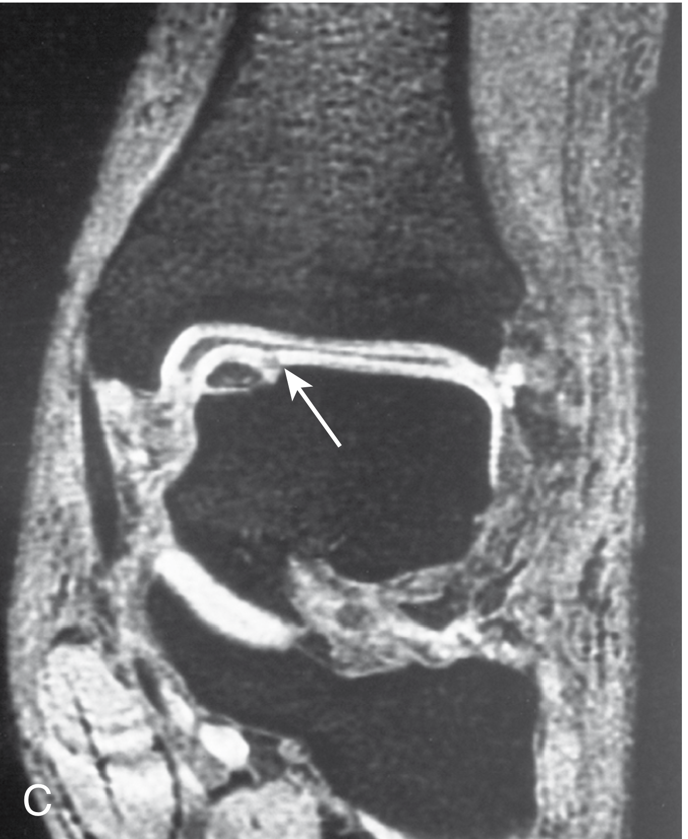
Arrow points to the area of low signal in the articular cartilage - Campbell's Operative Orthopaedics 15th Ed, Fig 3.8C
Summary Table
| Feature | Normal Cartilage | Injured Cartilage |
|---|---|---|
| Proteoglycan content | High | Depleted |
| Free water content | High (T2 bright) | Reduced |
| Collagen organization | Zonal, ordered | Disrupted |
| T2W signal | Intermediate-high (trilaminar) | Decreased / heterogeneous |
| Gradient-echo signal | Bright | Focally dark (defect) |
Key concept: MRI signal in cartilage depends primarily on mobile water content and collagen orientation. Injury disrupts proteoglycans (which hold water) and collagen architecture, both of which reduce T2 signal - making the injured area appear darker relative to the surrounding normal bright cartilage.
This is a shared conversation. Sign in to Orris to start your own chat.