
Generate a ct brain فلم اینڈ انڈسٹری will diagnose
آپ CT brain میں کون سی بیماری یا تشخیص دکھانا چاہتے ہیں؟ / Which diagnosis or condition should the CT brain image show?
You give me any and I will diagnose
CT brain axial scan with abnormal finding
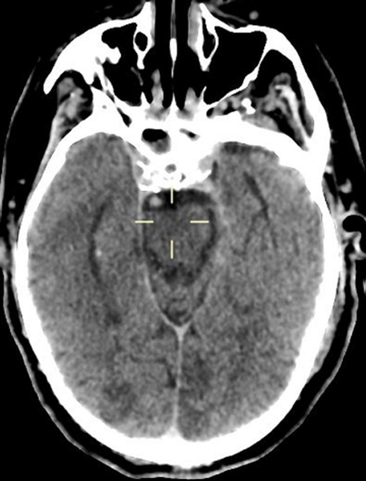
This diagnostic image is an axial non-contrast computed tomography (CT) scan of the brain. The scan reveals multiple areas of abnormal hypodensity located in the cortical and subcortical regions. These hypodense lesions are distributed bilaterally, predominantly involving the parietal and occipital lobes. The density of these areas is lower than that of the surrounding healthy brain parenchyma, which is characteristic of acute or subacute cerebral infarction. Anatomically, the image demonstrates the brain parenchyma, the midbrain/pons junction, and parts of the sphenoid sinus and orbits anteriorly. The falx cerebri is visible along the midline posteriorly. This finding is consistent with multifocal strokes, likely secondary to a systemic hypercoagulable state or cardioembolic event. The image serves as an educational example of identifying ischemic territories and evaluating bilateral cerebrovascular accidents in a critical care or emergency setting.
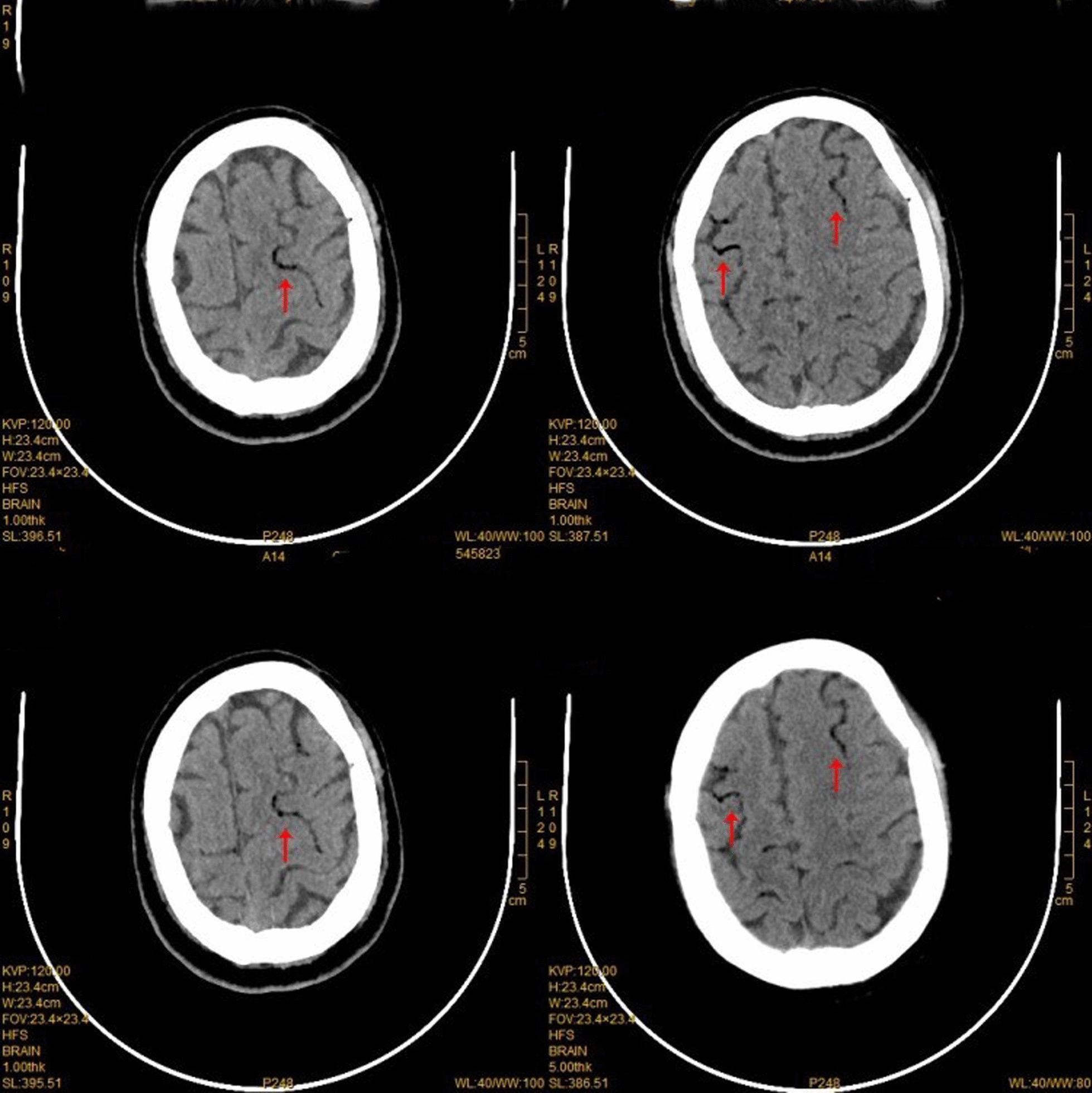
This diagnostic image consists of four non-contrast axial CT scan slices of the human brain, focusing on the superior frontal regions. The primary pathological finding is the presence of multiple, well-defined areas of abnormal lucency (low density) within the cortical blood vessels. These lucent pockets, marked by red arrows, follow the serpentine course of the sulci in the bilateral frontal lobes, appearing as dark, air-density streaks and bubbles contrasting sharply against the higher density of the blood and adjacent brain parenchyma. The distribution is predominantly cortical and peripheral. Clinically, these findings are pathognomonic for cerebral air embolism, demonstrating gas trapped within the distal cerebral vasculature. The image illustrates the disruption of normal anatomical density in a clinical emergency scenario, often occurring as a complication of thoracic procedures or trauma. The scans are useful for teaching the radiological identification of intravascular air and the diagnosis of iatrogenic or traumatic cerebral air embolism.
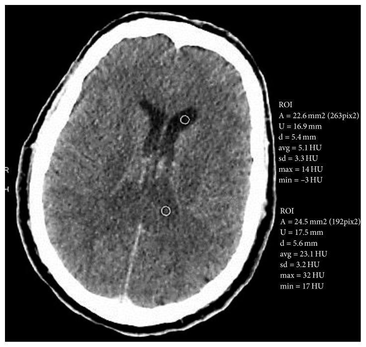
Diagnostic Imaging: This axial view of a cranial non-contrast computed tomography (CT) scan shows the internal structures of the brain with two defined regions of interest (ROI) for quantitative analysis. The primary finding is the presence of hyperdense material layered within the dependent portions of the posterior lateral ventricles. Quantitative ROI analysis reveals an average density of approximately 23.1 Hounsfield Units (HU) for this sediment (ROI #2), compared to 5.1 HU for normal cerebrospinal fluid (CSF) in the anterior horns (ROI #1). Morphologically, these hyperdense sedimentations demonstrate a fluid-debris level, a finding classically associated with intraventricular hemorrhage or the presence of inflammatory exudate (pus) in the context of ventriculitis. The brain parenchyma is visualized without obvious midline shift or mass effect. This image is medically significant for teaching the radiological identification of abnormal intraventricular densities and the use of Hounsfield Units to differentiate fluid types in neuroimaging, particularly in patients with suspected sepsis or coagulopathy.
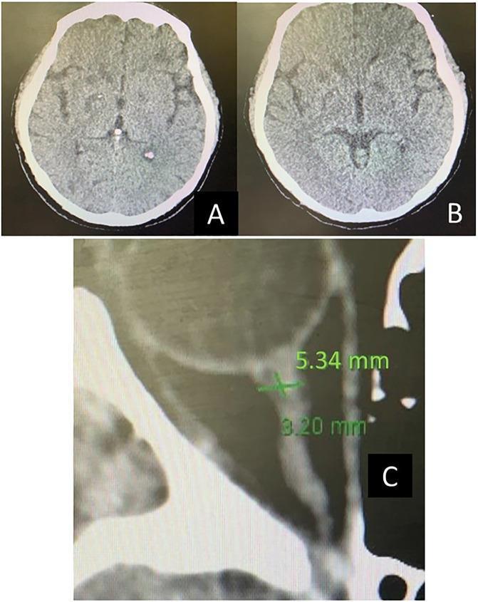
Educational clinical image panel consisting of axial non-contrast brain computed tomography (CT) scans and a diagnostic measurement of the ocular anatomy. Panel A shows an initial axial head CT with evidence of diffuse cerebral edema, characterized by a loss of gray-white matter differentiation and effacement of the cortical sulci. Panel B, a follow-up CT scan on day 4, demonstrates interval resolution of the edema, with improved visualization of brain parenchyma, sulcal spaces, and clearer gray-white matter boundaries. Panel C displays a zoomed diagnostic CT image of the orbit, illustrating a measurement of the optic nerve sheath diameter (ONSD). A transverse green caliper indicates an abnormal ONSD measurement of 5.34 mm, a finding clinically significant as an indirect marker for increased intracranial pressure (ICP). This set of images illustrates the radiological progression and monitoring of intracranial hypertension and brain compliance in an intensive care setting.
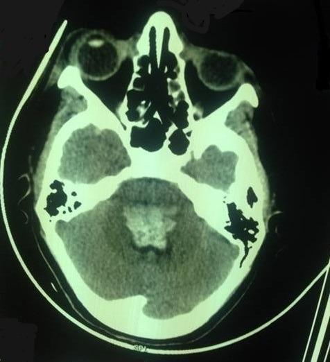
This diagnostic image is an axial non-contrast computerized tomography (CT) scan of the head at the level of the posterior fossa. The image displays the skull base, temporal bones, orbits, and paranasal sinuses. Key anatomical structures visible include the cerebellum and the brainstem (pons/medulla region). A significant abnormal finding is a centrally located, hyperdense area within the brainstem, consistent with an acute bulbo-protuberantial hematoma. The lesion measures approximately 32 x 21 x 29 mm and exerts a noticeable mass effect, resulting in the compression and effacement of the surrounding basal cisterns. The density variations clearly differentiate the high-attenuation bone of the skull, the intermediate-attenuation brain parenchyma, and the low-attenuation fluid within the remaining subarachnoid spaces. This clinical imaging is illustrative of severe neurological complications associated with conditions such as eclampsia, emphasizing the critical role of neuroimaging in evaluating sudden neurological deterioration in obstetric patients.
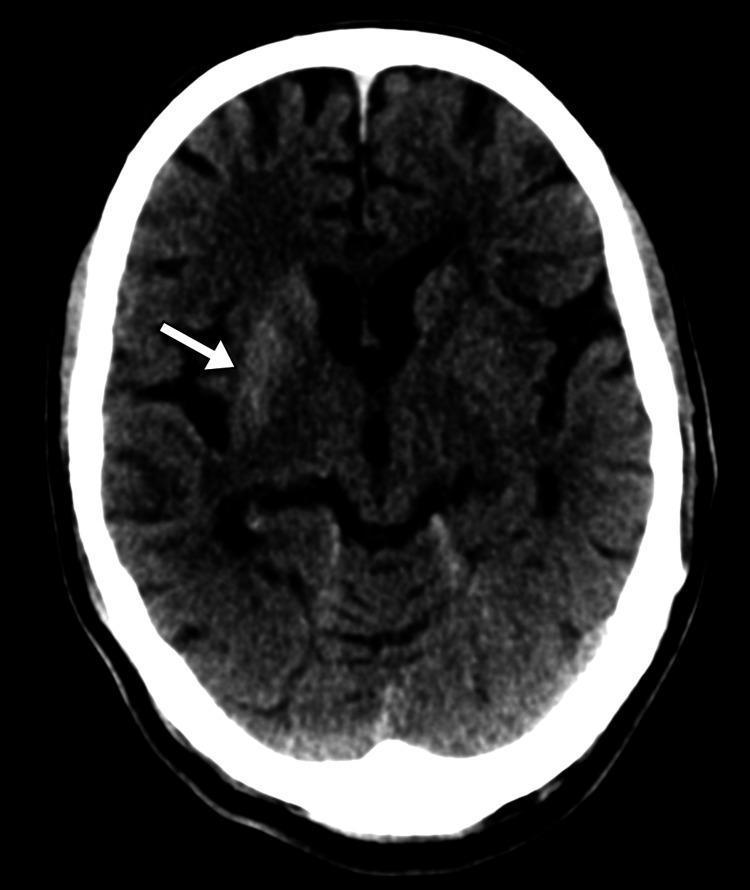
This diagnostic image is an axial non-contrast computed tomography (CT) scan of the brain. The primary finding is a localized area of hyperdensity within the right basal ganglia, specifically involving the caudate nucleus and putamen, as indicated by the white arrow. Despite this abnormal density, there is preserved gray-white matter differentiation elsewhere in the cerebral parenchyma. Crucially, there is no evidence of surrounding vasogenic edema, midline shift, or mass effect on the adjacent ventricular system. The intracranial space appears free of acute hemorrhage, and the skull vault remains intact. This radiological presentation is characteristic of non-ketotic hyperglycemic hemichorea (NHH), demonstrating the classic unilateral hyperdensity of the striatum on CT. The image serves as an educational example of how metabolic derangements can manifest as focal neuroradiological abnormalities without typical features of ischemia or space-occupying lesions.
- What is the density of the lesion - hyperdense (white) or hypodense (dark)?
- Where exactly is the abnormality located?
- Is there any midline shift, surrounding edema, or mass effect?
- Is there gray-white matter differentiation preserved or lost?
- What side is the lesion on?
Epidural haemorrhage
❌ Incorrect — The Diagnosis Was:
Non-Ketotic Hyperglycaemic Hemichorea (NHH)
- Hyperdense (bright white) lesion in the right basal ganglia - specifically the caudate nucleus and putamen (white arrow)
- No midline shift
- No surrounding edema
- No mass effect on the ventricles
- Preserved gray-white matter differentiation elsewhere
- No hemorrhage peripherally or along the skull
Why It's NOT Epidural Haemorrhage:
| Feature | Epidural Haemorrhage | This Scan |
|---|---|---|
| Location | Between skull & dura, peripheral | Deep basal ganglia, central |
| Shape | Biconvex / lenticular (lens-shaped) | Round, deep nucleus |
| Cause | Trauma / middle meningeal artery | Metabolic (hyperglycemia) |
| Edema | Often present | Absent |
| Midline shift | Common | None |
Now Here Is a Real Epidural Haemorrhage CT:
epidural hematoma CT scan biconvex lenticular hyperdense
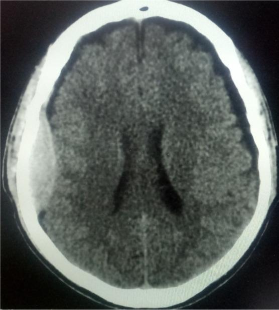
This diagnostic image is an axial slice from a non-contrast Computed Tomography (CT) scan of the head, representing a critical finding in neurosurgery and emergency medicine. The image demonstrates a classic biconvex (lenticular) hyperdense extra-axial collection in the right parietal region, characteristic of an acute epidural hematoma. The hyperdensity represents acute blood localized between the inner table of the skull and the dura mater. Notable mass effect is observed, evidenced by the inward compression of the underlying brain parenchyma and a subtle midline shift of the falx cerebri towards the left. The cerebral ventricles appear partially compressed. The high-density white outer rim represents the calvarium, while the brain tissue shows typical gray-white matter differentiation. This educational visual is used to teach the radiological hallmarks of intracranial hemorrhage following head trauma, specifically distinguishing the biconvex shape of an epidural hematoma from the crescent-shaped presentation of a subdural hematoma.
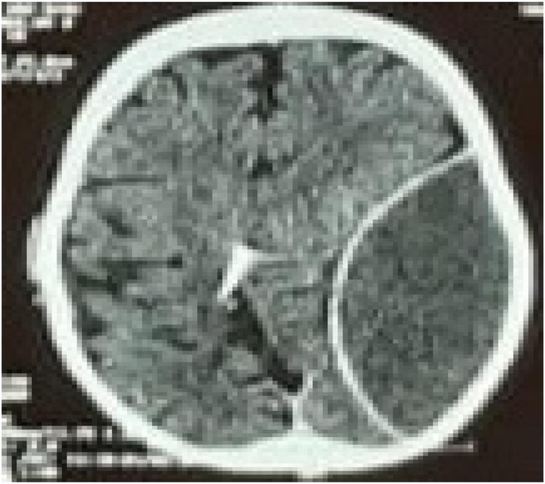
An axial non-contrast CT scan of the brain demonstrating a large, acute epidural (extradural) hematoma in the left hemisphere. The hematoma presents as a well-defined, biconvex (lenticular), hyperdense collection located between the skull and the dura mater. There is a significant mass effect evidenced by the compression of the left cerebral parenchyma, effacement of the adjacent cortical sulci, and midline shift toward the right. In the contralateral hemisphere, a radiopaque ventricular catheter is visible, extending into the posterior aspect of the right lateral ventricle near the occipital horn. This diagnostic image is representative of neurosurgical trauma emergencies, illustrating the classic appearance of arterial bleeding (typically from the middle meningeal artery) and secondary intracranial hypertension markers. The image serves as an educational resource for identifying acute traumatic intracranial hemorrhage and the presence of neurosurgical shunting devices.
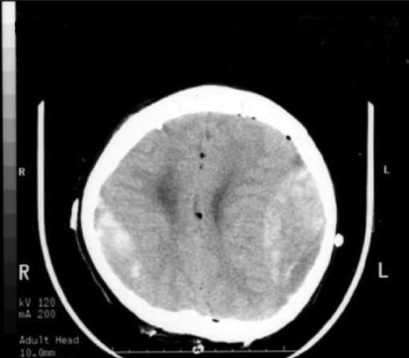
This diagnostic image is an axial non-contrast CT scan of the brain, demonstrating acute bilateral epidural hematomas (EDHs). In the frontoparietal regions, there are biconvex (lenticular) hyperdense extra-axial collections located adjacent to the inner table of the skull. The right-sided hematoma shows significant density, while the left-sided collection is also prominent, causing mild mass effect on the underlying brain parenchyma. Small punctate hypodensities are visible within the intracranial space, consistent with pneumocephalus (small air bubbles), which are typical in a postoperative clinical context following neurosurgical procedures like tumor resection. The lateral ventricles appear largely symmetric without significant midline shift or ventricular effacement at this level. This case illustrates a rare postoperative complication involving remote bilateral EDHs after surgical intervention for a pineal region tumor. The image is a critical educational resource for neurosurgery and radiology trainees to recognize acute intracranial hemorrhage and the importance of postoperative CT monitoring.
- Biconvex (lens-shaped) hyperdense collection hugging the inner skull
- Located peripherally between skull and dura
- Midline shift toward opposite side
- Caused by middle meningeal artery rupture after trauma
- Associated with lucid interval clinically
Key Learning Point:
When you see a deep, isolated basal ganglia hyperdensity with no edema and no midline shift - always think metabolic (hyperglycemia, hypernatremia) before hemorrhage. Epidural bleeds are always peripheral and lens-shaped.