
Collapse of lung lobes and thier findings in x ray and ct
Reading File
Lung lobe collapse (atelectasis) involves characteristic X-ray and CT findings that differ by lobe. The key signs include displacement of fissures, increased opacity, mediastinal/diaphragmatic shift, and compensatory hyperinflation of adjacent lobes.
Searching sources...
Finding Sources
Finding Sources
Finding Sources
Finding Sources
Finding Sources
Reading File
Reading File
Reading File
I have the main radiology content. Let me read the detailed lobe-by-lobe section and fetch the key images.
Reading File
Loading Image
Reading File
Loading Image
Reading File
Loading Image
I now have comprehensive material. Let me fetch a couple more key images to illustrate the answer well.
Loading Image
Loading Image
I have everything needed for a comprehensive, well-illustrated answer. Here it is:
Collapse of Lung Lobes: X-Ray and CT Findings
Source: Grainger & Allison's Diagnostic Radiology; Goldman-Cecil Medicine
General Principles of Lobar Collapse
Direct signs of volume loss:
- Displacement of fissures (the most reliable early sign)
- Crowding of pulmonary vessels and bronchi within the collapsed lobe
- Hilar elevation (upper lobe collapse) or hilar "smallness" (lower lobe collapse)
- Bronchial reorientation: the ipsilateral main bronchus becomes more horizontal in upper lobe collapse; more vertical in lower lobe collapse
Indirect signs:
- Compensatory hyperinflation of adjacent lobes (fewer, more widely spaced vessels)
- Mediastinal shift toward the collapsed lobe
- Diaphragmatic elevation (especially with lower lobe collapse)
- Displacement of the anterior junctional line contralaterally
A collapsed lobe appears dense due to retained secretions/fluid plus loss of aeration. Near-complete collapse is needed before the lobe becomes radiographically opaque.
Schematic Overview
This diagram shows the position of each collapsed lobe on frontal and lateral projections:
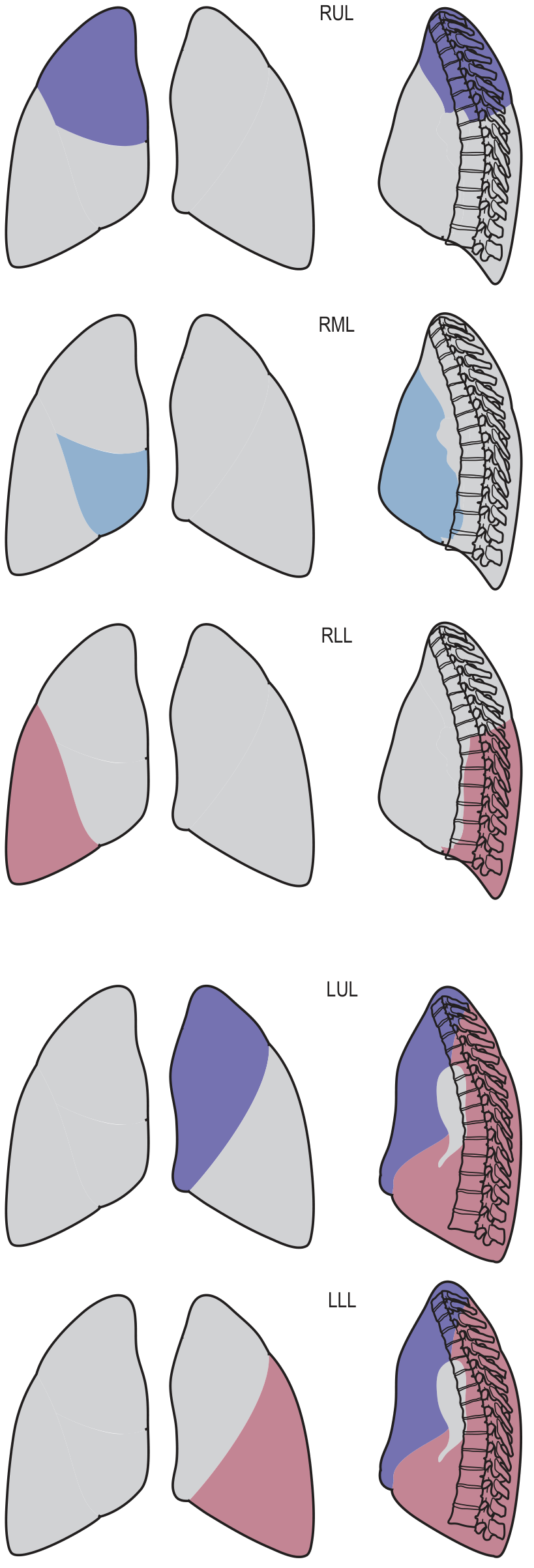
1. Right Upper Lobe (RUL) Collapse
X-Ray (PA):
- Upward displacement of the horizontal (minor) fissure
- Increased density in the right upper zone
- Right hilum elevation
- Tracheal deviation to the right
- Loss of right mediastinal border silhouette
- In "tight" collapse: a triangular wedge of soft tissue abuts the right mediastinum with a concave inferior margin (due to lower lobe hyperinflation) - the "Golden S-sign" when an underlying central mass causes the concavity
X-Ray (Lateral):
- Upward displacement of the horizontal fissure
- Anterior displacement of the upper part of the oblique fissure
CT:
- Triangular wedge of soft tissue anteriorly in the right hemithorax adjacent to the right mediastinum
- The collapsed lobe wraps around the superior mediastinum
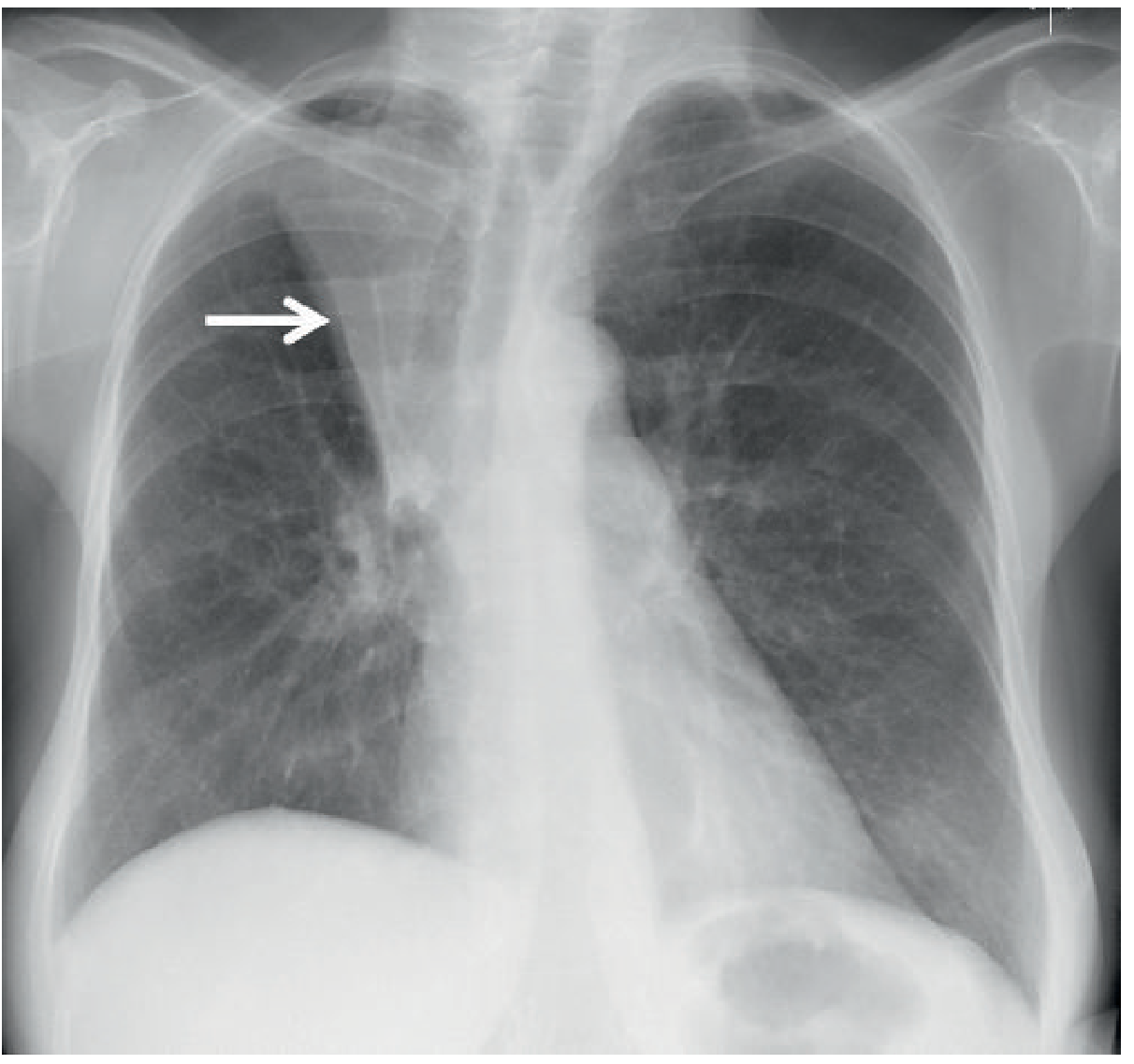
Fig. 7.19 - Tight Right Upper Lobe Collapse. The collapsed lobe (central bronchogenic carcinoma) results in increased right paramediastinal density (arrow).
2. Right Middle Lobe (RML) Collapse
X-Ray (PA):
- Ill-defined density at the right heart border (loss of right heart border silhouette - positive silhouette sign)
- Often subtle and easily missed
- Small triangular opacity in the right lower zone adjacent to the right heart border
X-Ray (Lateral):
- Wedge-shaped opacity between the horizontal and oblique fissures that converge anteriorly - the classic "bowtie" or "triangular" opacity with an anterior apex
CT:
- Band-like or wedge of soft tissue at the right cardiophrenic angle
- Fissures bow toward each other medially/anteriorly
3. Right Lower Lobe (RLL) Collapse
X-Ray (PA):
- Triangular density in the right lower zone medially, behind the heart
- Loss of silhouette of the medial right hemidiaphragm
- Right hilum "appears small" (interlobar artery obscured by opaque lobe)
- Right hemidiaphragm may be elevated
X-Ray (Lateral):
- Triangular density posteriorly
- Loss of the posterior right hemidiaphragm silhouette
- Vertebral column appears progressively denser inferiorly (important sign - normally the reverse is true)
- Posterior displacement of the oblique fissure
CT:
- Collapsed lobe lies in the posteromedial right hemithorax
- CT mucous bronchogram sign: tubular, low-attenuation branching airways within the enhancing collapsed lobe (on contrast-enhanced CT)
4. Left Upper Lobe (LUL) Collapse
This is distinctly different from RUL collapse because the left lung has no horizontal fissure.
X-Ray (PA):
- "Veil-like" increased density over the entire left hemithorax, greatest at the hilum (not a sharp margin)
- Loss of left heart border silhouette (lingula collapses anteriorly)
- Variable loss of the aortic knuckle silhouette
- Tracheal deviation to the left
- Left hilum elevation with increased angulation of the left main bronchus
- Luftsichel sign: a crescent of lucency (aerated superior segment of the left lower lobe) alongside the aortic knuckle, separating the aortic arch from the collapsed upper lobe
X-Ray (Lateral):
- Anterior displacement of the oblique fissure (toward the sternum)
- Increased retrosternal lucency (lower lobe hyperinflates anteriorly)
CT:
- Collapsed lobe forms a flat, broad band of soft tissue anteriorly against the chest wall and mediastinum
- Oblique fissure sweeps anteriorly
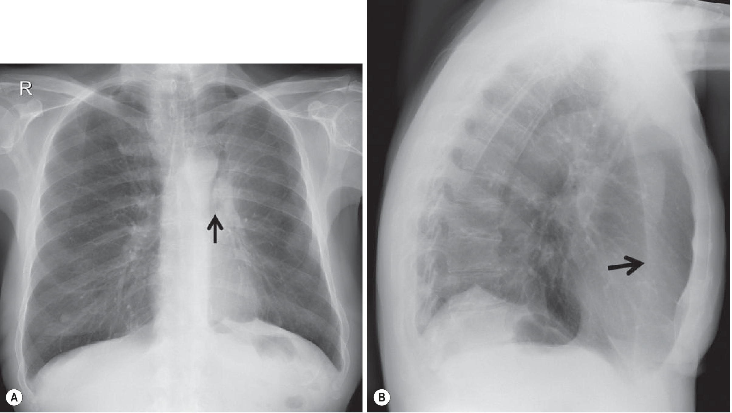
Fig. 7.21 - Left Upper Lobe Collapse. (A) Frontal view: increased angulation of left main bronchus (arrow). (B) Lateral view: anterior displacement of the oblique fissure (arrow) with increased retrosternal lucency.
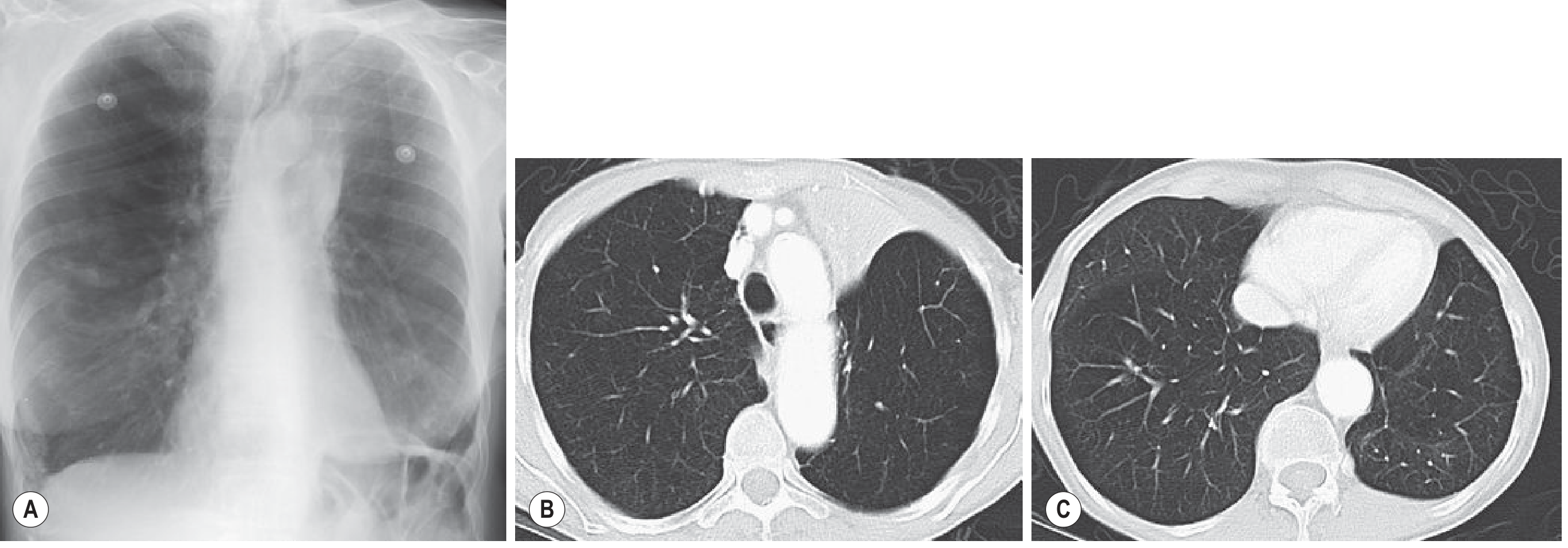
Fig. 7.22 - Atypical LUL Collapse on CXR and CT.
5. Left Lower Lobe (LLL) Collapse
Very similar in appearance to RLL collapse.
X-Ray (PA):
- Triangular density medially in the left lower zone (behind the heart)
- Loss of silhouette of the medial left hemidiaphragm
- Left hemidiaphragm may be elevated
- Loss of the descending aorta outline in severe collapse
X-Ray (Lateral):
- Triangular density posteriorly
- Loss of the posterior left hemidiaphragm silhouette
- Vertebral column appears progressively denser inferiorly
CT:
- Collapsed lobe in the posteromedial left hemithorax
- Oblique fissure displaced posteriorly and medially
6. Whole Lung Collapse
X-Ray:
- Complete opacification ("white-out") of the hemithorax
- Marked volume loss - ipsilateral mediastinal shift (this helps distinguish it from large pleural effusion, which shifts the mediastinum away)
- Contralateral lung expands across the midline anteriorly
- On lateral: marked anterior hyperlucency (contralateral lung displacement greatest anteriorly)
CT-Specific Signs Across All Lobes
| Sign | Significance |
|---|---|
| CT mucous bronchogram | Tubular low-attenuation airways in collapsed lobe - often indicates obstructive cause |
| Strong homogeneous enhancement | Favors atelectasis over consolidation/pneumonia (pneumonia shows less enhancement with air bronchograms) |
| Air bronchogram on CT | Does NOT exclude obstructive collapse (unlike plain film rules) |
| Fissure displacement | Best appreciated on coronal/sagittal reformats |
| Mediastinal anatomy | CT clarifies lymphadenopathy and causative masses |
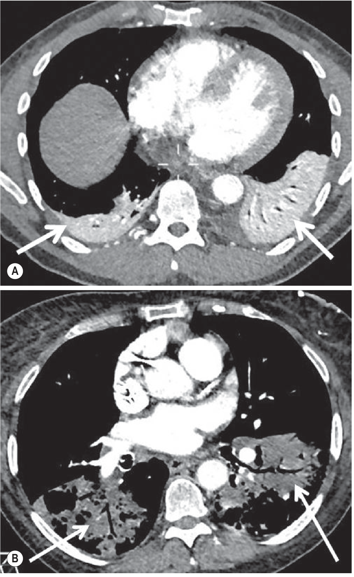
Fig. 7.12 - Enhancement pattern on contrast CT: Atelectasis (A) = dense homogeneous enhancement; Pneumonia (B) = relative lack of enhancement with air bronchograms.
Quick Summary Table
| Lobe | Fissure Movement | Opacity Location (PA) | Key Signs |
|---|---|---|---|
| RUL | Horizontal fissure - upward; upper oblique - anterior | Right upper zone / paramediastinal | Golden S-sign, hilar elevation, tracheal shift right |
| RML | Horizontal fissure - down; oblique - anterior/up | Right heart border (lateral wedge) | Right heart border silhouette loss, lateral wedge sign |
| RLL | Oblique fissure - posterior/medial | Behind heart, medial right lower zone | Medial diaphragm loss, vertebrae denser inferiorly |
| LUL | Oblique fissure - anterior | Veil over left hemithorax | Luftsichel sign, anterior fissure shift, retrosternal lucency |
| LLL | Oblique fissure - posterior/medial | Behind heart, medial left lower zone | Medial diaphragm loss, vertebrae denser inferiorly |
| Whole lung | All fissures invisible | Total hemithorax white-out | Ipsilateral mediastinal shift, anterior hyperlucency on lateral |
Grainger & Allison's Diagnostic Radiology, pp. 181-194; Goldman-Cecil Medicine, pp. 918-919
This is a shared conversation. Sign in to Orris to start your own chat.