
Neonate with jejunal atresia
Reading File
Finding Sources
Finding Sources
Searching PubMed
"jejunal atresia" AND management
Reading File
Reading File
Reading File
Reading File
Reading File
I have solid textbook coverage now. Let me get one relevant image to illustrate the classification/apple-peel appearance, then compose the answer.
Searching Images
jejunal atresia apple peel type radiograph triple bubble sign
Neonate with Jejunal Atresia
Overview and pathogenesis
Jejunoileal atresia is the most common gastrointestinal atresia, with an incidence of roughly 1 in 2,000-5,000 live births. Unlike duodenal atresia (a failure of recanalization), jejunoileal atresia is believed to result from a late intrauterine mesenteric vascular accident (segmental ischemic necrosis and resorption of a loop of bowel), rather than a primary defect in gut organogenesis - though disruption of signaling pathways (FGF, BMP, β-catenin) has also been implicated (Schwartz's Principles of Surgery, p. 171; Sabiston Textbook of Surgery, p. 3264).
Classification (Grosfeld/Louw system)
Jejunoileal atresias are divided into four (sometimes five) types (Sabiston, p. 3268):
- Type I - mucosal web/diaphragm with intact bowel wall and mesentery
- Type II - blind proximal and distal ends connected by a fibrous cord, mesentery intact
- Type IIIa - blind ends completely separated by a V-shaped mesenteric gap
- Type IIIb - "apple-peel" or "Christmas-tree" deformity: large mesenteric defect, absent segment, distal small bowel coiled around a single retrograde vessel from the ileocolic/right colic artery
- Type IV - multiple atresias ("string of sausages")
Type IIIb carries the highest risk of short bowel syndrome because of the tenuous retrograde blood supply and associated loss of bowel length.
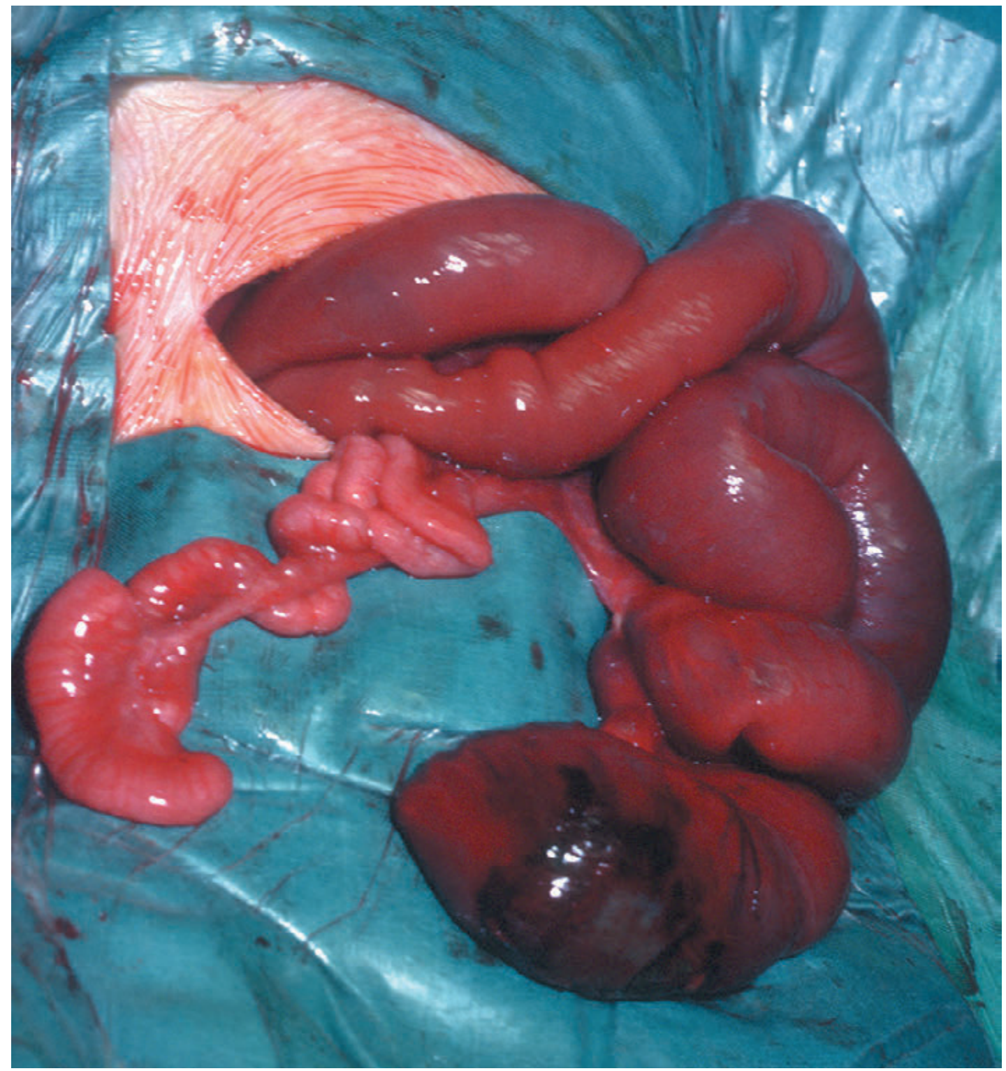
Clinical presentation
- Bilious vomiting is the dominant feature of jejunal atresia (more prominent than in ileal atresia, where abdominal distension predominates)
- Progressive abdominal distension
- A small amount of pale meconium may still be passed despite complete atresia
- Antenatally, maternal polyhydramnios is common with high (jejunal) obstruction, and prenatal ultrasound may show multiple dilated, fluid-filled bowel loops
- Multiple atresias occur in 10-15% of cases, so the entire bowel must be checked intraoperatively (with saline injection via a soft catheter distally)
Associated conditions/anomalies
Jejunoileal atresia is largely an isolated anomaly (unlike duodenal atresia, which associates with Down syndrome, cardiac and renal anomalies). The main association to screen for is cystic fibrosis, present in about 10% of cases. Meconium peritonitis can occur if in-utero perforation happens proximal to the atresia.
Diagnosis
- Plain abdominal radiograph: dilated, staggered air-fluid levels proximal to the obstruction (the higher the atresia, the fewer the loops)
- Contrast enema is not usually needed to diagnose jejunal atresia itself but helps exclude malrotation/other distal obstruction and typically shows an unused microcolon
- Prenatal ultrasound often first raises suspicion in the third trimester
Management
- Immediate stabilization: nasogastric/orogastric decompression, IV fluid resuscitation, correction of electrolytes, and NPO status
- Search for associated anomalies before surgery
- Operative repair once resuscitated:
- Resection of the dilated, dysmotile proximal bowel segment with primary end-to-end (or "end-to-back"/fish-mouthed) anastomosis
- If the proximal bowel is markedly dilated, a tapering enteroplasty is performed to match the caliber of the distal collapsed bowel and improve motility
- For multiple atresias (Type IV), multiple anastomoses may be required, sometimes over an endoluminal stent, with an emphasis on preserving maximal bowel length
- A temporary stoma is occasionally needed instead of primary anastomosis in unstable infants or heavily contaminated fields
Prognosis
Overall survival exceeds 90%. The main long-term risk, especially with Type IIIb (apple-peel) or Type IV disease, is short bowel syndrome from reduced intestinal length, requiring long-term nutritional support.
I ran a supplementary PubMed search for recent systematic reviews/meta-analyses specific to jejunal atresia management (last 3 years) and found no directly relevant high-tier evidence that would update or contradict the textbook-based management described above - the standard surgical approach (resection, tapering, primary anastomosis) remains current practice.
More information from neonatalogy side rather than surgical side
Finding Sources
Finding Sources
Finding Sources
Finding Sources
Finding Sources
Finding Sources
Finding Sources
Finding Sources
Reading File
Reading File
Reading File
Finding Sources
Searching PubMed
"intestinal atresia" AND "parenteral nutrition"
Searching the Web
jejunal atresia neonatal management NICU postoperative feeding TPN short bowel
Jejunal Atresia: The Neonatology Perspective
Shifting from the surgical repair itself to the medical/neonatal care around it - here is what matters from a neonatology standpoint, from antenatal counseling through NICU stabilization, perioperative medical management, and long-term follow-up.
1. Antenatal Recognition and Counseling
- Jejunal atresia is one of the few surgical anomalies reliably picked up on prenatal ultrasound - polyhydramnios plus multiple dilated, fluid-filled bowel loops with active peristalsis on third-trimester scans should raise suspicion (Creasy & Resnik's Maternal-Fetal Medicine, p. 2758; Pye's Surgical-Handicraft).
- MRI is not usually needed for jejunal/duodenal atresia the way it is for anorectal malformations, since ultrasound is already highly sensitive here.
- Antenatal identification allows the neonatal team to plan delivery at a tertiary center with NICU/pediatric surgery on site, arrange antenatal parental counseling (natural history, surgical plan, expected NICU course, risk of short bowel syndrome), and avoid an unplanned transfer of a vomiting, distended neonate.
- In-utero bowel perforation proximal to the atresia can cause meconium peritonitis, which may be visible antenatally as calcifications or ascites and changes the immediate postnatal risk profile (sepsis, adhesions).
2. Delivery Room / Immediate Postnatal Stabilization
- No specific mode of delivery is mandated by jejunal atresia alone; timing/route follow standard obstetric indications unless massive bowel dilation raises dystocia concerns.
- On the first feed or shortly after, the neonate develops bilious vomiting (the hallmark of jejunal-level obstruction, as opposed to the more distension-predominant picture of ileal atresia) - this is often the first clinical clue if antenatal diagnosis was missed.
- Immediate neonatal management once obstruction is suspected:
- NPO status
- Orogastric/nasogastric tube decompression on continuous or intermittent low suction to reduce aspiration risk and abdominal distension
- IV access and fluid resuscitation with attention to third-space losses and electrolyte derangements (vomiting/NG losses cause hypochloremic, hypokalemic metabolic alkalosis if prolonged)
- Temperature control/thermoregulation, as with any NICU admission
- Vitamin K, routine newborn screening, and standard NICU admission care in parallel
3. Diagnostic Workup (Neonatology-Driven)
- Plain abdominal radiograph is the first-line study - look for a limited number of dilated, air-fluid-filled loops (fewer, more proximal loops = more proximal/jejunal atresia; many loops = more distal disease).
- Contrast enema is used selectively to exclude distal causes (e.g., microcolon of unused bowel, Hirschsprung disease, meconium ileus) rather than to diagnose the atresia itself.
- Screen for associated conditions:
- Cystic fibrosis - present in roughly 10% of jejunoileal atresia cases; consider genetic testing/sweat testing once the infant is stable (Bailey and Love's Short Practice of Surgery, p. 7658).
- Look for maternal exposure history: jejunoileal atresia has been linked to maternal ergotamine or cocaine use and congenital rubella in pregnancy (Sleisenger and Fordtran's Gastrointestinal and Liver Disease, p. 1516).
- Unlike duodenal atresia, jejunoileal atresia is not strongly associated with Down syndrome or major cardiac/renal anomalies, but a general anomaly screen (cardiac exam/echo, renal ultrasound) is still reasonable given overlap in some series.
- Note the epidemiologic pattern: small bowel atresias are reported more often in Black infants, low-birth-weight infants, and twins (Sleisenger and Fordtran's, p. 1514).
4. Preoperative Medical Optimization
- The neonatology team's job before surgery is to correct dehydration and electrolyte abnormalities, ensure adequate urine output, treat hypoglycemia, and rule out sepsis (blood cultures/antibiotics if there are risk factors, given the possibility of in-utero perforation and meconium peritonitis).
- Coordinate anesthesia risk assessment - these are treated as urgent (not always emergent) cases once decompressed and resuscitated, with ongoing fluid/electrolyte correction continuing into the perioperative period (Barash, Cullen, and Stoelting's Clinical Anesthesia, p. 578).
5. Postoperative Neonatal/NICU Management
- Fluid and electrolyte management continues to be a primary neonatology responsibility postoperatively until GI losses resolve and enteral intake is established.
- Parenteral nutrition (TPN) is started early since the infant will be NPO for days to weeks:
- TPN calories are typically advanced to full goals over roughly 3-5 days
- IV lipid intake goal is around 3 g/kg/day, aiming for fat to supply around 40% of total calories
- Ongoing metabolic monitoring (electrolytes, triglycerides, liver function - watch for parenteral nutrition-associated cholestasis/liver disease with prolonged TPN) is essential
- Enteral feeding advancement begins once postoperative ileus resolves and bowel function returns (passage of stool, decreasing NG output). Feeds are advanced gradually and cautiously, with close monitoring for feeding intolerance, since the dysmotile dilated proximal bowel segment (even after tapering) may still have impaired peristalsis.
- Growth and nutritional surveillance: weight, length, head circumference trending, and micronutrient status (especially if TPN is prolonged).
6. Watching for Short Bowel Syndrome (SBS) - a Neonatology-Led, Long-Term Problem
- SBS is the dominant long-term neonatal medical concern after jejunal atresia repair, particularly with Type IIIb (apple-peel) or Type IV (multiple atresias) anatomy, where bowel length is already reduced at birth.
- SBS in the NICU is generally defined as a continued requirement for parenteral nutrition beyond about 3 months after bowel resection.
- Neonatology/nutrition teams manage this with graduated enteral feeds, consideration of continuous vs. bolus feeding strategies, and in some centers adjunct therapies (glutamine, modified diets, growth factors) to reduce TPN dependence, alongside close surveillance for TPN-associated liver disease, catheter-related bloodstream infections, and bacterial overgrowth in the residual bowel.
- Long-term follow-up should track growth, neurodevelopment (a known risk given prolonged NICU stay, TPN, and potential nutrient deficiencies), and gradual weaning from parenteral support if intestinal adaptation occurs over the following 1-2 years.
7. Family Counseling
Neonatologists (often together with pediatric surgery) counsel families about the expected NICU length of stay, the staged nature of feeding advancement, the possibility of a prolonged hospital course if SBS develops, and realistic expectations - overall survival for jejunoileal atresia exceeds 90%, but morbidity is meaningfully higher in apple-peel or multiple-atresia variants.
I also checked PubMed for recent (last 3 years) evidence specific to postoperative/nutritional management: a 2025 study on postoperative feeding patterns and GI reconstruction after neonatal intestinal atresia (PMID: 40291871) and a 2024 single-institution outcomes series (PMID: 39097495) are worth noting as current literature, though neither overturns the standard approach above - they support early, graduated enteral feeding advancement as favorable for GI functional recovery. No systematic review/meta-analysis-level evidence was found that contradicts this.
This is a shared conversation. Sign in to Orris to start your own chat.