
Treatment of fibrocystic disease
I'll read the medical library skill first, then search for fibrocystic disease treatment content in parallel.
Reading File
Now I'll search the library database and PubMed in parallel for fibrocystic disease treatment.
Finding Sources
Finding Sources
Searching PubMed
"fibrocystic breast disease" treatment management
Now I'll read the key sections from multiple textbooks in parallel.
Reading File
Reading File
Reading File
Now let me read the management section from Berek & Novak's and the answers section from Swanson's for treatment details.
Reading File
Reading File
Reading File
Let me also get the hormonal therapy section and the Bailey & Love section for drug details.
Finding Sources
Finding Sources
Reading File
Reading File
Reading File
Reading File
Reading File
I now have comprehensive information from multiple textbooks to compile a thorough answer on the treatment of fibrocystic disease. Let me synthesize it.
Treatment of Fibrocystic Disease (Fibrocystic Breast Changes)
Fibrocystic change represents a spectrum of benign breast conditions - cysts, stromal fibrosis, adenosis, epithelial metaplasia, and apocrine change - driven by an exaggerated breast response to hormonal cycling. The terms "fibrocystic disease," "mammary dysplasia," and ANDI (Aberrations of Normal Development and Involution) are used interchangeably in older and newer literature.
1. Reassurance First
The vast majority of patients require no specific treatment. Most patients need only reassurance that fibrocystic change is a transient phenomenon of aging, associated with hormonal effects on glandular breast tissue, that eventually subsides at menopause. Simple mastectomy or extensive breast tissue removal is not indicated.
- Berek & Novak's Gynecology, p. 982
- Sabiston Textbook of Surgery, p. 1384
2. Non-Pharmacological Measures
a. Supportive Brassiere
An appropriately sized, well-fitted support bra (including at night) is one of the most effective non-drug interventions. An ill-fitting bra is associated with a threefold risk of mastalgia. In a randomized trial, wearing a sports bra produced greater pain relief than hormonal modulation with danazol.
b. Caffeine Restriction
The role is controversial, but some studies suggest eliminating coffee, tea, and chocolate improves symptoms. Many patients self-report relief after caffeine withdrawal.
c. Diet and Weight
Maintaining a healthy body weight reduces estrogen load. Regular exercise (three times weekly) has been associated with improved quality of life in randomized controlled trials.
d. Cyst Aspiration
For symptomatic simple cysts, aspiration usually resolves the pain. Large cysts that rapidly recur after aspiration can be managed definitively with percutaneous vacuum-assisted excision. Cyst fluid does not need cytologic analysis unless it is bloody or a mass persists.
- Sabiston Textbook of Surgery, p. 1384
3. Vitamins and Supplements
These have been investigated but have insufficient evidence to draw clear conclusions:
| Supplement | Evidence |
|---|---|
| Vitamin E | Anecdotal benefit from biochemical changes in fibrocystic tissue; not confirmed in controlled trials |
| Vitamin B6 | Suggested due to biochemical effects on breast tissue; no robust clinical confirmation |
| Evening primrose oil (γ-linolenic acid, GLA) | Was studied for its effect on prostaglandin synthesis; failed to demonstrate efficacy over placebo in later trials, though safe and without significant side effects |
| Chamomile extract | Very small RCTs show symptom improvement; likely the safest non-hormonal supplement option, but true efficacy may be negligible |
- Berek & Novak's Gynecology, p. 984
4. Pharmacological Treatment (for Mastalgia)
Treatment is directed mainly at mastalgia (breast pain), which is the dominant symptom. A pain-score diary for at least 1 month helps classify pain as cyclic vs. non-cyclic and monitor response.
a. Hormonal Agents (First-Line for Moderate-Severe Pain)
| Drug | Mechanism | Dose | Notes |
|---|---|---|---|
| Danazol (Danocrine) | Synthetic androgen; suppresses pituitary gonadotropin, prevents LH surge, inhibits ovarian steroid formation | 100-200 mg twice daily, then taper to 100 mg/day | Only FDA-approved drug for mastalgia; significant androgenic side effects (acne, edema, voice change, weight gain, hirsutism, depression) - many patients discontinue |
| Tamoxifen | SERM (selective estrogen receptor modulator) | Low dose (10 mg/day); can be used topically | Effective for cyclic and noncyclic pain; off-label for mastalgia |
| Toremifene | SERM | - | Recognized treatment for mastalgia |
| Ormeloxifene | SERM | - | Recognized treatment; widely used in some regions |
| Bromocriptine | Dopamine agonist; inhibits prolactin (PRL) release | 2.5 mg twice daily for 3-6 months | Useful when TRH-induced elevated PRL is present; side effects: nausea, vomiting, headache |
| Depo-Provera (medroxyprogesterone) | Progestogenic; suppresses ovarian hormones | Injectable | Recognized for mastalgia; also reduces cyclic engorgement |
| OCP (combined oral contraceptive) | Suppresses cyclical hormonal fluctuation | Standard dosing | Reduces fibrocystic changes and fibroadenomas as a noncontraceptive benefit |
Note: GnRH analogs (e.g., Lupron/leuprolide) are not recommended for fibrocystic breast disease. They are effective for mastalgia but have significant side effects (bone loss, hypoestrogenism) and have no established role in fibrocystic disease management specifically.
- Berek & Novak's Gynecology, p. 990-991
- Sabiston Textbook of Surgery, p. 1384
- Bailey & Love's Short Practice of Surgery, 28th Ed., p. 941
b. NSAIDs
For extramammary or noncyclic chest-wall pain (e.g., Tietze syndrome, scapulothoracic bursitis), NSAIDs plus heat are recommended. Trigger point injections with local anesthetic and steroid along the medial scapular border are both diagnostic and therapeutic for referred scapulothoracic pain.
5. Surgical Intervention
Surgery is conservative and reserved for specific indications:
Biopsy is indicated when:
- No cyst fluid is obtained on aspiration
- Fluid is bloody or thick
- The cyst is complex or has an intracystic mass
- A mass persists after aspiration
- A suspicious mass fails to resolve at short-interval follow-up imaging
The primary surgical objective is to exclude carcinoma, not to treat fibrocystic change itself.
- Berek & Novak's Gynecology, p. 982
6. Cancer Risk Context
Treatment decisions must account for histologic subtype:
- Nonproliferative fibrocystic change - no increased cancer risk; reassurance is definitive treatment
- Proliferative change without atypia - ~2x cancer risk; surveillance
- Atypical ductal/lobular hyperplasia - ~5x cancer risk; closer follow-up warranted
- Family history + atypia - up to 11x increased risk
Cysts alone do not increase cancer risk. Hormonal therapies (danazol, tamoxifen) may offer secondary cancer risk reduction in high-risk groups.
Summary Algorithm
Fibrocystic Breast Changes
        |
        v
Exclude malignancy (exam + age-appropriate imaging ± biopsy)
        |
        v
Reassurance + lifestyle (bra support, caffeine restriction, exercise)
        |
        v
Mild-moderate pain → OCP / Vitamin E / supplements (anecdotal)
        |
        v
Moderate-severe cyclic mastalgia → Tamoxifen / Toremifene / Ormeloxifene
        |
        v
Severe, refractory → Danazol (FDA-approved) or Bromocriptine (if elevated PRL)
        |
        v
Symptomatic cysts → Aspiration (± excision if recurrent/complex)
        |
        v
Surgical biopsy only for indications above (never prophylactic mastectomy)
Sources:
- Berek & Novak's Gynecology (pp. 982-991) - Fibrocystic Change, Management of Fibrocystic Change, Mastalgia, Management of Mastalgia
- Sabiston Textbook of Surgery (pp. 1383-1385) - Fibrocystic Changes and Breast Pain
- Bailey & Love's Short Practice of Surgery, 28th Ed. (pp. 940-941) - ANDI, Mastalgia Treatment
- Swanson's Family Medicine Review - Fibrocystic breast changes Q&A
How to write clinical radiological and pathological correlation in a table in my study
Let me clarify what you need before diving in.
Asking for Preferences
Which condition or topic should the clinical-radiological-pathological correlation table cover? · What format do you want the table in?
The user skipped, so I'll provide a comprehensive answer: a well-structured clinical-radiological-pathological (CRP) correlation table for fibrocystic breast disease (continuing from the previous topic) in markdown format, plus a general guide on how to write such tables for any condition.
Clinical-Radiological-Pathological (CRP) Correlation Table
How to Structure a CRP Correlation Table
A CRP correlation table maps what the patient feels/shows → what imaging reveals → what the tissue looks like under the microscope. Each row represents a specific feature or finding, and columns run across all three domains so you can read horizontally and see the full picture of that one finding.
Standard Column Headers
| Feature | Clinical Finding | Radiological Finding | Pathological Finding |
|---|
You can add optional columns depending on depth needed:
- Significance / Implication (e.g., cancer risk, management trigger)
- Modality (which imaging - USG, mammography, MRI)
- Gross Pathology vs. Histology (split the pathology column if needed)
CRP Correlation Table: Fibrocystic Breast Disease
| Feature | Clinical Finding | Radiological Finding (Mammography / USG) | Pathological Finding (Gross + Histology) | Significance |
|---|---|---|---|---|
| Cyst formation | Soft, fluctuant, well-defined lump; may be tender; varies with menstrual cycle | USG: anechoic, well-defined, smooth-walled round/oval structure with posterior acoustic enhancement; Mammo: round/oval density | Gross: fluid-filled cavity (straw-colored, dark-green, or bloody fluid); lined by flattened cuboidal epithelium (type II) or apocrine epithelium (type I) | Cysts alone do NOT increase cancer risk; aspirate if symptomatic |
| Stromal fibrosis | Firm, diffuse breast nodularity; not well-defined; premenstrual tenderness | Mammo: dense, heterogeneous fibroglandular tissue; may obscure masses; USG: hyperechoic stromal tissue | Gross: firm white rubbery tissue; Histology: dense collagenous stroma replacing lobules | Causes dense breasts on mammography - increases false-negative rate |
| Adenosis | Diffuse nodularity; may be indistinguishable from carcinoma by palpation | Mammo: clustered microcalcifications (can mimic DCIS); USG: hypoechoic area or may be isoechoic | Histology: enlarged lobules with increased number of acini per lobule; myoepithelial cells preserved (key distinguishing feature from carcinoma) | Sclerosing adenosis: 1.5-2x cancer risk |
| Apocrine metaplasia | No distinct clinical finding; usually incidental | No specific imaging correlate | Histology: duct lining cells converted to large cells with abundant granular eosinophilic cytoplasm ("sweat-gland-like"); nuclei large with prominent nucleoli | Non-proliferative; no increased cancer risk |
| Epithelial hyperplasia (without atypia) | Not clinically palpable | May show calcifications; no specific imaging pattern | Histology: >2 cell layers lining ducts/acini; cells are regular, streaming pattern; no atypia | ~2x increased cancer risk |
| Atypical ductal hyperplasia (ADH) | Not clinically palpable | Mammo: clustered pleomorphic microcalcifications | Histology: proliferating ductal epithelium with some but not all features of low-grade DCIS; involves <2 duct spaces or <2 mm | ~5x cancer risk; ~11x if family history of breast cancer also present |
| Atypical lobular hyperplasia (ALH) | Not clinically palpable | No specific correlate; incidental finding at biopsy | Histology: cells filling and distending lobular acini; cells discohesive, small, uniform; similar to LCIS but involving <50% of acini in the lobule | ~5x cancer risk; bilateral risk |
| Ductal ectasia | Nipple discharge (thick, creamy, green); subareolar mass; nipple retraction in late stages | Mammo: tubular/rod-like calcifications (periductal, coarse); USG: dilated ducts with echogenic contents | Gross: dilated ducts filled with inspissated secretion; Histology: periductal chronic inflammation, plasma cells, fibrosis | May clinically mimic carcinoma; associated with smoking |
| Macromastia (severe) | Heavy, pendulous breasts; neck, shoulder, back, and breast pain | Not a radiological diagnosis; imaging to exclude pathology | Not a pathological entity per se | Pain is mechanical; reduction mammoplasty may be required |
| Cyclic mastalgia | Bilateral premenstrual breast pain, engorgement, tenderness; 3rd-4th decade | Normal imaging (no mass) - imaging done to exclude cancer | Histology: normal or shows stromal edema and epithelial proliferation in luteal phase | Benign; most resolves spontaneously; hormonal if severe |
Tips for Writing Your Own CRP Table (Any Disease)
-
Start with the pathology - list each histological subtype or lesion as a separate row. Pathology is the "ground truth" everything else maps to.
-
Work backwards clinically - for each pathological lesion, ask: what would the patient complain of? What would I feel on examination?
-
Map the imaging - for each lesion, ask: which modality best shows it (USG, CT, MRI, mammography, X-ray)? What is the specific imaging descriptor (echotexture, density, T1/T2 signal, enhancement pattern)?
-
Add a significance column - this is what makes the table useful for exams and clinical practice. Include: cancer risk, management trigger, differentials.
-
Use modality sub-columns when needed:
| Feature | Clinical | USG | Mammography | MRI | Histology | Risk |
|---|
- Color-code by risk tier if using Word/Excel:
- Green = nonproliferative (no increased risk)
- Yellow = proliferative without atypia (2x risk)
- Orange = atypical hyperplasia (5x risk)
- Red = carcinoma in situ (8-10x risk)
Key CRP Principle to Remember
Imaging is anatomy in vivo; pathology is anatomy ex vivo. The correlation works because both describe the same structural abnormality - one by its acoustic/magnetic/X-ray properties, the other by its cellular and architectural features.
If you want this table as a Word document, PDF, or Excel file to download, just let me know and I'll generate it for you.
Presentation of mastitis
Reading File
Now searching the library for mastitis presentation across multiple textbooks in parallel.
Finding Sources
Finding Sources
Excellent - multiple good sources found. Reading them all in parallel.
Reading File
Reading File
Reading File
Reading File
Good data so far. Now reading the Bailey & Love periductal mastitis section and Goldman-Cecil + granulomatous mastitis sections.
Reading File
Reading File
Reading File
Now reading the Bailey & Love periductal mastitis section which has the most detailed clinical content.
Reading File
Reading File
I now have comprehensive material from five textbooks. Let me fetch the clinical image of mastitis from Bailey & Love.
Loading Image
I have everything needed for a thorough, well-illustrated answer. Here it is:
Presentation of Mastitis
Mastitis is inflammation of the breast tissue, with or without infection. It is classified into distinct types based on setting, aetiology, and clinical behaviour - each with its own characteristic presentation.
Classification at a Glance
| Type | Who | Setting |
|---|---|---|
| Lactational (puerperal) mastitis | Breastfeeding mothers | Postpartum, most common |
| Non-lactational / periductal mastitis | Non-pregnant women, often smokers | Outside lactation |
| Granulomatous mastitis | Parous women, 4th-5th decade | Idiopathic / autoimmune |
| Tuberculous mastitis | Any, in endemic areas | Rare; secondary TB |
| Mastitis of infants | Neonates (both sexes) | First weeks of life |
1. Lactational (Puerperal) Mastitis
Who: Breastfeeding mothers. Incidence is 3-20% of lactating mothers. Most cases occur in the first month after delivery, particularly in first-time mothers, and again at weaning (due to engorgement and nipple trauma from baby's teeth).
Causative organisms:
- Staphylococcus aureus (most common) - causes abscess formation (single or multiple)
- Escherichia coli
- Streptococci - causes spreading cellulitis rather than abscess
- Hospital-acquired cases may be MRSA
Pathogenesis: Bacteria enter through cracked, fissured, or retracted nipples. Staphylococci cause milk to clot within the lactiferous ducts, then multiply within the clot. Duct blockage from epithelial debris causes milk stasis, further promoting infection.
Clinical Presentation:
| Feature | Description |
|---|---|
| Pain | Localized, severe, throbbing breast pain |
| Redness | Well-defined erythema, characteristically wedge-shaped (corresponding to a single lobe/segment) |
| Heat | Localized warmth and tenderness to touch |
| Swelling | Breast engorgement and swelling |
| Systemic symptoms | Flu-like illness - fever, chills, rigors, malaise |
| Progression to abscess | Fluctuant, tender lump; deep abscess may lack fluctuation; difficulty feeding |
| Axillary nodes | Enlarged, tender ipsilateral axillary lymph nodes |
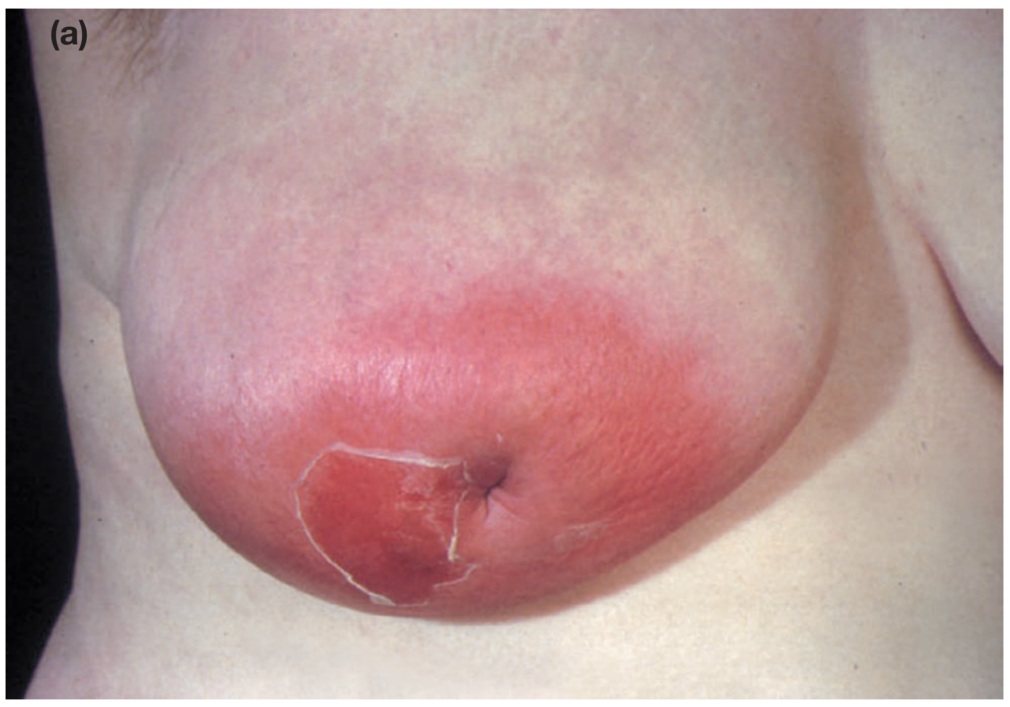
Acute mastitis showing erythema, swelling, and periareolar breakdown - Bailey & Love's Short Practice of Surgery, 28th Ed.
The initial stage is generalised cellulitis. If untreated, this progresses to suppuration and abscess formation.
2. Non-Lactational / Periductal Mastitis
Who: Non-lactating women; strongly associated with smokers (>90% of affected individuals are smokers). Also more common in women with large breasts, obesity, or previous breast surgery/radiation.
Pathogenesis: Chronic inflammation around the major subareolar milk ducts. Thought to be autoimmune in nature. Squamous metaplasia of lactiferous ducts can also cause a keratinous plug, ductal dilation, and eventual rupture with intense granulomatous periductal inflammation (Zuska disease / recurrent subareolar abscess).
Clinical Presentation:
| Feature | Description |
|---|---|
| Pain | Central, non-cyclical breast pain; located in the subareolar region |
| Mass | Tender, firm subareolar lump or abscess |
| Nipple discharge | Thick, purulent discharge; can be white, green, or bloody |
| Nipple retraction | Transverse, slit-like ("fish mouth") retraction - caused by fibrosis around major milk ducts |
| Abscess | Subareolar abscess; thick areolar muscle prevents perforation through areola, so pus tracks to areolar edge |
| Fistula | Mammary (milk) duct fistula - pus ruptures skin at the areolar edge forming a chronic fistulous opening |
| Mimics carcinoma | Chronic indurated subareolar mass with nipple retraction can be indistinguishable from cancer clinically |
Organisms: Staphylococci, enterococci, anaerobic streptococci, Bacteroides, and occasionally mycobacteria.
Key distinguishing sign: The transverse slit-like nipple retraction (like a fish's mouth) distinguishes periductal mastitis from carcinoma, which causes vertical or irregular nipple retraction.
3. Granulomatous (Idiopathic / Lobular) Mastitis
Who: Parous women, often in the 4th-5th decade. Associated with hyperprolactinaemia; otherwise idiopathic.
Clinical Presentation:
- Painful inflammatory phlegmon of the breast (indurated, poorly defined mass)
- Often tender, with overlying skin erythema and ulceration
- May present like multiple recurrent abscesses
- Frequently confused with carcinoma - both clinically and radiologically
- Occurs most often in parous women (96% association with prior pregnancy)
- Diagnosis requires core needle biopsy showing non-caseating granulomata surrounding breast lobules
- Culture must be sent to exclude tuberculosis
Management: Usually resolves within 9-12 months with observation alone. Steroids or methotrexate may be used. Surgical excision reserved for complicated/refractory cases.
4. Mastitis in Non-Lactating Women (Goldman-Cecil Summary)
The typical non-lactating presentation (usually in the 4th decade):
- Acute onset of severe breast pain and tenderness
- Followed by erythema and swelling, predominantly in the nipple-areolar area
- Cause: rupture of dilated subareolar ducts → inflammatory response to intraductal contents leaking into periductal tissue
Critical differential: The most important differential diagnosis for mastitis is inflammatory breast carcinoma. Failure to improve with antibiotics within 7-10 days mandates breast surgeon evaluation and imaging to exclude abscess or underlying cancer.
5. Tuberculous Mastitis
- Rare; occurs in areas where TB is endemic
- Presents as a cold, indurated breast mass with sinus formation
- Overlying skin may be discoloured or ulcerated
- Diagnosis: GeneXpert MTB/RIF testing on pus or biopsy; culture for AFB
- No acute inflammatory signs (non-tender, indolent course)
6. Mastitis of Infants
- Occurs in neonates (both boys and girls) in the first weeks of life
- Presents as warm, tender, erythematous enlargement of the neonatal breast bud
- Almost exclusively caused by S. aureus
- Rare but can progress to abscess
Comparison Table: Lactational vs. Periductal vs. Granulomatous
| Feature | Lactational Mastitis | Periductal Mastitis | Granulomatous Mastitis |
|---|---|---|---|
| Setting | Breastfeeding | Non-lactating | Non-lactating, parous |
| Age | Postpartum (any age) | Reproductive age | 4th-5th decade |
| Risk factors | Cracked nipple, poor latch | Smoking, obesity | Parity, hyperprolactinaemia |
| Pain | Severe, acute, localised | Central, non-cyclical | Chronic, diffuse phlegmon |
| Location | Wedge-shaped lobe | Subareolar | Peripheral/lobular |
| Nipple | Normal or cracked | Slit-like retraction, purulent discharge | May retract |
| Abscess | Common, often peripheral | Subareolar | Recurrent, may fistulate |
| Fistula | Milk fistula if I&D done | Mammary duct fistula at areolar edge | Skin ulceration |
| Systemic signs | Fever, flu-like, malaise | Minimal systemic upset | Minimal |
| Organism | S. aureus, streptococci | Mixed flora, anaerobes | Sterile (idiopathic) |
| Cancer mimic | Low risk | High (indurated mass + nipple retraction) | High (phlegmon) |
Key Investigations
- Ultrasonography: Cellulitis = increased echogenicity; abscess = hypoechoic collection with floating debris (changes with posture); thickened ducts in periductal mastitis
- Mammography: Usually deferred in acute phase; done to exclude malignancy once infection resolves
- Pus culture & sensitivity: Always; include GeneXpert for TB in endemic areas
- Core needle biopsy: Required when granulomatous mastitis or carcinoma is suspected
- Milk culture (midstream): For recurrent lactational mastitis
Sources:
- Bailey & Love's Short Practice of Surgery, 28th Ed. (pp. 947-949) - Lactational and periductal mastitis
- Creasy & Resnik's Maternal-Fetal Medicine (p. 252) - Lactational mastitis in breastfeeding
- Goldman-Cecil Medicine (p. 4499) - Mastitis in lactating and non-lactating women
- Robbins, Cotran & Kumar Pathologic Basis of Disease (p. 963) - Acute mastitis, squamous metaplasia of lactiferous ducts
- Berek & Novak's Gynecology (p. 983) - Granulomatous mastitis
- Current Surgical Therapy, 14th Ed. (p. 760) - Granulomatous mastitis management
This is a shared conversation. Sign in to Orris to start your own chat.