
Mucosal melanoma
Reading File
Now let me search the medical library and PubMed in parallel for comprehensive coverage.
Finding Sources
Searching PubMed
"mucosal melanoma"[MeSH Terms] AND treatment
Searching the Web
mucosal melanoma 2025 2026 treatment guidelines immunotherapy
Good hits. Let me now read the main textbook sources in parallel.
Reading File
Reading File
Reading File
Searching PubMed
"mucosal melanoma" AND immunotherapy
Let me get more content from the surgical and clinical sources, plus fetch the key clinical image.
Reading File
Reading File
Loading Image
Searching PubMed
"mucosal melanoma" AND (nivolumab OR pembrolizumab OR "anti-PD1")
Excellent - I have rich content. Let me get the key diagnostic/histology section from Fitzpatrick's and grab the abstract for the 2026 pembrolizumab + lenvatinib trial.
Finding Sources
Reading Article
PMID: 42140948
Reading Article
PMID: 39782303
Now let me read the AJCC staging table from Cummings to complete the staging section.
Reading File
I now have comprehensive content. Let me compose the full answer.
Mucosal Melanoma
Mucosal melanoma is a rare, aggressive subtype of melanoma arising from melanocytes residing in mucosal surfaces. It accounts for only ~1.3% of all melanomas but carries a disproportionately poor prognosis due to late presentation, lack of visible lesions, and a distinct molecular biology that limits response to therapies developed for cutaneous disease.
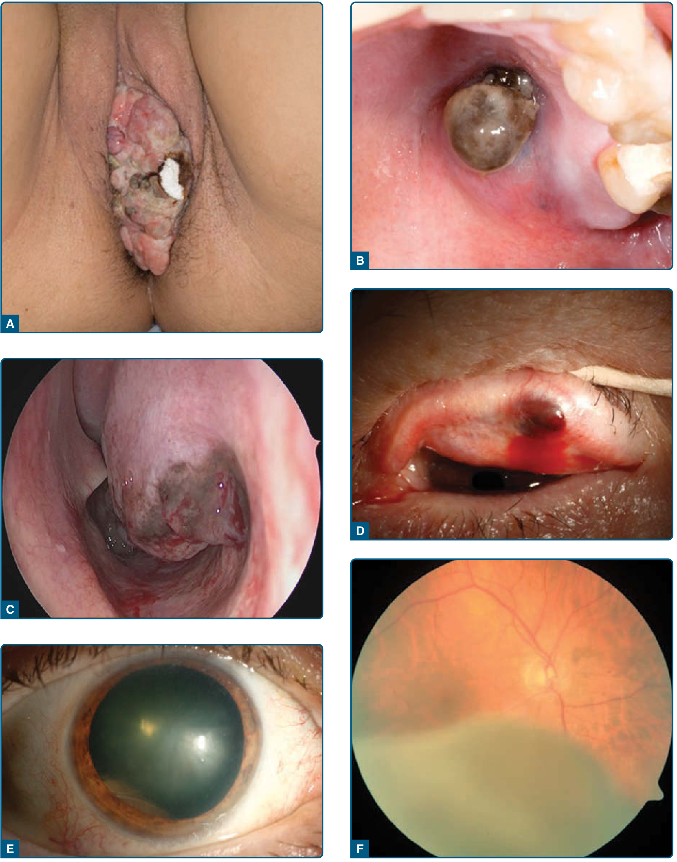
Sites of Origin
| Site | Notes |
|---|---|
| Head & neck (55% of cases) | Nasal cavity most common, then paranasal sinuses, oral cavity (palate most common), nasopharynx, larynx |
| Vulva / vagina | ~50% of all mucosal melanomas in women; highest BRAF/KIT mutation frequency |
| Anorectal | ~16.5% of mucosal melanomas; presents with bleeding, pain, rectal mass |
| Urethral | Rare |
| Conjunctiva | Visible early; higher frequency in patients with atypical nevi |
Head and neck prognosis: nasal cavity (best) > oral cavity > paranasal sinuses (worst). - K.J. Lee's Essential Otolaryngology, p. 810-812
Epidemiology and Risk Factors
- More frequent in women (largely due to vulvovaginal primaries)
- Affects a wide age range (20-80 years); no consistently identified environmental risk factors
- Rare in most populations; relatively higher incidence in East Asian and Sub-Saharan African populations compared to cutaneous melanoma
- No known environmental trigger analogous to UV radiation in cutaneous melanoma
- Represents 0.5-3% of all melanomas across all sites - Scott-Brown's Otorhinolaryngology, p. 488
Molecular Biology
Mucosal melanoma is molecularly distinct from cutaneous melanoma:
| Mutation | Frequency | Notes |
|---|---|---|
| BRAF | <10% overall; up to 26% vulvovaginal | Contrast with ~50% in cutaneous melanoma |
| NRAS / KRAS (RAS alterations) | 5-30% | Activate MAPK pathway similar to BRAF |
| NF1 | Most frequent driver overall | |
| c-KIT (receptor tyrosine kinase) | 5-20% | Higher in vulvovaginal and anorectal sites; targetable with imatinib, though mutation status does not predict survival |
Low BRAF mutation frequency means BRAF inhibitors (e.g., vemurafenib, dabrafenib) are generally not applicable for most mucosal melanomas. - Fitzpatrick's Dermatology, p. 2017
Clinical Presentation
- Typically presents as a pigmented, sessile or raised mass - colors range from light tan to black depending on melanin content
- ~40% are amelanotic (no pigment) - making clinical recognition difficult - Current Surgical Therapy 14e
- Common symptoms at specific sites:
- Nasal/sinonasal: epistaxis, nasal obstruction, facial pain, polyp-like mass
- Oral cavity: pigmented gingival or palatal mass, bleeding, loosening of teeth
- Anorectal: rectal bleeding, pain, change in bowel habits, palpable mass
- Vulvovaginal: bleeding, pruritus, pigmented lesion
- Disease is often indolent early with vague symptoms, leading to advanced-stage presentation at diagnosis
- Oral/sinonasal lesions: ulceration and bone infiltration are common; 20% are in situ lesions at diagnosis
Any suspicious mucosal pigmented lesion should be biopsied - the threshold should be low. - Cummings Otolaryngology, p. 1786
Histopathology
- Cells are epithelioid and/or spindle cell type; plasmacytoid, rhabdoid, small cell, giant cell, neurotropic, and desmoplastic variants are recognized
- Markedly pleomorphic nuclei with prominent nucleoli
- Intranuclear inclusions are typical
- Cells may contain melanin pigment (but may be absent in amelanotic tumors)
- Adjacent inflammatory infiltrate and necrosis common
- Background mucosa may show melanocytic atypia or melanoma in situ
- Immunohistochemistry: positive for S100, HMB-45, Melan-A/MART-1, SOX10 - Scott-Brown's Otorhinolaryngology, p. 488
Staging (AJCC 8th Edition - Head & Neck Mucosal Melanoma)
A unique feature: due to the inherently aggressive biology, all mucosal melanomas are classified T3 or higher; there is no T1 or T2 designation.
| Stage | T | Description |
|---|---|---|
| T3 | III minimum | Mucosa-limited disease |
| T4a | IVA | Moderately advanced: involvement of deep soft tissue, cartilage, bone, overlying skin |
| T4b | IVB | Very advanced: brain, dura, skull base, lower cranial nerves, masticator space, carotid artery, prevertebral space, mediastinal structures |
Distant metastasis = Stage M1 / Stage IVC.
Depth of invasion (as used in cutaneous melanoma Breslow thickness) has not been shown to predict prognosis in mucosal melanoma. - Cummings Otolaryngology; K.J. Lee's
Diagnosis and Workup
- Biopsy of any suspicious pigmented (or amelanotic) mucosal lesion
- Imaging: CT and/or MRI of primary site to assess bone erosion, deep tissue extension; PET-CT for systemic staging
- Molecular profiling: BRAF, NRAS, KIT mutation testing (guides targeted therapy eligibility)
- Sentinel lymph node biopsy (SLNB): used selectively; utility debated for head/neck mucosal primary
- Regional lymph node assessment: half of clinically palpable nodes are tumor-positive; ultrasound-guided FNA is useful
Worst histologic prognosis among paranasal sinus malignancies - worse than SCC, adenocarcinoma, or minor salivary gland tumors. - K.J. Lee's Essential Otolaryngology
Treatment
Surgery
- Wide surgical resection remains the primary treatment
- Negative margins are the goal but are often difficult to achieve anatomically
- Therapeutic neck dissection for clinically positive nodes
- Elective neck dissection is not recommended
- Endoscopic resection increasingly used for sinonasal disease at experienced centers
Radiation
- Adjuvant radiation at high dose (>54 Gy), standard fractionation, improves local-regional control but does not consistently improve OS
- Used postoperatively especially for positive/close margins and advanced T stage
- Hypofractionation protocols also employed
Systemic Therapy
Despite surgical control, most patients eventually develop distant metastases - the major cause of death.
Immunotherapy (checkpoint inhibitors):
- Anti-PD-1 agents (nivolumab, pembrolizumab) are the mainstay for advanced/metastatic disease
- Response rates are lower in mucosal melanoma (~20-30%) compared to cutaneous melanoma (~40-45%)
- Combination nivolumab + ipilimumab most commonly used in practice
- NCCN 2026 guidelines: adjuvant nivolumab is a category 2B option for resected mucosal melanoma; data are far fewer than for cutaneous disease
A 2025 retrospective study of 52 patients with sinonasal mucosal melanoma found overall survival at 1/2/5 years of 86.9%/74.1%/39.1%, with ~50% having a local response to immunotherapy but rare improvement at metastatic sites.
Anti-VEGFR / tyrosine kinase inhibitors:
- Combination of anti-PD-1 + lenvatinib (or axitinib) is gaining traction for mucosal melanoma given higher c-KIT and angiogenic signaling
- A 2026 phase II trial (Nat Commun) of neoadjuvant pembrolizumab + lenvatinib in resectable mucosal melanoma reported a 38.1% pathologic response rate (pCR 9.5%), median RFS 14.8 months; spatial profiling showed activated CD4+/CD8+ T cell signatures predicted response
- Axitinib + nivolumab studied in a phase 2 trial (ASCO 2025 data)
c-KIT inhibitors:
- Imatinib and other multi-kinase inhibitors are targetable against KIT-mutated mucosal melanoma, but KIT mutation status does not correlate with survival outcomes
Cytotoxic chemotherapy:
- Cisplatin/temozolomide: NCCN 2026 category 2B adjuvant option
- Lower efficacy compared to immunotherapy; used in refractory disease
Emerging:
- Tumor-infiltrating lymphocyte (TIL) therapy (lifileucel): 2025 data from C-144-01 shows activity in anti-PD-1 refractory mucosal melanoma
- Ceralasertib (ATR inhibitor) + durvalumab: promising in anti-PD-1-failed mucosal melanoma
- First-line lenvatinib + pembrolizumab (LEAP-003 China subset) has relevant mucosal melanoma data
Prognosis
| Setting | Approximate Outcome |
|---|---|
| Pharyngeal/oropharyngeal mucosal melanoma (5-year OS) | ~13% |
| Sinonasal mucosal melanoma (5-year OS) | ~20-35% |
| Stage 1 cutaneous melanoma (10-year OS) for comparison | >95% |
| Metastatic melanoma (all types, pre-immunotherapy era) | <20% |
Overall prognosis remains poor. Outcomes may be improving in the immunotherapy era but dedicated mucosal melanoma data are still limited. - Cummings Otolaryngology, Fitzpatrick's Dermatology, Current Surgical Therapy 14e
Key Differentiating Points vs. Cutaneous Melanoma
| Feature | Mucosal Melanoma | Cutaneous Melanoma |
|---|---|---|
| BRAF mutation | <10% (except vulvovaginal ~26%) | ~50% |
| KIT mutation | 5-20% | <5% |
| Staging starts at | T3/Stage III | T1/Stage I |
| UV exposure link | None | Primary cause |
| Immunotherapy response | Lower (~20-30%) | Higher (~40-45%) |
| Prognosis (stage-for-stage) | Worse | Better |
Recent evidence note: A 2026 review in Current Treatment Options in Oncology summarizes current status - key emerging strategies include anti-VEGFR combinations, ATR inhibition, and TIL therapy for this difficult-to-treat disease. NCCN 1.2026 guidelines recommend systemic therapy per the metastatic cutaneous melanoma algorithm for unresectable/metastatic mucosal disease, while adjuvant options remain category 2B.
This is a shared conversation. Sign in to Orris to start your own chat.