
is microfillare of wucheria bancroftei a larve
Microfilariae of Wuchereria bancrofti are not true larvae - they are actually pre-larval stage (sometimes called L1 stage) embryos.
Searching sources...
Reading File
Finding Sources
Reading File
Reading File
Loading Image
Finding Sources
Great - here is a thorough answer based on authoritative medical microbiology textbooks.
Are Microfilariae of Wuchereria bancrofti Larvae?
Short answer: Microfilariae are NOT true larvae. They are pre-larval embryos (sometimes called "pre-larvae" or the first embryonic stage).
Here is the breakdown:
What Are Microfilariae?
Microfilariae are the embryonated offspring produced by adult female W. bancrofti worms. The process works like this:
- The gravid female produces large numbers of embryonated eggs
- At oviposition (egg laying), the embryo uncoils to its full length (200-300 µm) inside the egg shell
- The egg shell elongates and is retained as a thin, flexible sheath
- This sheathed embryo is what we call a microfilaria ("small thread")
So a microfilaria is essentially an embryo inside a stretched egg shell (sheath) - not a true larva.
(Sherris & Ryan's Medical Microbiology, 8th Edition)
The Distinction: Embryo vs. Larva
| Feature | Microfilaria | True Larva |
|---|---|---|
| Stage | Pre-larval embryo | Post-embryonic developmental stage |
| Location | Peripheral blood (human host) | Mosquito (intermediate host) |
| Development | Not yet undergone larval molts | Has undergone ≥1 molt |
| Infective? | No - cannot directly infect humans | L3 stage is infective |
Life Cycle Context
The microfilariae circulate in the human bloodstream. To become true larvae, they must be taken up by a mosquito (the intermediate host/vector). Inside the mosquito:
- L1 larva - microfilaria develops after being ingested by mosquito
- L2 larva - first molt inside mosquito flight muscles
- L3 larva (infective stage) - migrates to mosquito's proboscis; this is the true infective larva that enters a new human host during a blood meal
So the progression is:
Embryo (egg) → Microfilaria (pre-larva in blood) → L1 → L2 → L3 (infective larva in mosquito)
Characteristics of W. bancrofti Microfilariae
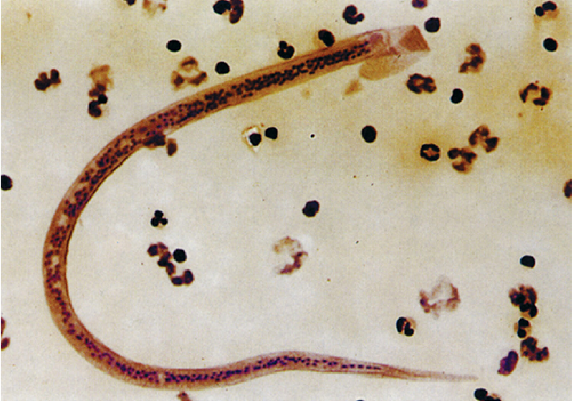
Microfilaria of W. bancrofti in blood film - Sherris & Ryan's Medical Microbiology, 8th Ed.
| Feature | Detail |
|---|---|
| Size | 245-295 µm long × 7-10 µm wide (in blood smear) |
| Sheath | Present (key distinguishing feature) |
| Tail nuclei | Absent - nuclei do NOT reach the tail tip |
| Periodicity | Nocturnal (peak in peripheral blood: 9 PM - 2 AM) |
| Location in human | Peripheral blood (and pulmonary vessels during daytime) |
(Medical Microbiology 9e; Sherris & Ryan's Medical Microbiology 8e)
Why the Confusion?
Some older texts loosely call microfilariae "larvae" or "first-stage larvae (L1)" because they are the first mobile offspring. However, strictly speaking:
- Microfilariae are pre-larval - they have not yet undergone any larval molt
- The true L1 stage only begins inside the mosquito after the microfilaria is ingested
- The term "larva" is more correctly reserved for post-embryonic, post-molt stages in the mosquito
Summary: Microfilariae of W. bancrofti are embryos (pre-larvae) circulating in human blood inside a sheath (stretched egg shell). They become true larvae only after being ingested by a mosquito, going through L1 → L2 → L3 stages. The L3 is the infective larval stage transmitted back to humans.
wucheria bancrofti complete microbiology
Searching sources...
Finding Sources
Finding Sources
Reading File
Reading File
Reading File
Reading File
Reading File
Reading File
Reading File
Loading Image
Loading Image
Finding Sources
Reading File
Loading Image
Here is the complete microbiology of Wuchereria bancrofti, compiled from multiple authoritative medical textbooks.
Wuchereria bancrofti - Complete Microbiology
1. Classification
| Feature | Detail |
|---|---|
| Kingdom | Animalia |
| Phylum | Nematoda (roundworms) |
| Superfamily | Filarioidea ("thread-like") |
| Class | Tissue nematode |
| Disease caused | Lymphatic filariasis (Bancroftian filariasis) |
2. Morphology
Adult Worms
- Long, slender, thread-like white worms coiled in lymphatic vessels
- Female: ~100 mm (10 cm) long
- Male: ~40 mm long
- Adults live coiled together in lymphatic vessels for up to 10 years
- Both sexes harbor endosymbiotic Wolbachia bacteria in their gut, which are required for normal reproduction and metabolism
Microfilariae
The gravid female produces large numbers of embryonated eggs. At oviposition, the embryo uncoils inside the elongating egg shell (retained as a thin flexible sheath) to become a microfilaria.
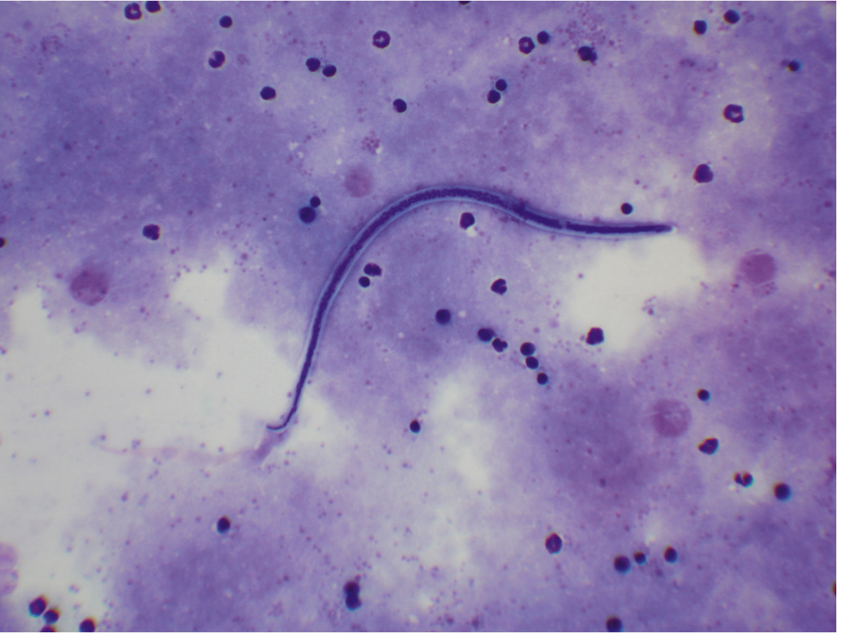
| Feature | W. bancrofti | B. malayi (for comparison) |
|---|---|---|
| Location | Blood | Blood |
| Sheath | Present | Present |
| Size | 245-295 µm × 7-10 µm | 180-230 µm × 5-6 µm |
| Tail nuclei | Absent (nuclei do NOT reach tip) | Present (2 distinct nuclei at tip) |
| Sheath staining (Giemsa) | Does not stain pink | Stains bright pink |
| Periodicity | Usually nocturnal | Nocturnal/subperiodic |
3. Life Cycle
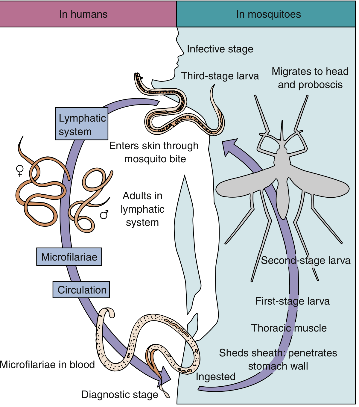
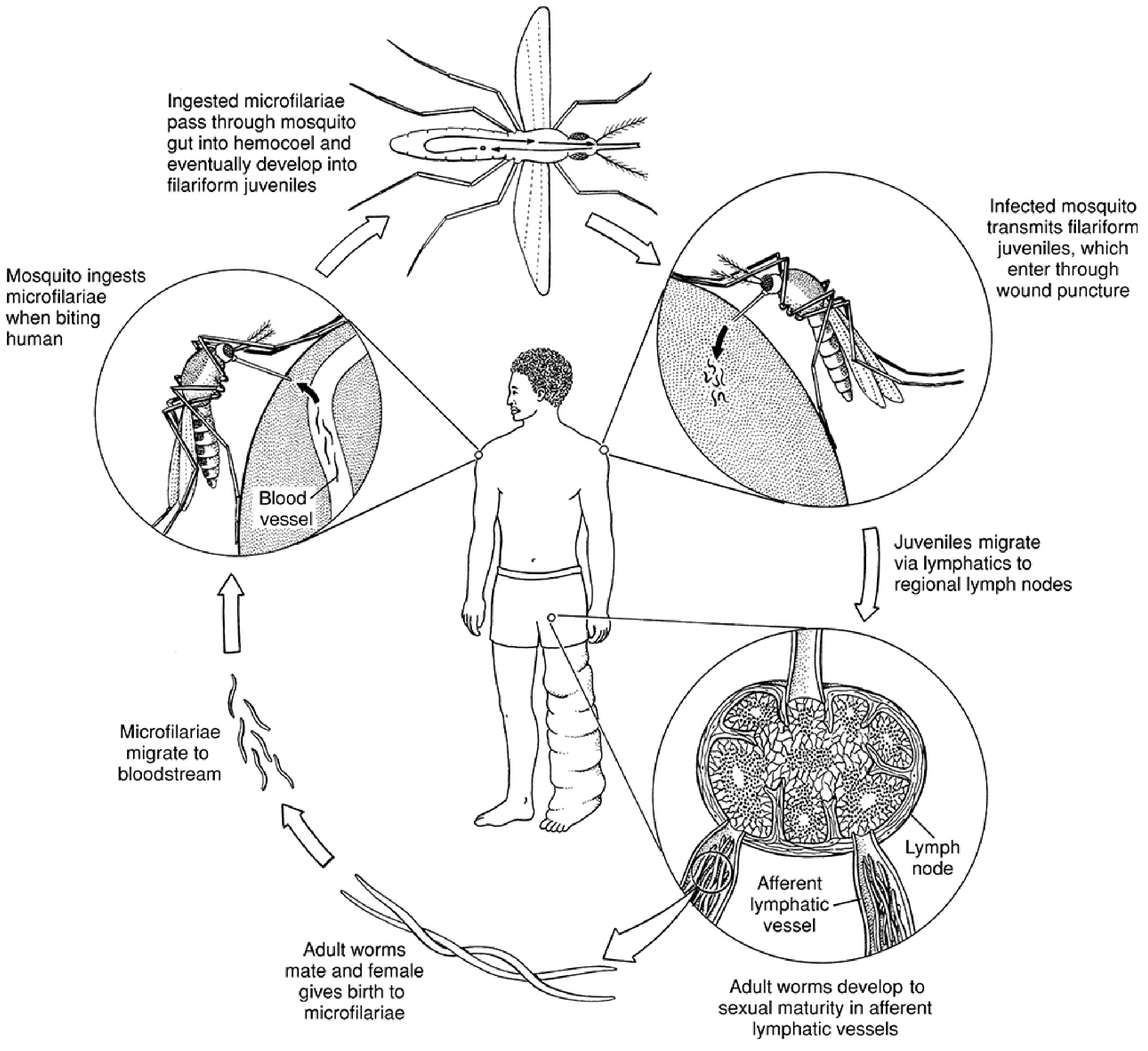
In the Human Host
- Infected mosquito bites → deposits L3 (infective) larvae into the skin wound
- L3 larvae migrate to the lymphatic vessels (arms, legs, groin)
- Larvae undergo molts and mature into adult worms over 6-12 months
- Adults mate; gravid females produce microfilariae → microfilariae enter lymph → reach peripheral blood
- Adults persist for up to 10 years
In the Mosquito (Intermediate Host/Vector)
- Mosquito ingests microfilariae during a blood meal
- Microfilariae shed their sheath and penetrate the mosquito stomach wall
- Develop in thoracic flight muscles: microfilaria → L1 (rhabditiform) → L2 → L3 (filariform/infective)
- L3 larvae migrate to the proboscis (mouthparts)
- Transmitted to a new human host during the next blood meal
Vector Mosquitoes
- W. bancrofti: transmitted by Culex, Anopheles, and Aedes mosquitoes
- No animal reservoir for W. bancrofti (humans are the only definitive host)
4. Epidemiology
- Infects approximately 120 million people in 73 countries
- Endemic in: central Africa, Mediterranean coast, Asia (India, China, Korea, Japan, Philippines, Malaysia), parts of Caribbean and South America (Haiti, Brazil, Trinidad)
- Concentrated in poorly sanitized, densely crowded urban and tropical/subtropical areas
- B. malayi (related species) has animal reservoirs (cats, monkeys); W. bancrofti does not
5. Pathology and Pathogenesis
Mechanism
Adult worms in lymphatic vessels are the primary cause of inflammatory and fibrotic reactions. Two phases:
Acute Phase
- Presence of molting adolescent worms and dying adults triggers:
- Dilatation of lymphatics
- Hyperplastic endothelial changes
- Lymphatic infiltration by lymphocytes, plasma cells, and eosinophils
- Thrombus formation (acute lymphangitis)
- Granuloma formation and fibrosis follow
- Recurring acute episodes of fever, lymphadenitis, lymphangitis, chills
Chronic Phase
- Repeated infections → permanent lymphatic obstruction
- Lymphedema, ascites, pleural effusion, hydrocele, joint effusion
- Elephantiasis: grotesque enlargement of extremities, scrotum, breasts from massive lymphatic blockade + skin thickening/fibrosis
- Dilated lymphatics may rupture → abscess, draining sinuses, chyluria (lymph in urine)
- Bacterial and fungal superinfections worsen tissue damage
Tropical Pulmonary Eosinophilia (TPE)
- Rare syndrome (< 1% of infected)
- Immune hyperresponsiveness to microfilariae trapped in the lungs
- Features: nocturnal paroxysmal cough/wheezing, weight loss, low-grade fever, marked blood eosinophilia (≥3000/µL), elevated IgE
- More common in young adult males
- Microfilariae usually absent from blood in TPE
- Responds rapidly to DEC
6. Clinical Syndromes
| Presentation | Features |
|---|---|
| Asymptomatic microfilaremia | Microfilariae present, no symptoms |
| Acute adenolymphangitis | Fever, tender lymph nodes, lymphangitis (centrifugally spreading down limbs) |
| Filarial fever | Low-grade fever, recurs over weeks/months |
| Orchitis / epididymitis | Bancroftian filariasis specifically involves testicular, spermatic cord lymphatics |
| Hydrocele | Common chronic manifestation |
| Elephantiasis | Massive limb, genital, breast enlargement (irreversible) |
| Chyluria | Lymph in urine (milky urine) from ruptured lymphatics |
| Tropical Pulmonary Eosinophilia | Nocturnal cough, wheeze, marked eosinophilia |
7. Laboratory Diagnosis
Direct Detection
| Method | Details |
|---|---|
| Peripheral blood film (Giemsa) | Gold standard - detects sheathed microfilariae; blood collected at night (10 PM - 2 AM) for nocturnal periodicity strains |
| Buffy coat examination | Concentrates microfilariae for better detection |
| Membrane filtration technique | Anticoagulated blood filtered through 5-µm membrane; washed, stained - detects small numbers |
| Thick blood film | More sensitive than thin film |
Indirect / Molecular
| Method | Details |
|---|---|
| Circulating antigen detection | ICT cards (immunochromatographic test) - sensitivity 96-100%, specificity ~98%; can be done any time of day; not FDA-approved in USA |
| Serology (antifilarial antibodies) | Available at reference labs; not species-specific |
| NAAT / PCR | Most sensitive; detects parasite DNA; no commercial platform yet |
| Eosinophilia | Usually present in acute episodes |
| Ultrasound | "Filarial dance sign" - live adult worms seen moving in dilated lymphatics |
Species Differentiation of Microfilariae
- W. bancrofti: sheathed, tail nuclei absent, sheath does not stain pink with Giemsa
- B. malayi: sheathed, 2 distinct nuclei in tail, sheath stains bright pink with Giemsa
8. Treatment
Drug of Choice
Diethylcarbamazine (DEC) - 6 mg/kg/day
- Mechanism: stimulates cholinergic receptors → muscle depolarization → worm paralysis; also enhances leukocyte adherence to microfilariae
- Kills microfilariae primarily; also has activity against adult worms
- Caution: Destruction of microfilariae releases Wolbachia antigens → inflammatory reaction (Mazzotti-like reaction); always screen for co-infection with Loa loa (risk of encephalopathy) and Onchocerca volvulus before starting DEC
Alternative/Adjunct Drugs
| Drug | Role |
|---|---|
| Ivermectin | Single dose reduces microfilariae for up to 6 months; fewer side effects than DEC |
| Albendazole | Used in combination for mass drug administration programs |
| DEC + Albendazole or DEC + Ivermectin | Used in WHO global elimination programs |
| Doxycycline | Targets Wolbachia endosymbionts → sterilizes/kills adult worms over weeks; used as adjunct |
Mass Drug Administration (MDA)
WHO Global Programme to Eliminate Lymphatic Filariasis (GPELF) uses annual MDA with DEC + albendazole (or ivermectin + albendazole in onchocerciasis co-endemic areas) to interrupt transmission.
9. Prevention and Control
- Vector control: insecticide spraying, larval source reduction (Culex breeding sites)
- Personal protection: mosquito nets, repellents, protective clothing
- Mass drug administration (MDA): annual single-dose treatment of entire at-risk populations
- No animal reservoir for W. bancrofti - human-to-mosquito-to-human transmission only
10. Key Distinguishing Facts (High-Yield Summary)
| Point | Detail |
|---|---|
| Causative agent of | Bancroftian filariasis (lymphatic filariasis) |
| Adult worm habitat | Lymphatic vessels |
| Adult worm lifespan | Up to 10 years |
| Diagnostic stage | Microfilariae in blood |
| Microfilaria features | Sheathed, no tail nuclei, 245-295 µm |
| Infective stage | L3 larva (in mosquito proboscis) |
| Vector | Culex, Anopheles, Aedes mosquitoes |
| Periodicity | Nocturnal (blood collected at night) |
| Animal reservoir | None (humans only) |
| Endosymbiont | Wolbachia bacteria |
| Drug of choice | DEC (diethylcarbamazine) |
| Classic chronic sign | Elephantiasis |
| Pulmonary complication | Tropical Pulmonary Eosinophilia |
Sources: Sherris & Ryan's Medical Microbiology 8e; Medical Microbiology 9e (Murray); Jawetz, Melnick & Adelberg's Medical Microbiology 28e; Katzung's Basic & Clinical Pharmacology 16e; Goldman-Cecil Medicine
electron transport chain
The electron transport chain (ETC) is a series of protein complexes in the inner mitochondrial membrane that transfer electrons from NADH/FADH2 to oxygen, driving ATP synthesis via oxidative phosphorylation. It consists of Complexes I-IV plus ATP synthase (Complex V).
Searching sources...
Finding Sources
Reading File
Reading File
Reading File
Reading File
Reading File
Finding Sources
Reading File
Reading File
Reading File
Loading Image
Loading Image
Loading Image
Finding Sources
Finding Sources
Loading Image
Excellent - I have all the material needed. Here is the complete, textbook-based answer on the Electron Transport Chain.
The Electron Transport Chain (ETC)
1. Overview and Location
The ETC is a series of protein complexes embedded in the inner mitochondrial membrane that transfer electrons from reduced coenzymes (NADH and FADH2) to molecular oxygen (O2), producing water. This electron flow releases free energy, which is used to pump H+ across the membrane, creating a gradient that drives ATP synthesis - a process called oxidative phosphorylation (OXPHOS).
"Energy-rich molecules such as glucose are metabolized by oxidation reactions yielding CO2 and H2O. Metabolic intermediates donate electrons to NAD+ and FAD to form NADH and FADH2. These reduced coenzymes donate electron pairs to the ETC. As electrons pass down the ETC, they lose free energy. This energy moves H+ across the inner mitochondrial membrane, creating a H+ gradient that drives ATP production."
- Lippincott Illustrated Reviews: Biochemistry, 8e
2. Mitochondrial Structure (Context)
| Compartment | Features |
|---|---|
| Outer membrane | Contains porin channels; freely permeable to small molecules and ions |
| Intermembrane space | Protons (H+) accumulate here after pumping; similar composition to cytosol |
| Inner membrane | Highly impermeable to H+, ATP, ADP, pyruvate; contains ETC complexes and ATP synthase; folded into cristae to increase surface area; >50% protein by mass |
| Matrix | Contains TCA cycle enzymes, β-oxidation enzymes, NAD+, FAD, ADP, Pi; site of ATP synthesis |
3. The Four Complexes of the ETC
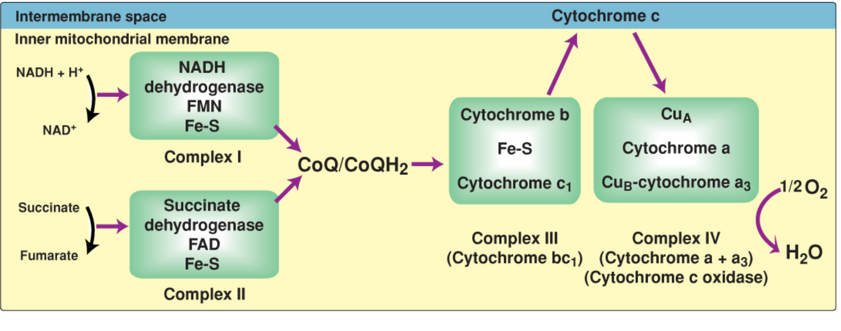
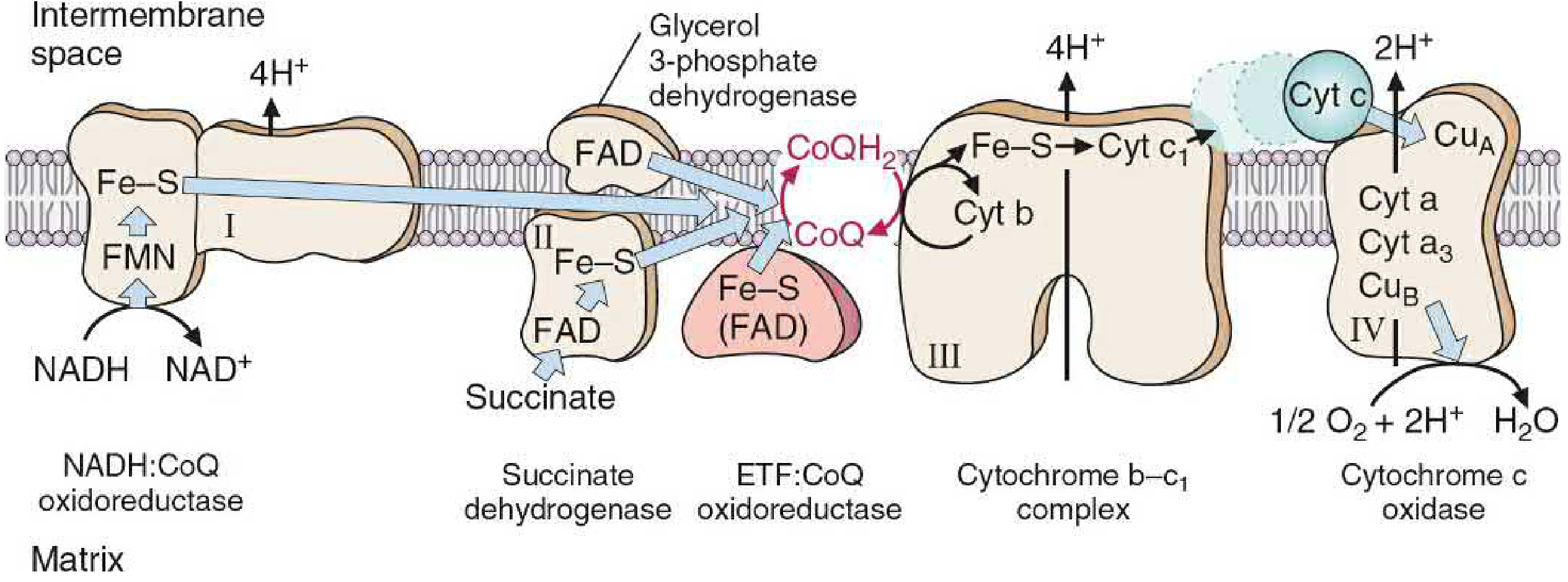
Complex I - NADH Dehydrogenase (NADH:CoQ Oxidoreductase)
- Electron donor: NADH (from TCA cycle, β-oxidation, glycolysis)
- Components: FMN (flavin mononucleotide), multiple Fe-S centers, ~45 subunits
- Reaction: NADH + H+ → NAD+ (oxidation) | FMN → FMNH2 → Fe-S → CoQ (reduced to CoQH2)
- Protons pumped: 4 H+ from matrix to intermembrane space per NADH
- Electron path: NADH → FMN → Fe-S centers → CoQ
Complex II - Succinate Dehydrogenase
- Electron donor: FADH2 (from oxidation of succinate → fumarate in TCA cycle)
- Components: FAD, Fe-S centers
- Reaction: Succinate → Fumarate; FADH2 transfers electrons to CoQ
- Protons pumped: 0 (no H+ pumping at Complex II - insufficient energy)
- Also accepts electrons from: glycerol-3-phosphate dehydrogenase, ETF:CoQ oxidoreductase (fatty acid oxidation)
- Electron path: FADH2 → Fe-S → CoQ
Coenzyme Q (CoQ / Ubiquinone) - Mobile Carrier
- A lipid-soluble quinone with a long hydrophobic isoprenoid tail (from cholesterol synthesis pathway)
- Freely diffuses within the inner membrane
- Accepts electrons from both Complex I and Complex II (and other flavoprotein dehydrogenases)
- Transfers electrons to Complex III
- Functions as a junction point linking flavoprotein dehydrogenases to cytochromes
- Accepts 2 electrons and 2 H+ from the matrix side → becomes CoQH2 (ubiquinol)
Complex III - Cytochrome bc1 (CoQ:Cytochrome c Oxidoreductase)
- Components: Cytochrome b, Fe-S protein, Cytochrome c1
- Reaction: CoQH2 donates electrons to cytochrome c via the Q cycle
- Protons pumped: 4 H+ per pair of electrons
- Passes electrons one at a time (single-electron transfers via Fe in cytochromes)
Cytochrome c - Mobile Carrier
- A small protein loosely associated with the outer face of the inner membrane (in the intermembrane space)
- Accepts single electrons from Complex III, carries them to Complex IV
- Iron in heme group cycles between Fe2+ (reduced) and Fe3+ (oxidized)
Complex IV - Cytochrome c Oxidase (Cytochrome a + a3)
- Components: CuA, Cytochrome a, Cytochrome a3, CuB
- The only complex that directly reacts with O2
- Reaction: 4 electrons from 4 cytochrome c molecules + 4H+ + O2 → 2 H2O
- Protons pumped: 2 H+ per pair of electrons (total 4 H+ per O2 molecule reduced)
- Electron path within Complex IV: cytochrome c → CuA → cyt a → cyt a3 (with CuB) → O2
4. Summary of Electron Flow and H+ Pumping
NADH → Complex I → CoQ → Complex III → Cyt c → Complex IV → O2 → H2O
↑
FADH2 → Complex II
| Complex | H+ Pumped | Electron Donor | Electron Acceptor |
|---|---|---|---|
| I (NADH dehydrogenase) | 4 H+ | NADH | CoQ |
| II (Succinate dehydrogenase) | 0 | FADH2 | CoQ |
| III (Cytochrome bc1) | 4 H+ | CoQH2 | Cytochrome c |
| IV (Cytochrome c oxidase) | 2 H+ | Cytochrome c | O2 |
| Total per NADH | 10 H+ | - | - |
| Total per FADH2 | 6 H+ | - | - |
5. ATP Synthesis - Chemiosmotic Hypothesis (Mitchell Hypothesis)
The pumping of H+ from matrix to intermembrane space creates:
- A chemical gradient (pH gradient) - intermembrane space is more acidic
- An electrical gradient - intermembrane space is more positive
Together these form the proton-motive force (PMF), which drives H+ back into the matrix through ATP synthase (Complex V).
ATP Synthase (Complex V / F1F0-ATPase)
- F0 domain - spans the inner membrane; contains a ring of c-subunits; has the H+ channel
- F1 domain - protrudes into matrix; contains 3 α and 3 β subunits; β subunits catalyze ATP synthesis
- As H+ flows through F0, it rotates the c ring, causing conformational changes in the 3 β subunits of F1
- Each rotation of the c ring: binds ADP + Pi → phosphorylates ADP to ATP → releases ATP
- One full rotation produces 3 ATP (one per β subunit)
ATP Yield
| Substrate | H+ pumped | ATP synthesized |
|---|---|---|
| NADH | 10 H+ | ~2.5 ATP |
| FADH2 | 6 H+ | ~1.5 ATP |
(Based on ~3 H+ per ATP synthesized + 1 H+ for phosphate transport into matrix)
6. Inhibitors of the ETC
| Inhibitor | Site of Action | Mechanism |
|---|---|---|
| Rotenone (rat poison), Amytal (barbiturate) | Complex I | Block electron transfer from Fe-S to CoQ |
| Carboxin | Complex II | Blocks electron transfer from FADH2 |
| Antimycin A | Complex III | Blocks electron transfer from cytochrome b to CoQ |
| Cyanide (CN-), Carbon monoxide (CO), Azide (N3-) | Complex IV | Bind to heme iron of cytochrome a3; prevent O2 reduction |
| Oligomycin | ATP synthase (F0) | Blocks H+ channel in F0; inhibits ATP synthesis |
| 2,4-Dinitrophenol (DNP) | Uncoupler (not an ETC inhibitor) | Lipophilic H+ carrier; dissipates proton gradient as heat without making ATP |
| Aspirin (high dose) | Uncoupler | Uncouples OXPHOS at toxic doses → fever |
| Thermogenin (UCP1) | Physiologic uncoupler | Allows H+ reentry without ATP synthesis → heat (brown fat thermogenesis) |
"All electron carriers before the block are fully reduced; those after the block are oxidized. Inhibition of the ETC also inhibits ATP synthesis because these processes are tightly coupled."
- Lippincott Illustrated Reviews: Biochemistry 8e
7. Cytoplasmic NADH - Shuttle Systems
NADH produced in the cytoplasm (e.g., during glycolysis) cannot directly cross the inner mitochondrial membrane. Two shuttle systems exist:
| Shuttle | Transfers to | ATP Yield |
|---|---|---|
| Malate-aspartate shuttle | Mitochondrial NADH → Complex I | 2.5 ATP |
| Glycerol-3-phosphate shuttle | Mitochondrial FADH2 → Complex II | 1.5 ATP |
8. Reactive Oxygen Species (ROS)
Electron leakage from the ETC (mainly at Complexes I and III) produces:
- Superoxide (O2•-)
- Hydrogen peroxide (H2O2)
- Hydroxyl radical (OH•)
Cellular defenses against ROS:
- Superoxide dismutase (SOD) - converts O2•- to H2O2
- Catalase - converts H2O2 to H2O + O2
- Glutathione peroxidase - reduces H2O2 using glutathione
9. Clinical Relevance
| Condition | ETC Connection |
|---|---|
| Myocardial ischemia | O2 deficit stops ETC → no ATP → cell injury/death |
| Cyanide/CO poisoning | Complex IV blocked → cells cannot use O2 → histotoxic hypoxia |
| Iron deficiency | Fe-S centers and cytochromes depleted → ETC impaired (not just anemia) |
| Mitochondrial diseases (MELAS, MERRF) | Mutations in mitochondrial DNA encoding ETC subunits |
| Thermogenesis in brown fat | UCP1 uncouples ETC → generates heat (important in neonates and cold exposure) |
Sources: Lippincott Illustrated Reviews: Biochemistry 8e; Basic Medical Biochemistry: A Clinical Approach 6e; Harper's Illustrated Biochemistry 32e; Fischer's Mastery of Surgery 8e
porphyria
Your credits are exhausted. You can purchase a top-up or subscribe to a plan to continue using Orris. If you're already subscribed, your daily credits will reset tomorrow.
This is a shared conversation. Sign in to Orris to start your own chat.