
Myethenia gravis
myasthenia gravis ptosis clinical features neuromuscular junction
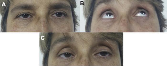
A clinical photograph series consisting of three panels (A, B, and C) demonstrating the Simpson test in a patient with suspected myasthenia gravis. Panel A shows the patient at rest with asymmetric palpebral ptosis, where the upper eyelids droop over the iris, particularly on the right side. Panel B depicts the patient performing a sustained upward gaze for approximately two minutes; the irises are positioned superiorly with significant inferior scleral show and visible compensatory elevation of the eyebrows. Panel C shows the patient returning to a neutral primary gaze immediately after the maneuver, revealing a positive test result marked by a visible increase in bilateral eyelid ptosis (fatigability) compared to the baseline in Panel A. This diagnostic maneuver is used in clinical neurology to assess neuromuscular junction dysfunction by demonstrating objective muscle fatigue. Key anatomical features include the palpebral fissures, iris, and sclera.
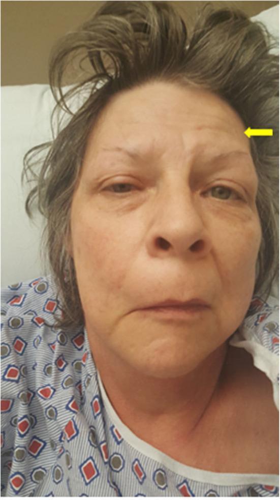
This clinical photograph displays a front-facing view of a middle-aged female patient presenting with significant facial asymmetry, primarily affecting the right side. Key visual findings include unilateral facial droop and mild ptosis of the right eyelid. There is a notable contrast in forehead furrowing: the right side of the forehead appears smooth and unfurrowed, while the left side shows visible transverse wrinkles. A yellow arrow points to the patient's left forehead (anatomical left), highlighting the frontalis sign where the muscle is recruited to compensate for ptosis. The nasolabial fold on the right is flattened, and the right corner of the mouth exhibits a downward droop. These features are clinically representative of a lower motor neuron facial nerve (CN VII) palsy, though the context identifies this specific case as a manifestation of Myasthenia Gravis mimicking Bell's palsy. The image is a valuable educational resource for distinguishing between central and peripheral facial weakness and understanding ocular and bulbar manifestations of neuromuscular junction disorders.
"myasthenia gravis"[MeSH Terms] AND treatment
Myasthenia Gravis (MG)
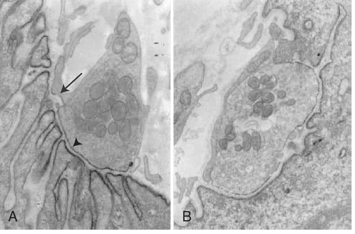
Definition and Pathophysiology
| Antibody | Frequency | Clinical Notes |
|---|---|---|
| Anti-AChR (nicotinic acetylcholine receptor) | 80-90% of generalized MG; 50% of ocular MG | Main immunogenic region on the two α-subunits of the heteropentameric receptor complex |
| Anti-MuSK (muscle-specific tyrosine kinase) | ~5% of AChR-negative patients | More prominent facial/bulbar involvement, more severe weakness |
| Anti-LRP4 (lipoprotein receptor-related protein 4) | 1-3% of all patients | Associated with mild-to-moderate symptoms |
- Complement activation - causing lysis of the muscle endplate membrane
- Cross-linking AChRs - accelerating receptor internalization and degradation
- Direct blockade of ACh binding sites
Epidemiology
-
US prevalence: approximately 20 per 100,000 (~60,000 patients)
-
Prevalence has been increasing over the past 50 years (partly due to better diagnosis in the elderly)
-
Women affected ~3x more than men before age 40; incidence roughly equal after age 50
-
Mean age of onset: women 28-35 years; men 42-49 years
-
The majority of MG patients in the US are now over age 50
-
A thymoma is found in 10% of MG patients; one-third of thymoma patients develop MG
-
Bradley and Daroff's Neurology in Clinical Practice, Chapter 108
Clinical Features
Cardinal Feature
Ocular Symptoms (initial in ~65% of patients)
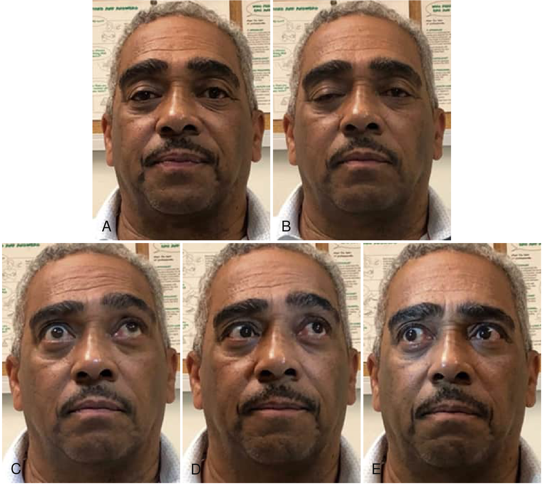
- Ptosis - often asymmetric, may shift from eye to eye (this shifting is virtually pathognomonic of MG)
- Diplopia - from weakness of extraocular muscles without pupillary involvement
- Lid overshoot after downgaze then upgaze ("lid twitch sign" or Cogan's sign)
- Ocular MG: remains confined to ocular muscles in ~10-15% (up to 58% in Asian pediatric populations)
Bulbar Symptoms (initial in ~16% of patients)
- Dysphagia - can occur at oral, pharyngeal, or esophageal levels; silent aspiration in >35% of those with dysphagia
- Dysarthria - nasal or slurred speech worsening with prolonged talking
- Jaw fatigue - worsening during chewing of tough foods; patient may hold jaw closed with hand ("jaw prop" sign)
- Facial weakness - "myasthenic snarl" instead of a smile, flat affect, mask-like facies
Neck and Limb Weakness
- Neck extensors commonly affected - "dropped head syndrome"
- Proximal > distal limb weakness
- Rarely, initial weakness in neck, finger/elbow extensors, hip flexors, or ankle dorsiflexors
Simpson Test (Fatigability Test)
Important Negative Findings
- Tendon reflexes: normal
- Sensation: normal
- Pupils: unaffected (key differentiator from other NMJ disorders and third nerve palsy)
Disease Course
- Maximum weakness in the first year in two-thirds of patients
- Ocular symptoms usually generalize within 2 years if untreated
- Three historical stages (pre-immunosuppression era):
- Active stage: fluctuating, worsening symptoms
- Inactive stage: fluctuations occur but tied to identifiable triggers
- Burnt-out stage: fixed weakness, muscle atrophy (rare today)
- Factors that worsen MG: emotional upset, infection (especially viral respiratory), thyroid dysfunction, pregnancy, menstruation, surgery, drugs affecting NMT, fever
Diagnosis
1. Clinical Recognition
- Characteristic pattern of fatigable, fluctuating weakness in specific muscle groups (ocular, bulbar, neck, proximal limbs)
2. Serology
- Anti-AChR antibodies (binding, blocking, modulating) - most sensitive and specific initial test
- Anti-MuSK antibodies if AChR-negative
- Anti-LRP4 antibodies for double-seronegative cases
- Thymoma-associated MG: also test for anti-titin and anti-ryanodine receptor antibodies
3. Edrophonium (Tensilon) Test
- IV edrophonium (short-acting AChE inhibitor) causes transient improvement of weakness within 30-60 seconds if MG
- Also used to distinguish myasthenic crisis (weakness improves with edrophonium) from cholinergic crisis (weakness worsens with edrophonium)
- Requires atropine on standby (risk of bradycardia/bronchospasm)
4. Electrodiagnostics
- Repetitive nerve stimulation (RNS) at 2-3 Hz: decremental response (>10% decrement) in compound muscle action potential - characteristic of postsynaptic NMJ defect
- Single-fiber EMG (SFEMG): increased jitter - most sensitive test for NMJ dysfunction
5. Imaging
- CT chest (contrast-enhanced): mandatory in all newly diagnosed MG patients to evaluate for thymoma
Classification (Osserman/MGFA)
| Class | Description |
|---|---|
| I | Ocular MG only |
| IIA | Mild generalized, predominantly limb/axial |
| IIB | Mild generalized, predominantly bulbar/respiratory |
| III | Moderate generalized |
| IV | Severe generalized |
| V | Intubation required (myasthenic crisis) |
Treatment
1. Symptomatic Treatment
- Pyridostigmine (Mestinon) - AChE inhibitor, first-line symptomatic therapy; 60 mg every 4-6 hours. Does NOT modify disease course. Longer-acting than neostigmine (onset 60-120 min, duration 3-6 hours).
2. Rapid Immunomodulation (for crisis or acute worsening)
- Plasma exchange (PLEX/plasmapheresis) - removes circulating antibodies; fastest onset (days)
- IV Immunoglobulin (IVIG) - equivalent efficacy to PLEX; 2 g/kg over 5 days; preferred when vascular access is difficult
3. Long-term Immunosuppression
- Corticosteroids (prednisone) - mainstay; note that high-dose steroids may transiently worsen MG before improvement ("steroid dip") - typically start low and titrate up
- Azathioprine - most commonly used steroid-sparing agent; onset delayed 12-18 months
- Mycophenolate mofetil - alternative steroid-sparing agent
- Ciclosporin / Tacrolimus - for refractory cases
- Methotrexate - also used as an alternative
4. Biological Therapies (newer agents)
- Eculizumab (anti-C5 complement inhibitor) - approved for generalized AChR+ MG
- Ravulizumab - longer-acting anti-C5
- Efgartigimod (anti-FcRn) - reduces IgG antibody levels including anti-AChR
- Rozanolixizumab (anti-FcRn)
- A 2025 network meta-analysis (PMID 40346603) comparing novel biologics found significant differences in efficacy and tolerability across agents in generalized MG.
5. Thymectomy
- Indicated in all MG patients with thymoma (mandatory)
- Recommended in non-thymomatous AChR+ generalized MG patients aged 18-65 years
- The majority of generalized MG patients will undergo thymectomy, which improves long-term outcomes
- Surgical approaches include transsternal and video-assisted thoracoscopic surgery (VATS)
- Thymoma-associated MG almost invariably has AChR antibodies (not MuSK or LRP4)
6. Drugs to Avoid in MG
- Aminoglycoside antibiotics (gentamicin, tobramycin)
- Fluoroquinolones, macrolides
- Neuromuscular blocking agents (non-depolarizing > depolarizing)
- Beta-blockers
- Magnesium (blocks NMJ)
- Chloroquine / hydroxychloroquine
- D-penicillamine (can induce MG de novo)
Myasthenic Crisis
- Definition: severe exacerbation of MG weakness requiring respiratory support (intubation/ventilation)
- Precipitants: infection (especially respiratory), aspiration, surgery, drugs, rapid steroid taper, stress
- Dysphagia is a precipitant in ~56% of crises
- Management: ICU admission, intubation if needed, PLEX or IVIG, hold/reduce pyridostigmine (excess ACh can worsen secretions), treat underlying trigger
| Feature | Myasthenic Crisis | Cholinergic Crisis |
|---|---|---|
| Cause | Undertreated MG / trigger | AChE inhibitor overdose |
| Response to edrophonium | Improves | Worsens |
| Secretions | Normal | Excessive (SLUDGE) |
| Pupil | Normal/dilated | Miotic |
MG vs. Lambert-Eaton Myasthenic Syndrome (LEMS) - Key Distinction
| Feature | MG | LEMS |
|---|---|---|
| Site of defect | Postsynaptic | Presynaptic |
| Antibody | Anti-AChR, Anti-MuSK | Anti-VGCC (P/Q-type) |
| Muscles mainly affected | Ocular, bulbar, proximal | Proximal lower limb |
| Eye signs | Ptosis + diplopia common | Ptosis mild; diplopia rare |
| Reflexes | Normal | Reduced (enhance post-exercise) |
| Autonomic features | Absent | Present (dry mouth, impotence) |
| Association | Thymoma | Small cell lung cancer |
| RNS | Decremental at 3 Hz | Incremental at high frequency (>10 Hz) |
Special Populations
- Neonatal MG: Transient (weeks) from maternal IgG crossing placenta; ~12% of infants born to MG mothers; hypotonia, poor feeding, weak cry; self-limiting
- Congenital myasthenic syndromes: NOT autoimmune; genetic mutations in NMJ proteins; AChE inhibitors helpful for many subtypes
- Pediatric MG: Ocular form predominates, especially in Asian children (up to 58% remain ocular)
- Bradley and Daroff's Neurology in Clinical Practice, 8th ed., Chapter 108
- Ganong's Review of Medical Physiology, 26th ed.
- Morgan and Mikhail's Clinical Anesthesiology, 7th ed.
- Tintinalli's Emergency Medicine, p. 587
- Recent evidence: PMID 40346603 - 2025 network meta-analysis on novel biologics in MG treatment