
Kartagener syndrome
Reading File
Now let me search the medical library and PubMed in parallel.
Finding Sources
Finding Sources
Searching PubMed
Kartagener syndrome primary ciliary dyskinesia
Good results. Now let me read the relevant textbook sections in parallel.
Reading File
Reading File
Reading File
Reading File
Excellent content. Let me now fetch the key images and also read more from Murray & Nadel's for the clinical features section, plus get genetics info from Goldman-Cecil.
Loading Image
Loading Image
Loading Image
Reading File
Reading File
I now have comprehensive information to write a detailed answer. Let me also fetch the PCD diagnostic algorithm image.
Loading Image
I now have everything needed for a complete, well-illustrated answer.
Kartagener Syndrome
Kartagener syndrome is a clinical variant of Primary Ciliary Dyskinesia (PCD), defined by the classic triad:
- Situs inversus (complete mirror-image reversal of thoracic and abdominal organs)
- Chronic sinusitis (and/or rhinosinusitis)
- Bronchiectasis
It is also called the Kartagener triad or immotile cilia syndrome (when the broader ciliary dysfunction is emphasized).
- K J Lee's Essential Otolaryngology, p. 48
- Scott-Brown's Otorhinolaryngology Head & Neck Surgery Vol. 1, p. 1087
Genetics and Epidemiology
-
Inheritance: Autosomal recessive
-
Prevalence: PCD affects approximately 1 in 20,000 individuals at birth; Kartagener syndrome accounts for ~50% of all PCD cases (i.e., those with situs inversus)
-
Gene defects: Mutations in genes encoding dynein arm components (e.g., DNAI1, DNAI2, DNAAF1/KTU, DNAAF2, and >50 other PCD-associated genes). No single gene accounts for all cases.
-
Histology: A Text and Atlas, p. 784
Pathophysiology
The root cause is a structural defect in the axonemal dynein arms of cilia and flagella. Normal cilia have a 9+2 microtubule arrangement with outer and inner dynein arms that generate the ATP-powered sliding force for ciliary beating.
In Kartagener/PCD:
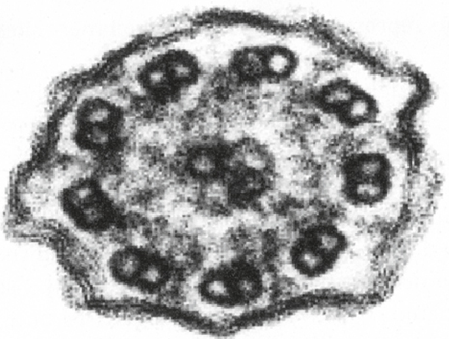
- The dynein arms are absent (outer, inner, or both) from the A-tubules of peripheral microtubule doublets
- Basal bodies are often misoriented (feet point in random directions rather than a uniform orientation)
- Result: absent or severely reduced ciliary beat frequency and coordination → failure of mucociliary transport
Why situs inversus occurs: During embryonic development, nodal cilia in the embryonic node normally beat in a directional manner to establish left-right asymmetry. When these cilia are dysfunctional, laterality is determined randomly, giving a ~50% chance of situs inversus in affected individuals - which is why only half of PCD patients develop Kartagener syndrome.
- Histology: A Text and Atlas, pp. 784-787
- Junqueira's Basic Histology, p. 350
Electron Micrograph - Absent Dynein Arms
This cross-section EM of a cilium from a PCD patient shows the 9+2 doublets with absent dynein arms (compare to normal cilia where arm projections are clearly visible on each doublet):
Clinical Features
The clinical manifestations reflect where motile cilia are distributed:
Respiratory Tract
- Recurrent sinusitis / rhinosinusitis - year-round, beginning in infancy
- Nasal polyps - present in ~27% of Kartagener patients
- Bronchiectasis - progressive airway damage from recurrent infections; basal predominant
- Chronic productive cough - from early childhood (<6 months of age)
- Recurrent otitis media - from Eustachian tube ciliary dysfunction
- Neonatal respiratory distress - a common early clue
Reproductive System
- Male infertility (virtually universal) - sperm flagella share the same 9+2 dynein arm structure; immotile flagella = azoospermia/asthenospermia. Spermatogenesis is normal, but the sperm cannot swim.
- Female subfertility - fallopian tube cilia are also impaired. Females may occasionally be fertile if residual ciliary movement is sufficient, but there is an increased risk of ectopic pregnancy due to impaired ovum transport.
CNS
- Hydrocephalus internus (rare) - ependymal cells lining the CSF spaces possess motile cilia; dysfunction may impair CSF circulation through narrow inter-ventricular spaces.
Chest X-Ray and CT Findings
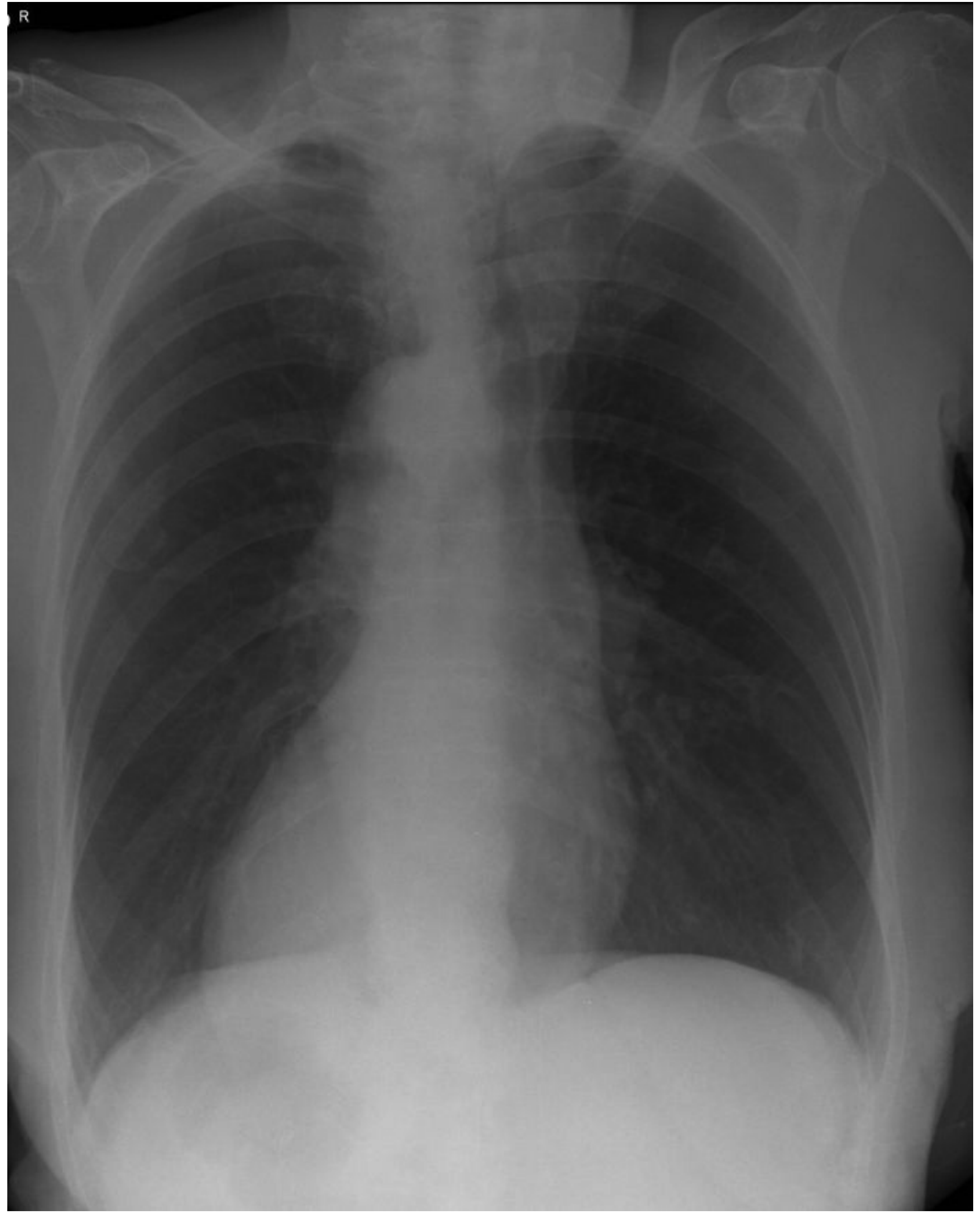
Chest radiograph showing dextrocardia (heart on the right) - one of the hallmarks of situs inversus:
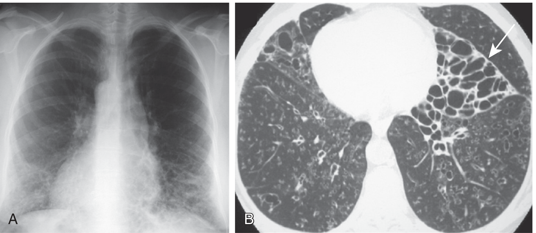
Frontal CXR + Axial CT in Kartagener syndrome: dextrocardia with basal linear opacities (bronchial wall thickening) on CXR (A); severe cystic bronchiectasis on CT, particularly on the left, with the left lung showing a morphologic configuration resembling a right middle lobe (arrow in B):
- Murray & Nadel's Textbook of Respiratory Medicine, p. 1596
- Goldman-Cecil Medicine, p. 3739
Diagnosis
Clinical suspicion is triggered by the presence of ≥2 key features (per the ATS PCD guidelines):
- Unexplained neonatal respiratory distress
- Year-round daily cough beginning before 6 months of age
- Year-round daily rhinosinusitis beginning before 6 months
- Abnormal organ laterality (situs inversus/heterotaxy)
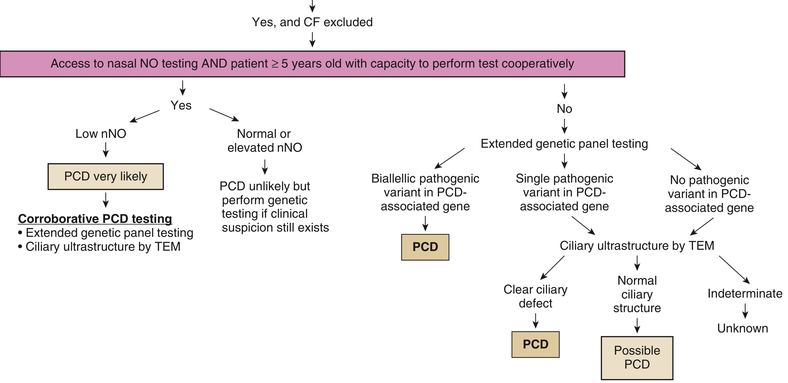
Diagnostic Algorithm (ATS 2018)
Key tests:
| Test | Details |
|---|---|
| Nasal nitric oxide (nNO) | Markedly low in PCD (nNO is produced by sinuses and is impaired in ciliary dysfunction). Requires patient ≥5 years old. First exclude CF (significant minority of CF patients also have low nNO). |
| Extended genetic panel testing | Biallelic pathogenic variants in a PCD-associated gene = diagnostic. >50 genes known. |
| Transmission electron microscopy (TEM) | Gold standard for showing dynein arm defects, but ~30% of PCD patients have normal or near-normal TEM - poor sensitivity. |
| High-speed video microscopy | Assesses ciliary beat frequency/pattern; suffers from low specificity (chronic infection can secondarily impair cilia). |
- Murray & Nadel's Textbook of Respiratory Medicine, pp. 1596-1597
Management
There is no cure; management is supportive and aimed at minimizing airway damage:
- Airway clearance techniques - chest physiotherapy, positive expiratory pressure devices, mucolytics (hypertonic saline); a 2024 systematic review (PMID 39269762) found airway clearance is a mainstay of therapy but evidence on optimal techniques remains limited.
- Antibiotic therapy - prompt treatment of respiratory exacerbations; long-term low-dose macrolides for their anti-inflammatory effects (used similarly to bronchiectasis management).
- Sinus management - endoscopic sinus surgery for refractory sinusitis and polyposis.
- Fertility counseling - for both male (assisted reproduction via ICSI using morphologically normal sperm) and female patients.
- Surveillance - regular pulmonary function tests, CT chest monitoring, hearing assessments (for otitis media sequelae).
- Lung transplantation in end-stage disease.
Key Summary Table
| Feature | Detail |
|---|---|
| Classic triad | Situs inversus + Chronic sinusitis + Bronchiectasis |
| Inheritance | Autosomal recessive |
| Core defect | Absent dynein arms on ciliary A-tubules |
| % of PCD with situs inversus | ~50% |
| Male fertility | Almost always infertile (immotile sperm) |
| Female fertility | Subfertile; increased ectopic pregnancy risk |
| Nasal polyps | ~27% |
| Screening test | Nasal nitric oxide (low) |
| Gold standard | TEM (but 30% false negative) |
| Confirmatory | Biallelic PCD gene mutations |
Acute appendicitis
Reading File
Searching sources...
Finding Sources
Finding Sources
Searching PubMed
"appendicitis"[MeSH Terms] AND "diagnosis" AND "management"
Excellent hits. Now let me read the core sections from Bailey & Love (surgery), Sleisenger (GI), Robbins (pathology), and Rosen's (EM) in parallel.
Reading File
Reading File
Reading File
Reading File
Good content. Now let me read the detailed Bailey & Love section on signs, investigations, scoring, and management, plus get radiology content and Schwartz's surgery section.
Reading File
Reading File
Reading File
Let me now get the clinical signs, management sections from Bailey & Love, and the CT/imaging details, plus read the Schwartz surgery clinical diagnosis section.
Reading File
Reading File
Excellent content. Now let me fetch key images and get the management/treatment section from Bailey & Love and Schwartz.
Loading Image
Loading Image
Loading Image
Reading File
Now let me get the management/treatment section from Bailey & Love and the investigations section.
Reading File
I now have comprehensive, well-sourced content to write a complete answer.
Acute Appendicitis
Overview and Epidemiology
Acute appendicitis is inflammation of the vermiform appendix and is the most common cause of emergency abdominal surgery worldwide. It accounts for approximately 17% of emergency department presentations for acute abdominal pain and 30% of acute surgical abdominal emergencies in patients under 50.
-
Lifetime risk: 8.6% in males, 6.7% in females
-
Peak incidence: Teens and early twenties; equal sex incidence before puberty; male-to-female ratio rises to 3:2 at age 25
-
Rare in infants (<5 years); more dangerous due to underdeveloped omentum - inflammation less well-contained, leading to higher perforation rates
-
Bailey and Love's Short Practice of Surgery, 28th ed., pp. 1358-1359
-
Sleisenger and Fordtran's Gastrointestinal and Liver Disease, p. 172
Aetiology and Pathophysiology
The central mechanism is luminal obstruction leading to progressive intraluminal pressure rise:
-
Obstruction - in 50-80% of cases, caused by:
- Faecolith / appendicolith (most common in adults) - inspissated faecal material, calcium phosphates, bacteria, and epithelial debris
- Lymphoid hyperplasia (most common in children/young adults)
- Tumour (e.g. caecal carcinoma - important in middle-aged/elderly)
- Foreign body, parasites (Enterobius vermicularis)
- Fibrotic stricture (from previous resolved appendicitis)
-
Consequences of obstruction:
- Continued mucus secretion raises intraluminal pressure
- Lymphatic drainage obstructed → oedema and mucosal ulceration
- Bacterial translocation to the submucosa (mixed aerobic/anaerobic flora; E. coli and Bacteroides fragilis predominate in perforation)
- Progressive venous obstruction → ischaemia of appendix wall
- Gangrene → perforation → peritonitis
Note: Progression to perforation is not inevitable - spontaneous resolution is recognised, suggesting uncomplicated and perforated appendicitis may represent different disease processes.
- Schwartz's Principles of Surgery, 11th ed., pp. 1359-1360
- Robbins & Kumar Basic Pathology, p. 587
Pathology (Morphological Stages)
| Stage | Features |
|---|---|
| Early/Catarrhal | Congested subserosal vessels, perivascular neutrophilic infiltrate; dull, granular erythematous serosa. Diagnosis requires neutrophil infiltration of muscularis propria |
| Suppurative | Focal abscesses within the wall; pus-filled lumen |
| Gangrenous | Haemorrhagic ulceration + gangrenous necrosis extending to serosa |
| Perforated | Rupture → suppurative/faecal peritonitis or peri-appendiceal abscess |
Histology - acutely inflamed appendix with pus-filled lumen (L) and inflammation extending to serosa (S):
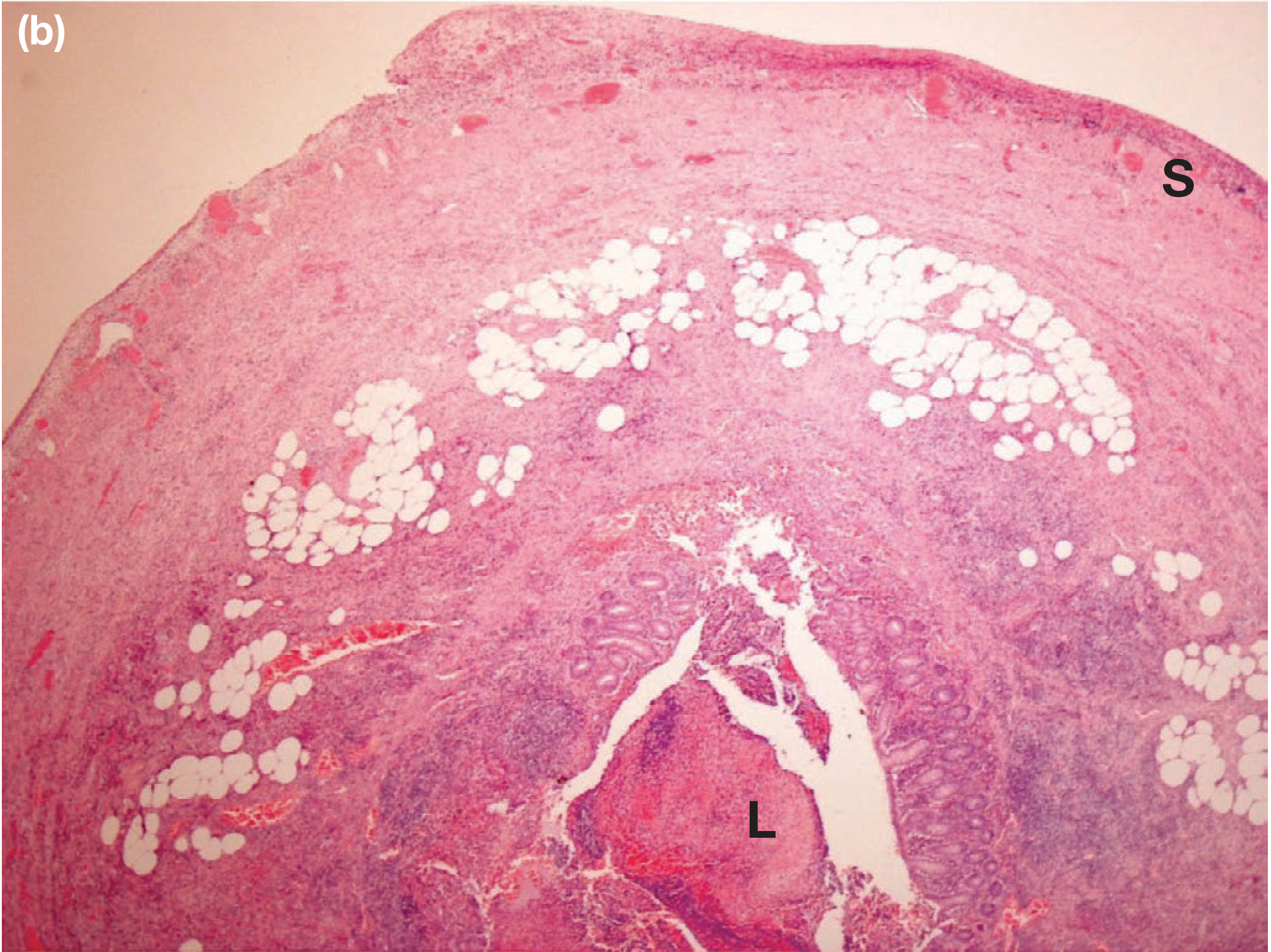
- Robbins & Kumar Basic Pathology, pp. 587-588
Clinical Features
Symptoms - Classic Sequence
- Periumbilical/central colic (visceral pain from appendix - T10 dermatome)
- Pain migrates to the right iliac fossa (RIF) within 6-8 hours, becomes constant and somatic as parietal peritoneum becomes inflamed
- Anorexia (very characteristic - nearly universal)
- Nausea ± vomiting (usually follow onset of pain, not precede it)
- Low-grade fever 37.2-38.4°C
The classic visceral-to-somatic pain migration is present in only ~50% of patients. Atypical presentations are more common in the elderly, young children, and with malpositioned appendix.
Signs
| Sign | Description | Appendix Position |
|---|---|---|
| McBurney's point tenderness | Maximal tenderness 1/3 of the way from ASIS to umbilicus | Normal position |
| Rovsing's sign | RIF pain on palpation/release of LIF | Normal position |
| Dunphy's sign | Pain on coughing | Retrocaecal |
| Psoas sign | Pain on hip extension (stretches iliopsoas) | Retrocaecal |
| Obturator sign | Pain on internal rotation of flexed hip | Pelvic |
| Rectal tenderness | Right-sided tenderness in pouch of Douglas | Pelvic |
Position-specific presentations:
- Retrocaecal (~65%): Rigidity may be absent ("silent appendix"), deep loin tenderness, psoas spasm causing hip flexion
- Pelvic: No abdominal rigidity; suprapubic pain; diarrhoea; bladder symptoms (frequency); tenesmus; only tender on rectal exam
- Postileal: Pain may not shift; diarrhoea; ill-defined tenderness near umbilicus
Vital signs
-
First 6 hours: Often no fever or tachycardia
-
After 6 hours: Mild pyrexia 37.2-37.7°C, pulse 80-90 bpm
-
High fever (>38.5°C) in a child suggests alternative diagnosis (e.g. mesenteric adenitis)
-
High fever + high WBC in adults suggests perforation/abscess
-
Bailey and Love's Surgery, pp. 1359-1362
-
Schwartz's Principles of Surgery, pp. 1400-1401
Investigations
Laboratory
| Test | Finding | Notes |
|---|---|---|
| WBC | Leukocytosis ~10,000-14,000/mm³ | ~17,000 in gangrenous/perforated; normal WBC does NOT exclude appendicitis |
| CRP | Elevated | More useful later in disease course; also helps predict perforation |
| Urinalysis | May show microscopic pyuria/haematuria (pelvic appendix near ureter/bladder) | Rules out UTI/nephrolithiasis |
| Pregnancy test | Mandatory in women of childbearing age | Rules out ectopic pregnancy |
| Procalcitonin, IL-6 | Elevated in complicated disease | Useful predictors of perforation |
Scoring Systems
Alvarado Score (MANTRELS) - most widely used:
| Feature | Points |
|---|---|
| Migration of pain to RIF | 1 |
| Anorexia | 1 |
| Nausea | 1 |
| Tenderness in RLQ | 2 |
| Rebound tenderness | 1 |
| Elevated temperature | 1 |
| Leukocytosis | 2 |
| Shift (left WBC shift) | 1 |
| Total | 10 |
- Score 5-6: Suggestive → surgical review or imaging
- Score 7-8: Probable appendicitis
- Score 9-10: Almost certainly appendicitis
Imaging
Ultrasound (first-line, especially in children and women)
- Sensitivity ~80%, specificity ~90%
- Technique: high-frequency linear transducer with graded compression
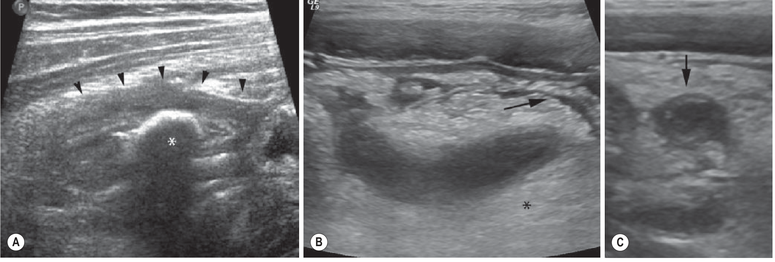
- Primary criteria: tubular, blind-ending, non-compressible structure with outer diameter >6 mm
- Additional findings: wall hyperaemia, hyperechoic periappendiceal fat, appendicolith (with posterior acoustic shadowing)
Ultrasound images showing thickened appendix with appendicolith (A), necrotic appendix with surrounding inflamed mesenteric fat suggesting perforation (B, C):
CT scan (best for adults, complicated cases)
- Sensitivity ~94-98%, specificity ~95%
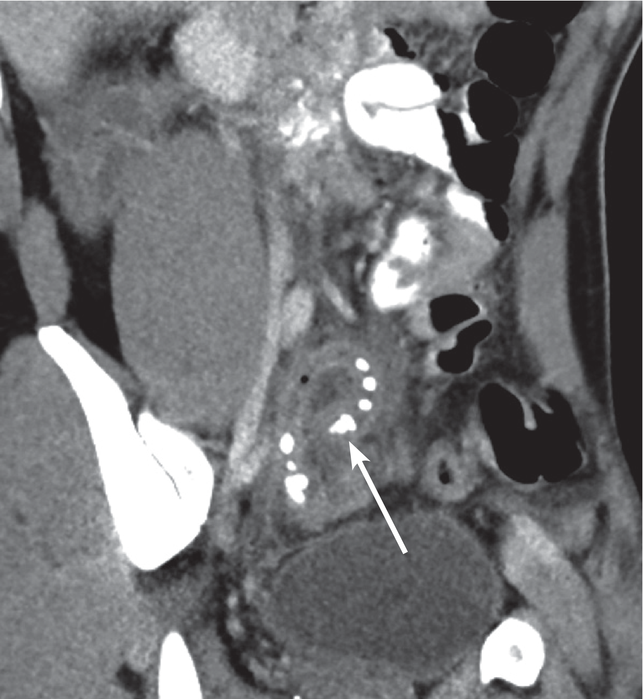
- Shows: enlarged enhancing appendix, periappendiceal fat stranding, appendicoliths, perforation, abscess
- Rarely necessary in children if USS is adequate
CT image - coronal reformat showing inflamed, enlarged appendix curling toward the pelvis (arrow) with multiple radiopaque appendicoliths and extensive periappendiceal fat stranding:
- Grainger & Allison's Diagnostic Radiology, pp. 1801-1802
Differential Diagnosis
| Children | Adults | Adult Females | Elderly |
|---|---|---|---|
| Gastroenteritis | Regional enteritis (Crohn's) | Mittelschmerz | Diverticulitis |
| Mesenteric adenitis | Ureteric colic | Pelvic inflammatory disease | Colonic carcinoma |
| Meckel's diverticulitis | Perforated peptic ulcer | Pyelonephritis | Intestinal obstruction |
| Intussusception | Torsion of testis | Ectopic pregnancy | Mesenteric infarction |
| Henoch-Schönlein purpura | Pancreatitis | Torsion/rupture of ovarian cyst | Leaking AAA |
| Lobar pneumonia | Rectus sheath haematoma | Endometriosis | Torsion of appendix epiploicae |
Key clinical pitfalls:
-
Loose stools misdiagnosed as gastroenteritis (pelvic appendicitis)
-
Pyuria/dysuria misdiagnosed as UTI (appendix adjacent to ureter/bladder)
-
Antibiotics masking/mollifying signs and delaying presentation
-
RLQ pain from right lower lobe pneumonia (always consider chest exam)
-
Bailey and Love's Surgery, p. 1362
Management
General Resuscitation (all patients)
- IV access, IV fluids, analgesia (opioids are safe and do not mask signs)
- NBM (nil by mouth)
- Broad-spectrum IV antibiotics covering aerobic and anaerobic organisms (e.g. cefuroxime + metronidazole; or co-amoxiclav; or piperacillin-tazobactam for perforated disease)
- Pregnancy test, cross-match if needed
Uncomplicated Appendicitis
Standard treatment: Appendicectomy (laparoscopic preferred)
- Laparoscopic appendicectomy is now the standard of care
- Advantages over open: less pain, faster recovery, lower wound infection rate, better cosmesis, allows concurrent diagnosis of alternative pathology
- No further antibiotics needed post-operatively for uncomplicated disease
Antibiotic-first (non-operative) management - evolving alternative:
- Multiple RCTs (APPAC, CODA trials) show ~70% of patients with uncomplicated appendicitis can be successfully treated with antibiotics alone in the short term
- ~30% require appendicectomy within 1 year due to recurrence
- Patients with appendicoliths have significantly higher failure rates with antibiotics alone
- The 2024 SAGES guidelines and 2026 WSES Jerusalem Guidelines support offering antibiotics-first as an option for selected patients with uncomplicated appendicitis (shared decision-making)
Complicated Appendicitis
Perforated appendicitis:
- Septic patient: Immediate surgery (laparoscopic or open), though associated with higher risk of abscesses and enterocutaneous fistulae due to dense adhesions
- Stable patient with abscess/phlegmon: Percutaneous CT-guided drainage + IV antibiotics; interval appendicectomy can be considered at 6-12 weeks (though evidence for mandatory interval appendicectomy is limited)
- Post-operative antibiotics for 3-7 days
Appendix mass (phlegmon):
- Often managed non-operatively with antibiotics initially
- In children with unobstructed appendix mass, conservative management is appropriate; interval appendicectomy considered 6 weeks later but not mandated
Post-operatively:
-
Routine histology of all appendicectomy specimens - to detect unexpected pathology (carcinoid, carcinoma, Crohn's)
-
~10-20% of appendicectomies find a normal appendix ("negative appendicectomy") - acceptable rate to avoid missed perforation; the 2023 systematic review (PMID 37914953) confirmed imaging integration reduces this rate significantly
-
Bailey and Love's Surgery, pp. 1363-1366
-
Schwartz's Principles of Surgery, pp. 1401-1405
Special Populations
| Population | Key Points |
|---|---|
| Children <5 years | Underdeveloped omentum → rapid perforation; often present after perforation; higher diagnostic difficulty |
| Elderly | Atypical presentation common; RIF localisation unusual; higher risk of caecal malignancy as underlying cause; get CT before OR |
| Pregnant women | Appendix displaced superiorly by gravid uterus; RIF tenderness may be in right flank; perforation risk much higher → low threshold for surgery; MRI preferred over CT in first trimester |
| Immunocompromised | Blunted inflammatory response; minimal signs; higher perforation rate at presentation |
Key Summary
| Feature | Detail |
|---|---|
| Most common cause | Faecolith obstruction (adults); lymphoid hyperplasia (children) |
| Classic triad | Periumbilical pain migrating to RIF + anorexia + low fever |
| Hallmark sign | McBurney's point tenderness |
| Diagnostic histology | Neutrophilic infiltration of muscularis propria |
| Best scoring tool | Alvarado score (max 10) |
| First-line imaging | USS (children/women); CT (adults, complicated) |
| Ultrasound criterion | Outer diameter >6 mm, non-compressible |
| Treatment (uncomplicated) | Laparoscopic appendicectomy OR antibiotics-first (shared decision) |
| Treatment (perforated, septic) | Emergency appendicectomy + antibiotics |
| Key organisms (perforation) | E. coli + Bacteroides fragilis |
| Lifetime risk | 8.6% (M), 6.7% (F) |
Recent guidelines update (2026): The WSES Jerusalem Guidelines 2025 (PMID 41604201) published in JAMA Surgery now formally endorse antibiotic-first therapy as a valid alternative to surgery in carefully selected uncomplicated appendicitis, with the caveat that patients with appendicoliths on imaging have substantially higher failure rates and should be offered surgery as first choice.
This is a shared conversation. Sign in to Orris to start your own chat.