
patients with degen- erative neurological symptoms, a diagnosis of adrenoleukodystro- phy should be excluded.
adrenoleukodystrophy diagnosis neurological symptoms
adrenoleukodystrophy MRI brain white matter demyelination
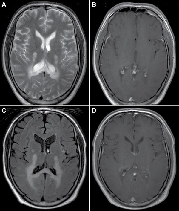
This composite diagnostic image presents axial brain MRI scans from a patient with adrenomyeloneuropathy (AMN) transitioning to cerebral adrenoleukodystrophy (cALD). Panels A (T2-weighted) and C (FLAIR) demonstrate progressive white matter disease. Initially, hyperintense signals are localized in the splenium of the corpus callosum and periventricular parieto-occipital white matter. Follow-up imaging in Panel C shows significant spatial extension of these lesions, appearing more confluent and diffuse. Panels B and D provide T1-weighted sequences with gadolinium contrast. While the initial scan (B) shows no pathological enhancement, the follow-up scan (D) reveals faint rim enhancement at the leading edge of the demyelinating lesion. This contrast enhancement is a critical clinical marker of active neuroinflammation and blood-brain barrier disruption. The series illustrates the radiologic hallmarks of X-linked adrenoleukodystrophy progression, including the characteristic posterior-to-anterior spread of demyelination and the transition to an active inflammatory stage.
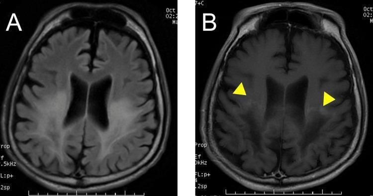
This diagnostic image set consists of two axial brain MRI scans from a 27-year-old male with adolescent cerebral-type adrenoleukodystrophy (ALD). Image (A) is a Fluid-Attenuated Inversion Recovery (FLAIR) sequence showing symmetric, diffuse, high-intensity signals in the posterior deep white matter, specifically involving the parieto-occipital lobes. This represents demyelination or vasogenic edema. Image (B) is a post-gadolinium contrast-enhanced T1-weighted sequence of the same region. It highlights linear border enhancement (indicated by yellow arrowheads) at the leading edges or anterior periphery of the white matter lesions. This peripheral enhancement is characteristic of active inflammatory demyelination and breakdown of the blood-brain barrier. These findings are clinically significant for monitoring disease progression in X-linked adrenoleukodystrophy, illustrating the classic radiographic 'zonal' pattern of the condition.
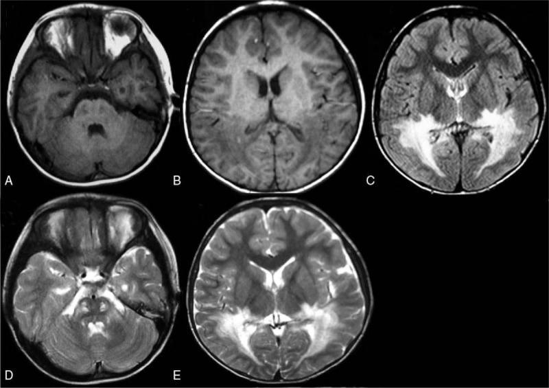
This diagnostic imaging panel consists of axial brain MRI sequences (A-E) illustrating characteristic white matter lesions associated with X-linked adrenoleukodystrophy (X-ALD). The panel includes T1-weighted (A, B), Fluid-Attenuated Inversion Recovery (FLAIR) (C), and T2-weighted (D, E) sequences. There is prominent bilateral, symmetrical signal abnormality involving the periventricular white matter, primarily within the posterior regions including the temporal, parietal, and occipital lobes. On T1-weighted images, these areas appear as regions of decreased signal intensity (hypointense). In contrast, the FLAIR and T2-weighted sequences demonstrate significant hyperintensity in the same distribution, indicating demyelination and abnormal fluid accumulation. Lesions are also visible within the brainstem. The distribution follows a characteristic 'butterfly' pattern in the posterior cerebrum, which is a classic radiographic sign for pediatric cerebral X-ALD. This imaging is used for medical education to demonstrate neurodegenerative metabolic disorders and the radiographic evolution of leukodystrophies.
Adrenoleukodystrophy (ALD)
Overview and Genetics
Clinical Phenotypes
| Phenotype | Population | Onset | Key Features | % of Cases |
|---|---|---|---|---|
| Childhood cerebral ALD | Males | 4-11 years | Cognitive decline, behavioral change, cortical blindness, seizures, progressive to decerebration | ~45-50% |
| Adolescent/adult cerebral | Males | Teens-adult | Similar to childhood form, slower progression | ~5-8% |
| Adrenomyeloneuropathy (AMN) | Males | 3rd-5th decade | Slowly progressive spastic paraparesis, peripheral neuropathy, sphincter disturbances, hypogonadism, variable adrenal insufficiency | ~25% |
| Female carrier myelopathy | Heterozygous females | 3rd-4th decade | Mild-moderate spastic paraparesis, gait ataxia, urinary incontinence; can mimic MS | ~10-20% |
| Isolated Addison disease | Males | Variable | No neurologic involvement; only adrenal insufficiency | ~10% |
Why ALD Must Be Excluded in Degenerative Neurological Presentations
-
Mimics common conditions: AMN in adults closely mimics multiple sclerosis, hereditary spastic paraplegia, or other spinocerebellar degenerations. Up to 20% of female carriers have white matter changes on MRI, making MS the primary differential. Rare presentations include olivopontocerebellar atrophy and adult-onset spinocerebellar ataxia patterns.
-
Adrenal involvement may be subtle: Overt Addison disease may not be apparent, particularly in female carriers or in adult AMN. The neurology can precede recognizable adrenal signs.
-
The diagnosis is biochemically confirmable: Plasma VLCFA levels (C26 absolute level; C26:C22 ratio; C24:C22 ratio) are reliably elevated. Combined plasma and fibroblast testing detects ~93% of female carriers.
-
Treatment exists: Adrenal replacement therapy prolongs life. Early hematopoietic stem cell transplantation or gene therapy (lentiviral ABCD1 transduction of autologous CD34+ cells) can stabilize or prevent progression if started early. Missing the diagnosis delays these interventions.
MRI Findings
Pathology
Diagnostic Workup
- Plasma VLCFAs: Elevated C26, C26:C22, and C24:C22 ratios - the primary biochemical marker
- ABCD1 gene sequencing: Confirms the diagnosis
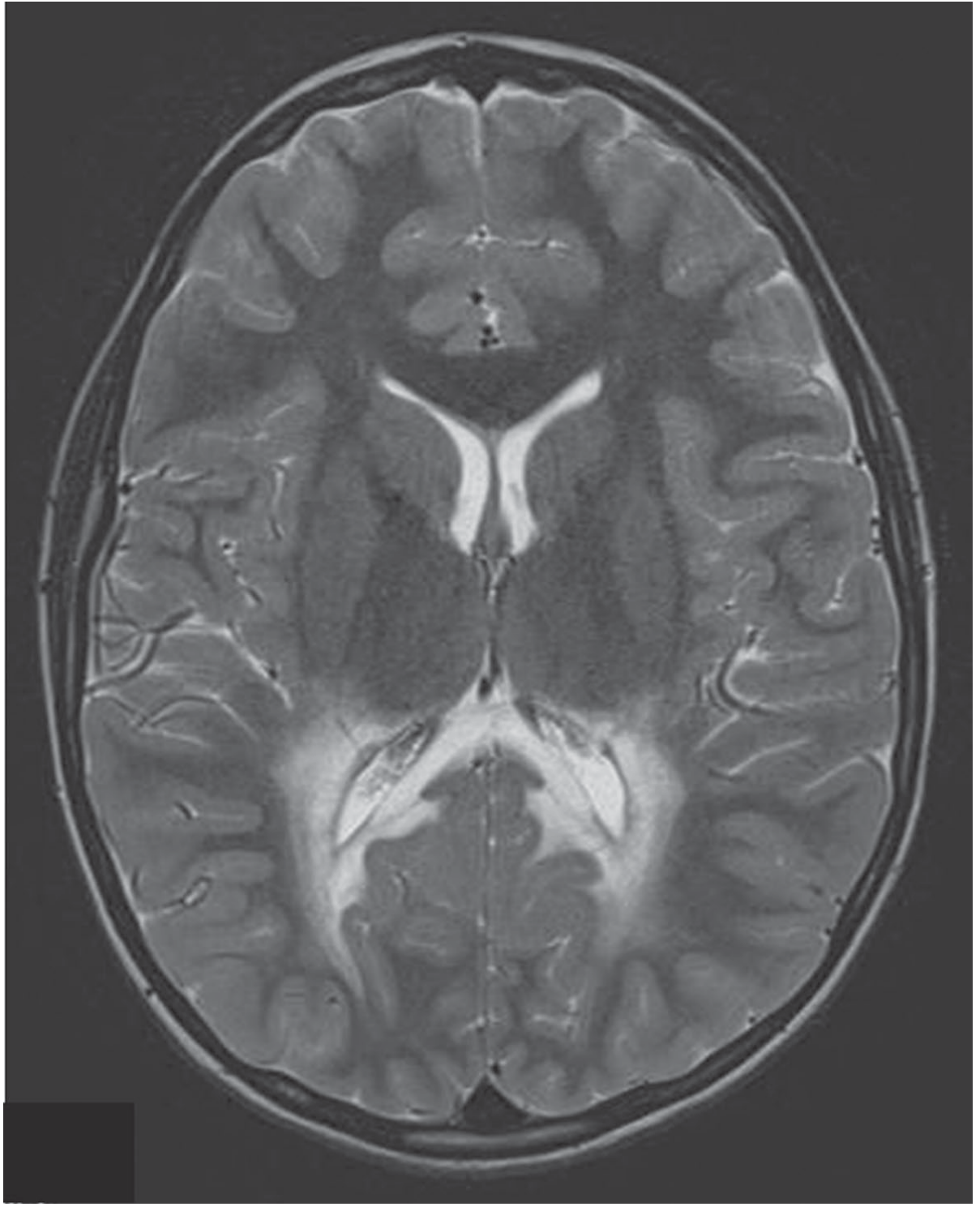
- Brain MRI: Posterior periventricular hyperintensity on T2/FLAIR; gadolinium enhancement at active lesion margins
- Adrenal function: Low serum sodium, elevated potassium, low cortisol, absent rise in 17-hydroxyketosteroids post-ACTH stimulation
- Nerve biopsy (in AMN): Loss of myelinated and unmyelinated fibers with lamellar inclusions in Schwann cell cytoplasm
- CSF: May show elevated protein
Treatment
| Modality | Indication | Notes |
|---|---|---|
| Adrenal replacement therapy | Adrenal insufficiency | Prolongs life; may produce partial neurologic remission |
| Lorenzo's oil (erucic + oleic acids) | Normalizes plasma VLCFAs | Large open-label trials failed to show neurologic efficacy |
| Allogeneic bone marrow transplantation | Early cerebral ALD | Stabilizes disease; reverses some MRI changes if done early |
| Gene therapy (lentiviral ABCD1 into CD34+ cells) | Early disease, pre-symptomatic | Prevents symptom emergence; most promising if instituted early |
- Harrison's Principles of Internal Medicine, 22nd Ed.; Adams and Victor's Principles of Neurology, 12th Ed.