
Role of PBS for diagnosis of parasitic Infection diagnosis
peripheral blood smear malaria Plasmodium Giemsa stain
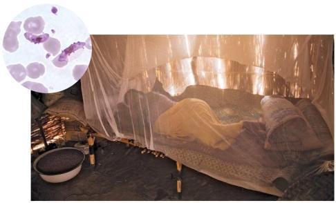
This composite educational image illustrates public health interventions and the pathophysiology of malaria. The main clinical photograph depicts a person in a rural setting in eastern Chad sleeping under a white, semi-transparent insecticide-treated bed net (ITN), a primary preventative measure against vector-borne diseases. The environment reflects a high-risk tropical region with simple housing structures. An inset diagnostic micrograph shows a Giemsa-stained thin blood smear at high magnification, highlighting Plasmodium falciparum. Visible are pale purple erythrocytes, several of which contain characteristic banana-shaped or crescentic gametocytes, diagnostic of P. falciparum infection. The visual contrast between the bedside preventative measure and the microscopic evidence of infection serves as a teaching tool for infectious disease management, epidemiology, and the importance of vector control in endemic regions. This material is suitable for medical students and public health professionals studying parasitology and global health strategies.
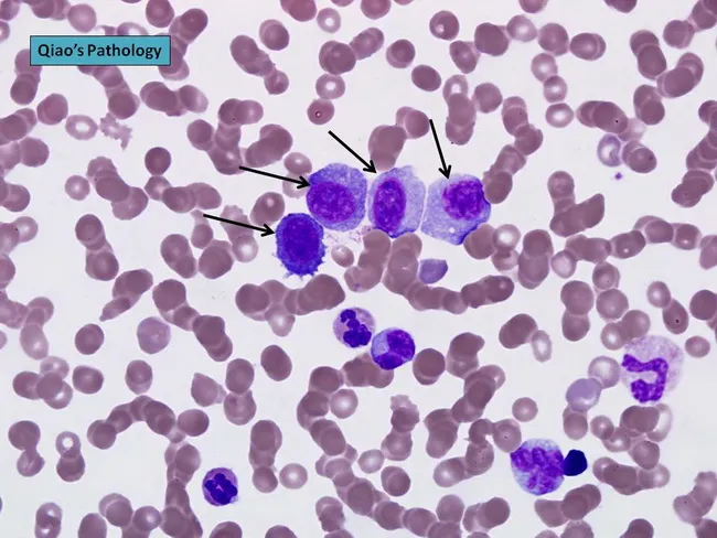
Peripheral blood smear prepared from a patient with suspected plasma cell leukemia (PCL) was examined by bright‑field light microscopy after Wright‑Giemsa staining. The smear shows several circulating plasma‑cell–like elements, including four conspicuous proplasmacytes with dispersed nuclear chromatin and prominent nucleoli, as indicated by arrows in the original figure. In addition, background erythrocytes exhibit rouleaux formation, a common feature in paraproteinemic states. The cells display eccentrically placed nuclei with pronounced basophilic cytoplasm and a coarse chromatin pattern; occasional cells show a perinuclear clearing (Golgi rim) suggestive of plasmacytic differentiation. Immunoglobulin light‑chain restriction is not determined on this smear alone but flow cytometry or immunohistochemistry would typically confirm clonal plasma cells. Relative proportions indicate that neoplastic plasma cells constitute a substantial fraction of leukocytes, consistent with plasma cell leukemia rather than reactive plasmacytosis. PCL is an aggressive myelomatous process often presenting with cytopenias, organomegaly, and sometimes lymphadenopathy; prognosis is poor, and treatment responsiveness is limited. This image illustrates characteristic morphologic features used for differential diagnosis against multiple myeloma, Waldenström macroglobulinemia, and other causes of plasmacytosis. Clinically relevant keywords include plasma cells, rouleaux, Wright‑Giemsa stain, 100X oil, peripheral blood, leukemia, myeloma, and hematologic malignancy.
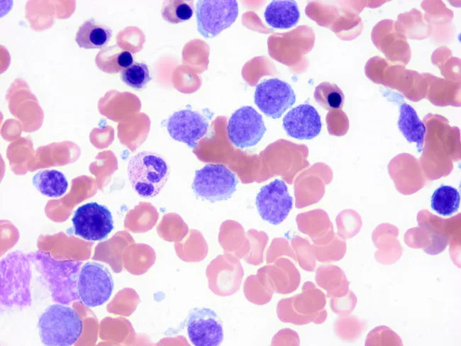
This image depicts a peripheral blood smear prepared with Wright-Giemsa stain and examined under light microscopy at high magnification. The predominant features are small, mature-appearing lymphocytes with scant cytoplasm and discrete basophilic nuclei interspersed among erythrocytes. A characteristic subset of circulating lymphocytes displays short, polarity-restricted cytoplasmic villi (finger-like projections) consistent with splenic marginal zone lymphoma (SMZL) involvement of peripheral blood. The villi are typically slender and localized to one pole of the cell, unlike the longer, circumferential villous extensions seen in hairy cell leukemia, which aids in differential diagnosis. Some lymphocytes appear slightly irregular or irregular nuclear contours; occasional larger atypical cells may be present but are less common. The background shows normocytic red cells with normal distribution; platelets are not prominent. This cytomorphology supports SMZL in the context of known splenomegaly or lymphoproliferative disease and is often corroborated by immunophenotyping and molecular studies. Clinically, peripheral blood involvement occurs in roughly half to two-thirds of SMZL cases and helps establish disease burden. The image illustrates the diagnostic utility of meticulous peripheral smear review for small-vessel lymphocytosis and villous lymphocytes, informing differential diagnoses, guiding flow cytometry panels, and contributing to monitoring and prognosis.
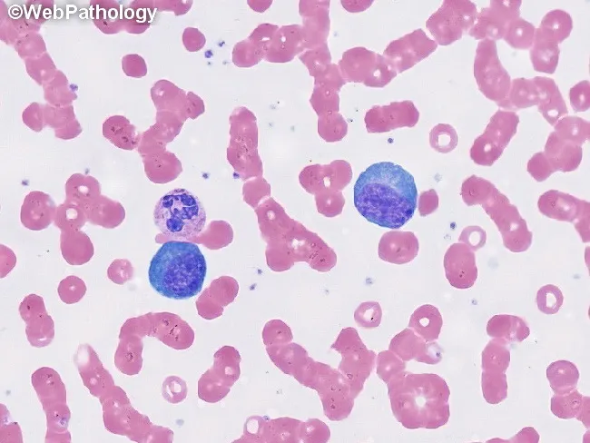
This is a peripheral blood smear prepared with Wright-Giemsa stain and examined under brightfield illumination at high magnification (approximately 1000x total, 100x objective with oil immersion). The smear displays predominantly erythrocytes with uniform pink cytoplasm and characteristic biconcave morphology, arranged singly with occasional rouleaux. Interspersed among the red cells are leukocytes with visible nuclei. Notably, two large lymphocyte-like cells or mononuclear leukocytes appear conspicuously larger than surrounding erythrocytes; these cells have round to oval, deeply basophilic nuclei with dense chromatin and scant, lightly basophilic cytoplasm. No visible granulocytic cytoplasm granules or multiple lobes are clearly resolved in this field, and there are no obvious nucleoli or cytoplasmic inclusions evident in these cells. The background lacks abnormal pigment or reticulocytosis. Overall cell morphology is consistent with a normal or mildly reactive hematologic smear; in a clinical context, additional data such as complete blood count, differential, and patient history would be needed to determine whether there is leukocytosis or blasts. This image serves as an educational reference for recognizing basic peripheral blood components, evaluating leukocyte size variation, and assessing RBC morphology for hematology training and diagnostic differential diagnoses. Additional cases and image contexts can reinforce recognition of hematologic normal variants and patterns.
blood smear trypanosoma microfilaria Babesia parasites microscopy
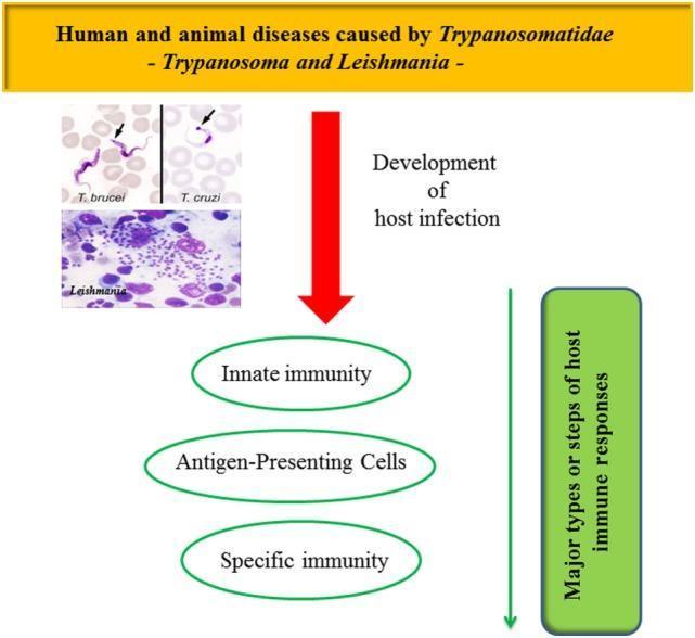
Educational infographic and clinical microscopy composite titled 'Human and animal diseases caused by Trypanosomatidae - Trypanosoma and Leishmania'. The visual includes three Giemsa-stained clinical photographs: Trypanosoma brucei (showing an elongated, undulating trypomastigote in a blood smear), Trypanosoma cruzi (showing a characteristic C-shaped trypomastigote with a large kinetoplast indicated by an arrow), and Leishmania (demonstrating numerous intracellular amastigotes within a host macrophage, appearing as small purple-stained dots). A central red arrow labeled 'Development of host infection' points toward a conceptual flowchart of the host immune response. This flowchart identifies three major stages: Innate immunity, Antigen-Presenting Cells (APCs), and Specific immunity. A vertical green bar on the right summarizes these as 'Major types or steps of host immune responses'. The content illustrates the intersection of clinical parasitology and immunology, focusing on the pathophysiology of African Trypanosomiasis, Chagas disease, and Leishmaniasis.
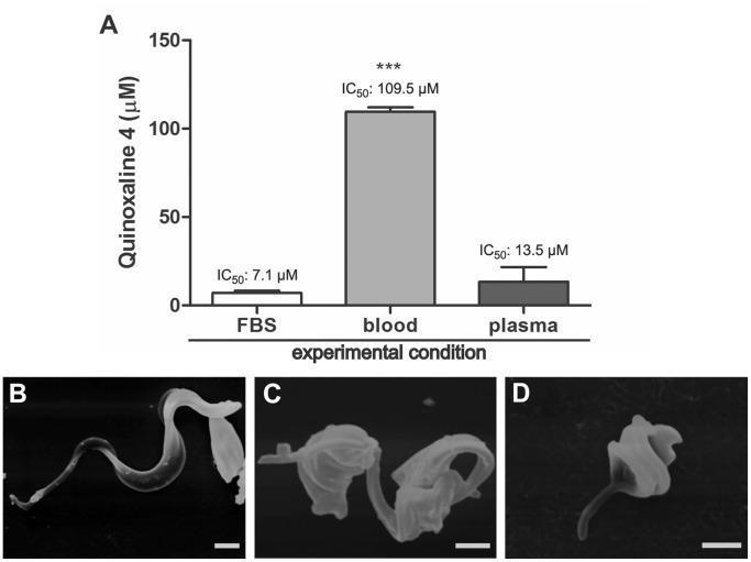
Educational figure regarding the anti-parasitic activity of quinoxaline 4 against Trypanosoma cruzi trypomastigotes. Panel A is a comparison bar chart illustrating the effective concentration (EC50) of the compound under three experimental conditions: fetal bovine serum (FBS), whole mouse blood, and mouse plasma. The Y-axis measures concentration in micromolar (µM). Results show a significant increase in EC50 when blood is present (109.5 µM) compared to FBS (7.1 µM) or plasma (13.5 µM), suggesting compound interaction with blood cells. Panels B-D are scanning electron microscopy (SEM) images demonstrating morphological changes in the parasite. Panel B shows a control trypomastigote with a characteristic elongated, slender body and prominent flagellum. Panels C and D show parasites treated with IC50 and IC90 concentrations, respectively, exhibiting marked pathological alterations including cell body shortening, widening, and loss of typical undulating morphology. Scale bars represent 1 µm. This content is relevant to parasitology and infectious disease pharmacology research.
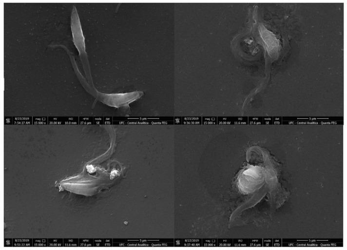
Scanning electron microscopy (SEM) images displaying the morphology of Trypanosoma cruzi epimastigotes under varying experimental conditions. The upper-left panel (A) serves as a control, showing a healthy parasite with a characteristic elongated cell body, smooth membrane integrity, and a prominent emerging flagellum. Panels B, C, and D demonstrate ultrastructural changes following treatment with an experimental compound at IC50 and 2 % IC50 concentrations. These treated parasites exhibit significant morphological distortions, including loss of typical spindle shape, membrane degradation with visible surface irregularities, and apparent leakage of cytoplasmic content. The images highlight the dose-dependent pathological effects of the treatment on the parasite's external structures, specifically the disruption of the plasma membrane. Scale bars indicate 5 μm. This diagnostic-style visual is used in parasitology and pharmacology to study the efficacy of trypanocidal agents and the mechanisms of cell death, such as necrosis, in Chagas disease research.
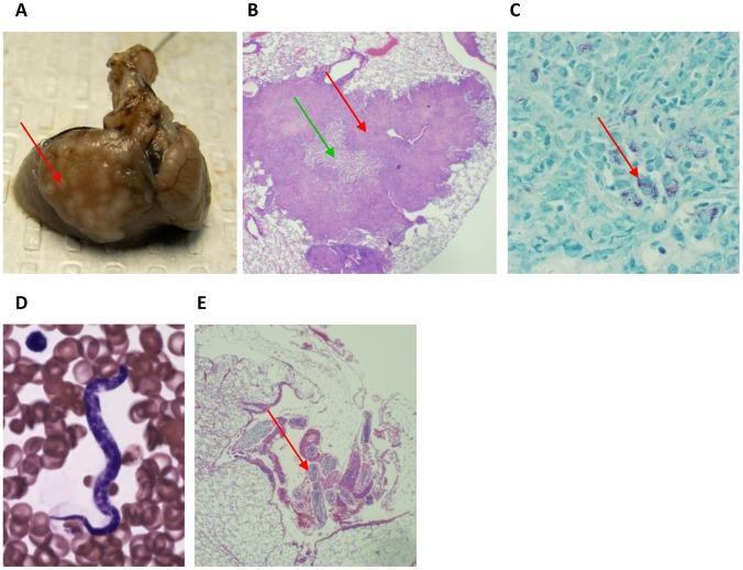
Educational histological and clinical composite illustrating coinfection with Mycobacterium tuberculosis and Litomosoides sigmodontis. Panel A: Macroscopic clinical photograph of lung tissue featuring distinct superficial granulomatous lesions (red arrow). Panel B: H&E stained light microscopy (40x) of a pulmonary granuloma (red arrow) exhibiting a characteristic necrotic core (green arrow) and surrounding inflammatory infiltrate. Panel C: High-magnification acid-fast stain (100x) identifying acid-fast bacilli (M. tuberculosis) appearing as slender red rods (red arrow) within the tissue. Panel D: Peripheral blood smear (Eosin-Y Azure A Methylene Blue, 100x) showing a slender, basophilic microfilaria of L. sigmodontis amidst erythrocytes. Panel E: H&E stained lung cross-section (40x) demonstrating an adult L. sigmodontis nematode (red arrow) situated within the pleural space. This collection highlights diagnostic features of tuberculosis granulomas, acid-fast staining techniques, and the morphological differentiation between microfilarial and adult stages of filarial parasites in various anatomical compartments.
Role of Peripheral Blood Smear (PBS) in Diagnosis of Parasitic Infections
Types of Blood Smear
Thick Smear
- A small drop (~5 µL) of blood is spread to ~1 cm diameter, air-dried, and stained without methanol fixation (RBCs lyse during staining)
- Multiple layers of lysed RBCs concentrate parasites - detects parasitemia as low as ~40 parasites/µL
- Best for: detection - more sensitive, especially at low parasitemia
- Count is reported as parasites per oil-immersion field or per 200 WBC nuclei
- Limitation: RBC morphology is lost, so species identification is harder
Thin Smear
- A standard hematologic smear fixed with methanol and stained with Giemsa
- Single intact layer of RBCs - preserves both parasite and RBC morphology
- Sensitivity lower (~1000 parasites/µL); but superior for species identification
- Useful for counting exact % parasitemia and monitoring treatment
- If thin smear is negative, a thick smear must be done; if still negative, repeat smears every 12-24 hours
From Tintinalli's Emergency Medicine: "A long and careful search for parasites is necessary before a film is declared negative." - Tintinalli's Emergency Medicine, p. 1101
Parasites Identifiable on PBS
1. Malaria (Plasmodium spp.) - Most Important
"The diagnostic test for malaria infection is examination of a Giemsa-stained peripheral blood smear, which permits the asexual stages of the parasite to be identified within infected red cells." - Robbins Pathologic Basis of Disease
- Appearance of infected RBCs (size, shape, stippling)
- Appearance of parasites (ring forms, trophozoites, schizonts, gametocytes)
- Stages present
Species Differentiation on PBS
| Feature | P. falciparum | P. vivax | P. ovale | P. malariae |
|---|---|---|---|---|
| RBC size | Normal | Enlarged | Enlarged, often oval/fimbriated | Normal to small |
| Stippling | Maurer's clefts (coarse) | Schuffner's dots (fine, pink) | Schuffner's dots | Ziemann dots (rarely seen) |
| Ring forms | Multiple rings per RBC; "appliqué" / "headphone" forms; accolé (marginal) | Single large ring | Single ring | Single thick ring |
| Trophozoite | Delicate, scanty cytoplasm; only rings seen in peripheral blood | Amoeboid, irregular | Compact | Band-form (diagnostic) |
| Schizont | Rarely seen in peripheral blood (sequesters) | 12-24 merozoites | 4-12 merozoites | "Rosette" / "daisy" - 6-12 merozoites |
| Gametocyte | Banana/crescent-shaped (diagnostic) | Round | Round | Round |
| % Parasitemia | Can exceed 2-5% | Usually <2% | Usually <2% | Usually <2% |
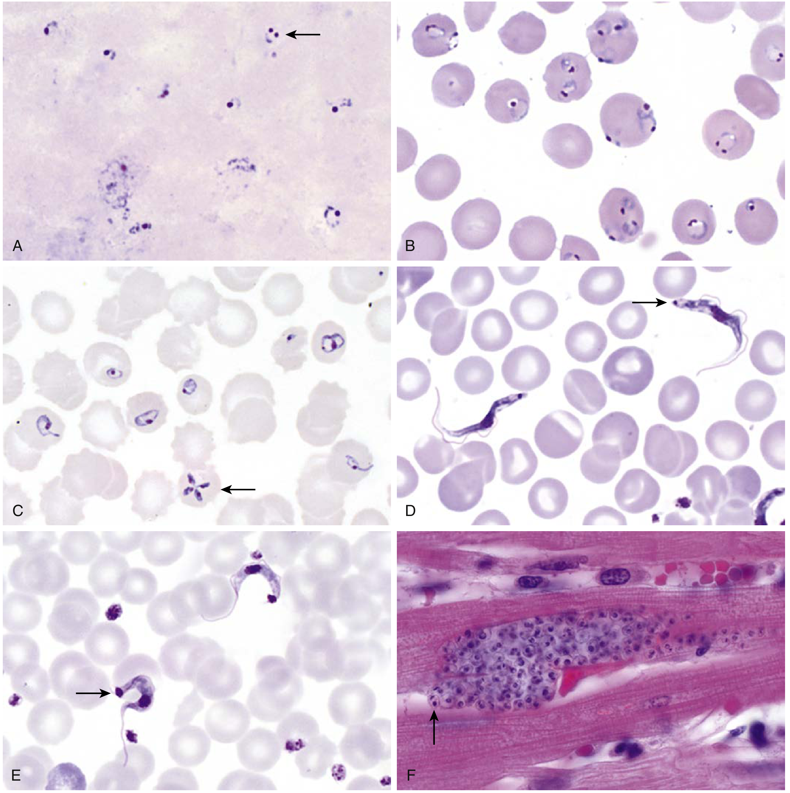
Important: P. knowlesi is morphologically identical to P. malariae on light microscopy - any patient from Asia with high parasitemia resembling P. malariae should be assumed to have P. knowlesi and PCR used for differentiation. - Tintinalli's Emergency Medicine
- Hemozoin (malarial pigment) inside RBCs and within phagocytes
- Leukocytes containing hemozoin
- Enlarged platelets (rapid turnover from splenic sequestration)
2. Babesiosis (Babesia spp.)
| Feature | Babesia | P. falciparum |
|---|---|---|
| Tetrad (Maltese cross) | Present (pathognomonic) | Absent |
| Hemozoin pigment | Absent | Present |
| Morphology | Heterogeneous - round, oval, spindled, "racket" forms | More uniform rings |
| Extracellular forms | Seen in heavy infections | Absent |
| Large amoeboid trophozoites/gametocytes | Absent | Present |
"Babesia species usually have a heterogeneous appearance with round, oval, spindled, and racket forms coexisting on the same peripheral blood smear. Babesia-infected cells lack hemozoin pigment, which is present in Plasmodium-infected cells." - Henry's Clinical Diagnosis and Management by Laboratory Methods
3. Trypanosomiasis
- Trypomastigotes are seen as extracellular, elongated organisms with:
- A central nucleus
- A kinetoplast (small in T. brucei, large in T. cruzi)
- An anterior flagellum and undulating membrane
- T. cruzi has a characteristic C-shape or U-shape due to its large kinetoplast
- Blood film is best examined during the febrile phase
4. Microfilariae (Filariasis)
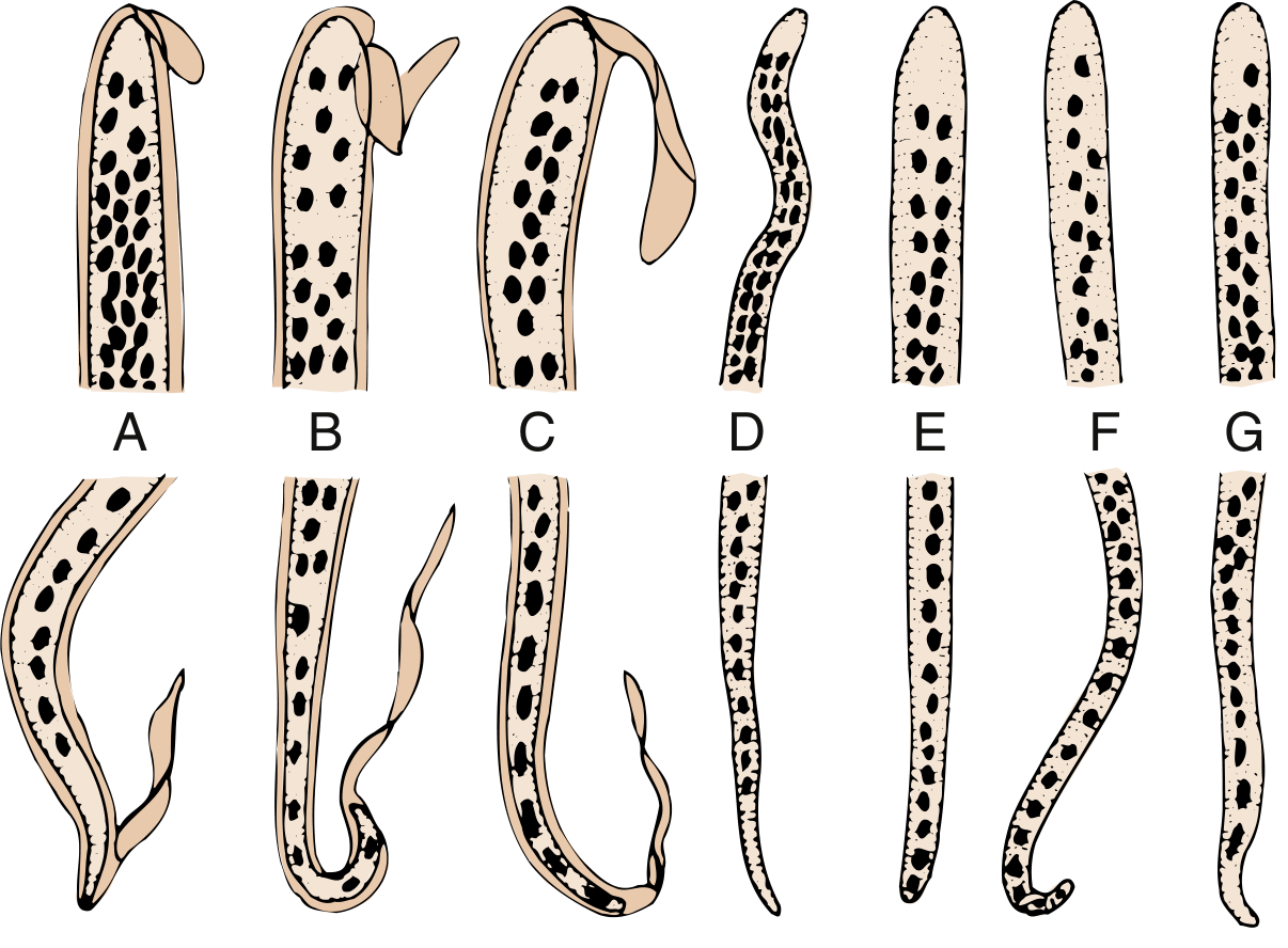
| Species | Sheath | Size | Key PBS features |
|---|---|---|---|
| Wuchereria bancrofti | Present (does not stain with Giemsa) | 245-295 µm | No nuclei in tail tip |
| Brugia malayi | Present (stains bright pink with Giemsa) | 180-230 µm | Two distinct nuclei in tail |
| Loa loa | Present (does not stain) | 250-300 µm | Nuclei extend to tail tip |
| Mansonella perstans | Absent | 150-200 µm | Blunt tail with nuclei |
"W. bancrofti, B. malayi, and Loa loa demonstrate a sheath on their microfilariae. The B. malayi sheath stains bright pink with Giemsa, whereas the W. bancrofti and L. loa sheaths tend not to stain." - Medical Microbiology 9e
- W. bancrofti and B. malayi - nocturnal periodicity (midnight sample required)
- Loa loa - diurnal periodicity (midday sample)
- M. perstans - non-periodic (any time)
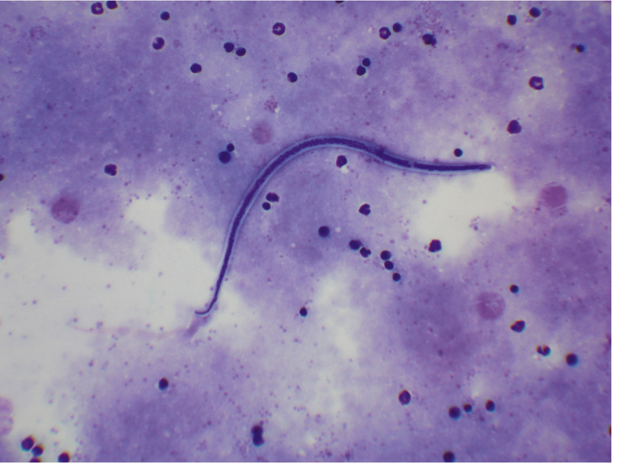
"Eosinophilia is common and a nocturnal peripheral blood smear may show the immature forms, or microfilariae." - Bailey & Love's Short Practice of Surgery
5. Leishmania (Visceral Leishmaniasis / Kala-azar)
- Leishmania amastigotes (Leishman-Donovan bodies) can be seen in peripheral blood, but are more reliably found in buffy coat smears or bone marrow/splenic aspirates
- On PBS: tiny oval organisms (2-4 µm) with a nucleus and kinetoplast, seen within monocytes/macrophages
- LD bodies appear as purple-stained round/oval forms inside mononuclear cells
Summary Table: PBS Findings per Parasite
| Parasite | PBS Finding | Stain | Notes |
|---|---|---|---|
| P. falciparum | Intraerythrocytic rings, banana gametocytes | Giemsa | High parasitemia possible; no enlarged RBCs |
| P. vivax | Enlarged RBCs, Schuffner's dots, amoeboid trophozoites | Giemsa | RBC enlargement key feature |
| P. malariae | Band-form trophozoites, rosette schizonts | Giemsa | Normal RBC size |
| P. ovale | Fimbriated/oval RBCs, Schuffner's dots | Giemsa | Smallest number of merozoites |
| Babesia | Maltese cross tetrad, no hemozoin | Giemsa | Mimics P. falciparum |
| T. brucei | Extracellular trypomastigote with small kinetoplast | Giemsa | Undulating membrane, anterior flagellum |
| T. cruzi | C/U-shaped trypomastigote, large kinetoplast | Giemsa | Amastigotes in tissue, not blood |
| W. bancrofti | Sheathed microfilaria (unsheathed sheath with Giemsa) | Giemsa | Nocturnal periodicity |
| B. malayi | Sheathed microfilaria - pink sheath with Giemsa | Giemsa | Nocturnal periodicity |
| Loa loa | Sheathed microfilaria | Giemsa | Diurnal periodicity |
| Leishmania | Amastigotes (LD bodies) inside monocytes | Giemsa | Better on buffy coat/bone marrow |
Limitations of PBS and Adjuncts
| Limitation | Solution |
|---|---|
| Low sensitivity at parasitemias <40/µL | Thick smear + repeat smears at 12-24h intervals |
| Requires skilled microscopist | QBC (quantitative buffy coat), RDTs |
| Cannot differentiate P. knowlesi from P. malariae | PCR |
| May miss Leishmania in blood | Buffy coat / bone marrow smear |
| Wrong timing for microfilaria | Collect at time of peak periodicity |
"PCR and other molecular assays are more sensitive than the smear, but the detection of low levels of parasitemia does not improve identification of those with significant symptoms. Molecular assays are useful for confirmation of species, which is important for appropriate therapy." - Robbins Pathologic Basis of Disease
5 marks answer
Role of Peripheral Blood Smear (PBS) in Diagnosis of Parasitic Infections
Introduction
Two Types of Smear
| Thick Smear | Thin Smear | |
|---|---|---|
| Purpose | Detection (screening) | Species identification |
| Sensitivity | Detects ~40 parasites/µL | Detects ~1000 parasites/µL |
| RBC morphology | Lost (RBCs lyse) | Preserved |
| Use | Low parasitemia | % parasitemia, morphology |
Parasites Diagnosed on PBS
1. Malaria (Plasmodium spp.)
| Feature | P. falciparum | P. vivax | P. ovale | P. malariae |
|---|---|---|---|---|
| RBC size | Normal | Enlarged | Enlarged, oval/fimbriated | Normal/small |
| Stippling | Maurer's clefts | Schuffner's dots | Schuffner's dots | Ziemann dots (rare) |
| Rings | Multiple/RBC; appliqué; headphone forms | Single, large | Single | Single, thick |
| Trophozoite | Delicate (sequesters) | Amoeboid | Compact | Band-form |
| Gametocyte | Banana/crescent-shaped | Round | Round | Round |
| Schizont | Rarely seen | 12-24 merozoites | 4-12 merozoites | Rosette (6-12) |
2. Babesiosis (Babesia spp.)
- Intraerythrocytic rings resembling P. falciparum
- Maltese cross (tetrad) form - pathognomonic
- No hemozoin pigment (unlike Plasmodium)
- Heterogeneous forms: round, oval, spindled, "racket"
3. Trypanosomiasis
- Extracellular trypomastigotes with nucleus, flagellum, undulating membrane
- T. brucei - small kinetoplast
- T. cruzi - C/U-shaped, large kinetoplast
4. Microfilariae (Filariasis)
- Seen as elongated larvae in blood
- Differentiated by sheath and tail nuclear pattern
- B. malayi sheath stains bright pink with Giemsa; W. bancrofti sheath does not stain
- Timing of collection is critical: nocturnal for W. bancrofti/B. malayi; diurnal for Loa loa
5. Visceral Leishmaniasis
- Leishman-Donovan (LD) bodies - amastigotes seen inside monocytes/macrophages
- Better visualized on buffy coat smear
Limitations
- Low sensitivity at very low parasitemia → repeat smears required
- Cannot differentiate P. knowlesi from P. malariae → PCR needed
- Requires trained microscopist
- Supplemented by: RDTs, PCR, serology, QBC