
Femoral triangle
Reading File
The femoral triangle is a subfascial space in the upper thigh bounded by the inguinal ligament superiorly, the sartorius laterally, and the adductor longus medially. It contains the femoral nerve, artery, vein, and lymphatics (NAVEL from lateral to medial).
Searching sources...
Finding Sources
Searching Images
femoral triangle anatomy boundaries contents
Reading File
Reading File
Loading Image
Loading Image
Reading File
Loading Image
Reading File
Here is a complete account of the femoral triangle, drawn from Gray's Anatomy for Students and the THIEME Atlas of Anatomy.
Femoral Triangle
Overview
The femoral triangle is a wedge-shaped subfascial depression at the junction of the anterior abdominal wall and the upper thigh. Its apex points inferiorly and is continuous with the adductor canal, which carries the femoral vessels distally to the popliteal fossa via the adductor hiatus in adductor magnus.
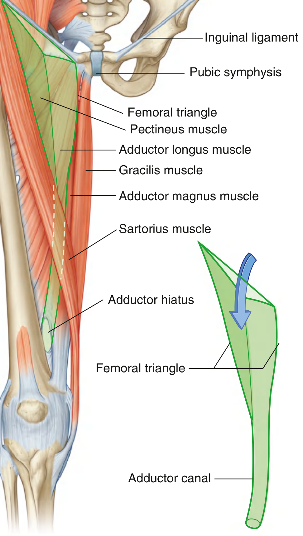
Boundaries
| Structure | Role |
|---|---|
| Inguinal ligament | Base (superior border) - spans ASIS to pubic tubercle |
| Medial margin of sartorius | Lateral border |
| Medial margin of adductor longus | Medial border |
| Apex | Inferior - where sartorius and adductor longus converge |
Roof: Fascia lata (with the saphenous opening / cribriform fascia where the great saphenous vein pierces it)
Floor (from lateral to medial):
- Iliopsoas (lateral)
- Pectineus (medial)
- Adductor longus (most medial)
Contents (lateral to medial: NAVEL)
N - A - V - E - L
| Structure | Notes |
|---|---|
| Nerve (femoral) | Lateral to the femoral sheath; divides rapidly into anterior and posterior divisions + saphenous nerve |
| Artery (femoral) | Continuation of external iliac below inguinal ligament; pulsation palpable midway between ASIS and pubic symphysis |
| Vein (femoral) | Medial to artery; receives the great saphenous vein at the saphenous opening |
| Empty space (femoral canal) | Most medial compartment of femoral sheath; contains lymphatics |
| Lymphatics | In the femoral canal; femoral ring is the superior opening - site of femoral hernias |
Note: The femoral nerve is outside the femoral sheath; only the artery, vein, and lymphatics (within the femoral canal) are enclosed by it.
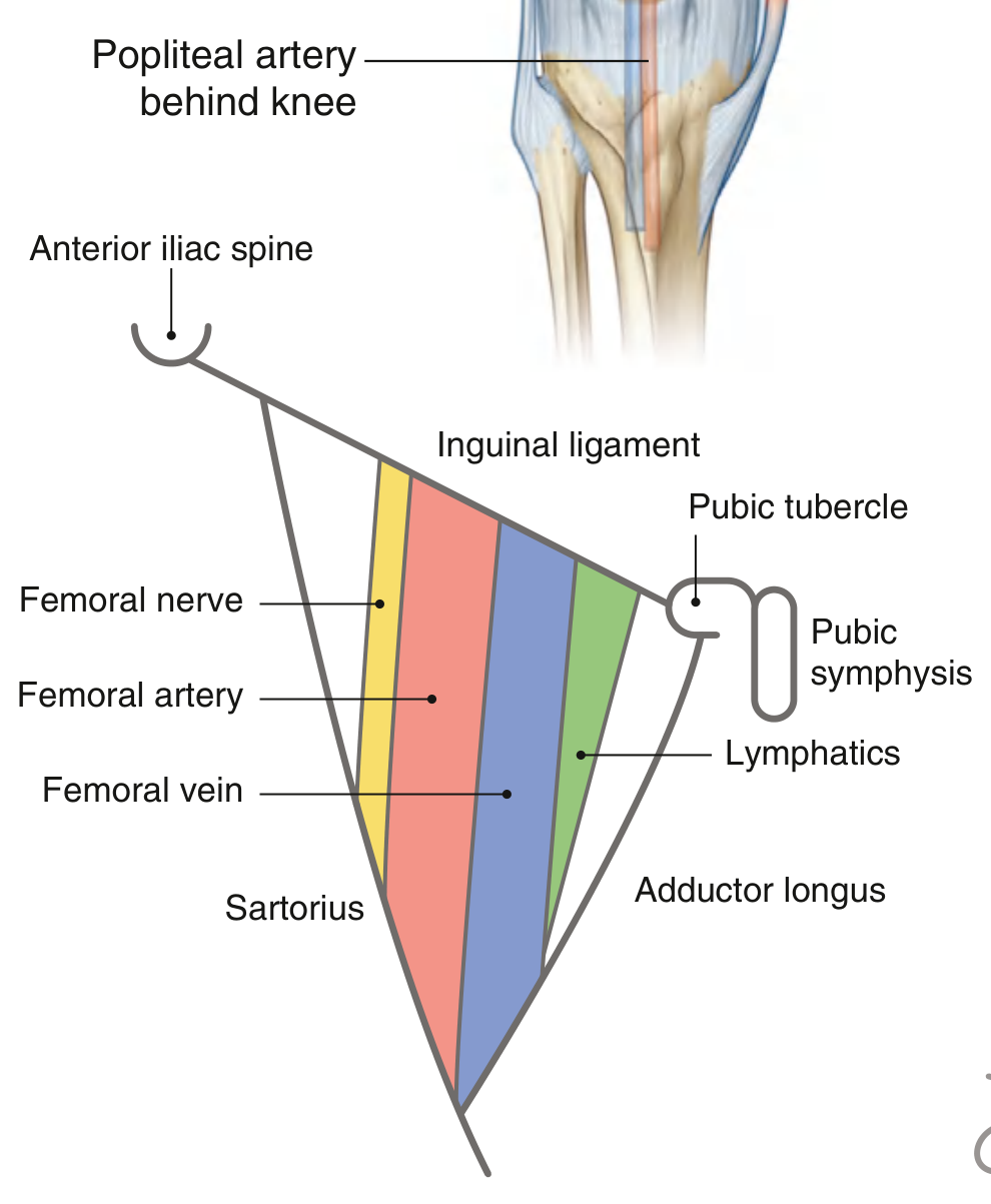
Femoral Sheath
The femoral sheath is a funnel-shaped fascial sleeve that surrounds the femoral artery, vein, and lymphatics. It is continuous superiorly with the transversalis fascia and iliac fascia. It has three compartments:
- Lateral - femoral artery
- Intermediate - femoral vein
- Medial - femoral canal (lymphatics; conical shape; opens superiorly at the femoral ring)
The femoral nerve lies lateral to and outside the sheath.
Detailed view (THIEME Atlas)
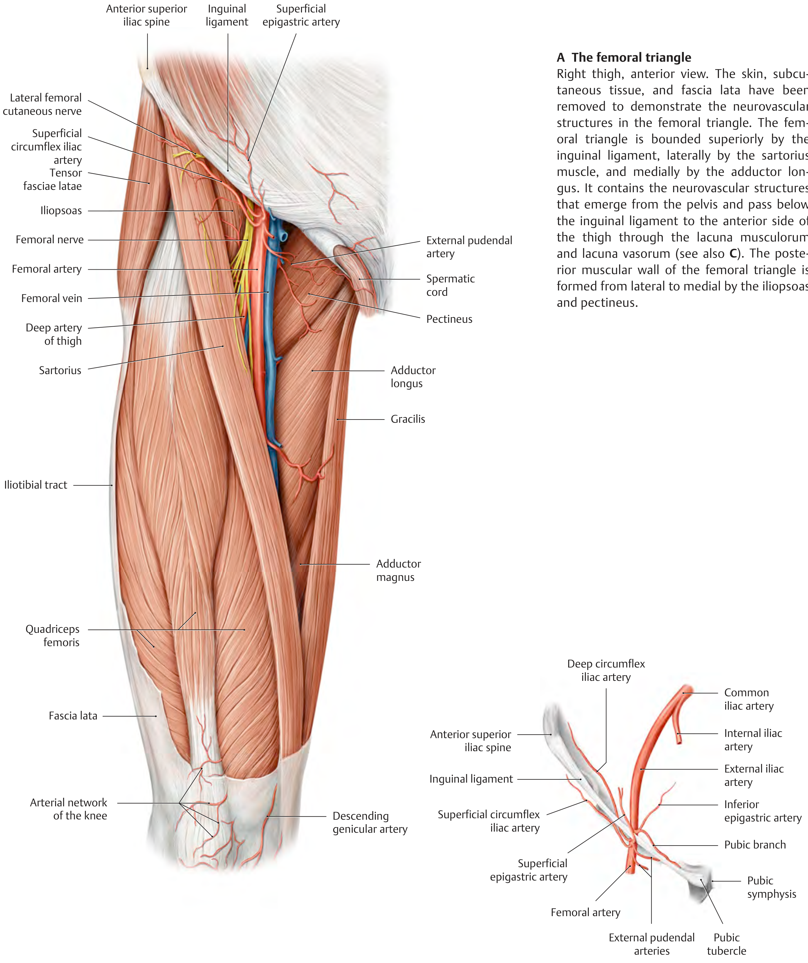
The iliopectineal arch divides the space below the inguinal ligament into:
- Muscular compartment (lateral): transmits iliopsoas, femoral nerve, and lateral femoral cutaneous nerve
- Vascular compartment (medial): transmits femoral artery, vein, and deep inguinal lymphatics
The most medial part of the vascular compartment (medial to the femoral vein) is the femoral ring, covered by the femoral septum. This commonly contains the Rosenmüller node (a deep inguinal lymph node) and is the site of femoral hernias.
Femoral Artery - Surface Marking
The femoral artery is palpable just inferior to the inguinal ligament, at the midpoint between the ASIS and pubic symphysis (the "mid-inguinal point"). This is a key landmark for:
- Femoral pulse assessment
- Femoral artery/vein cannulation
- Cardiac catheterization access (coronary angiography, pulmonary artery catheterization)
- Peripheral vascular procedures
Continuation - Adductor Canal
From the apex of the femoral triangle, the femoral artery and vein enter the adductor canal (Hunter's canal), a fascial tunnel in the medial thigh. They pass through the adductor hiatus in adductor magnus to emerge in the popliteal fossa as the popliteal vessels.
Clinical Relevance
| Condition | Mechanism |
|---|---|
| Femoral hernia | Bowel/omentum protrudes through the femoral ring (medial to femoral vein); more common in women due to wider pelvis |
| Femoral vascular access | Femoral artery/vein cannulated in triangle for cardiac cath, angioplasty, central venous access |
| Femoral nerve injury | Causes weakness of quadriceps (knee extension), loss of patellar reflex, sensory loss over anteromedial thigh and medial leg |
| Femoral artery aneurysm | Presents as pulsatile groin mass |
| Lymphadenopathy | Inguinal/femoral nodes enlarged in lower limb infection, STIs, melanoma |
Sources: Gray's Anatomy for Students, p. 675-676; THIEME Atlas of General Anatomy and Musculoskeletal System, p. 573
This is a shared conversation. Sign in to Orris to start your own chat.