
SLE notes
Reading File
Searching sources...
Finding Sources
Searching PubMed
"systemic lupus erythematosus"[MeSH Terms] AND management
Finding Sources
Good - I have multiple rich sources. Let me pull from the most comprehensive ones simultaneously.
Reading File
Reading File
Reading File
Reading File
Reading File
Reading File
Reading File
Reading File
I now have comprehensive content. Let me compile the full SLE notes.
Loading Image
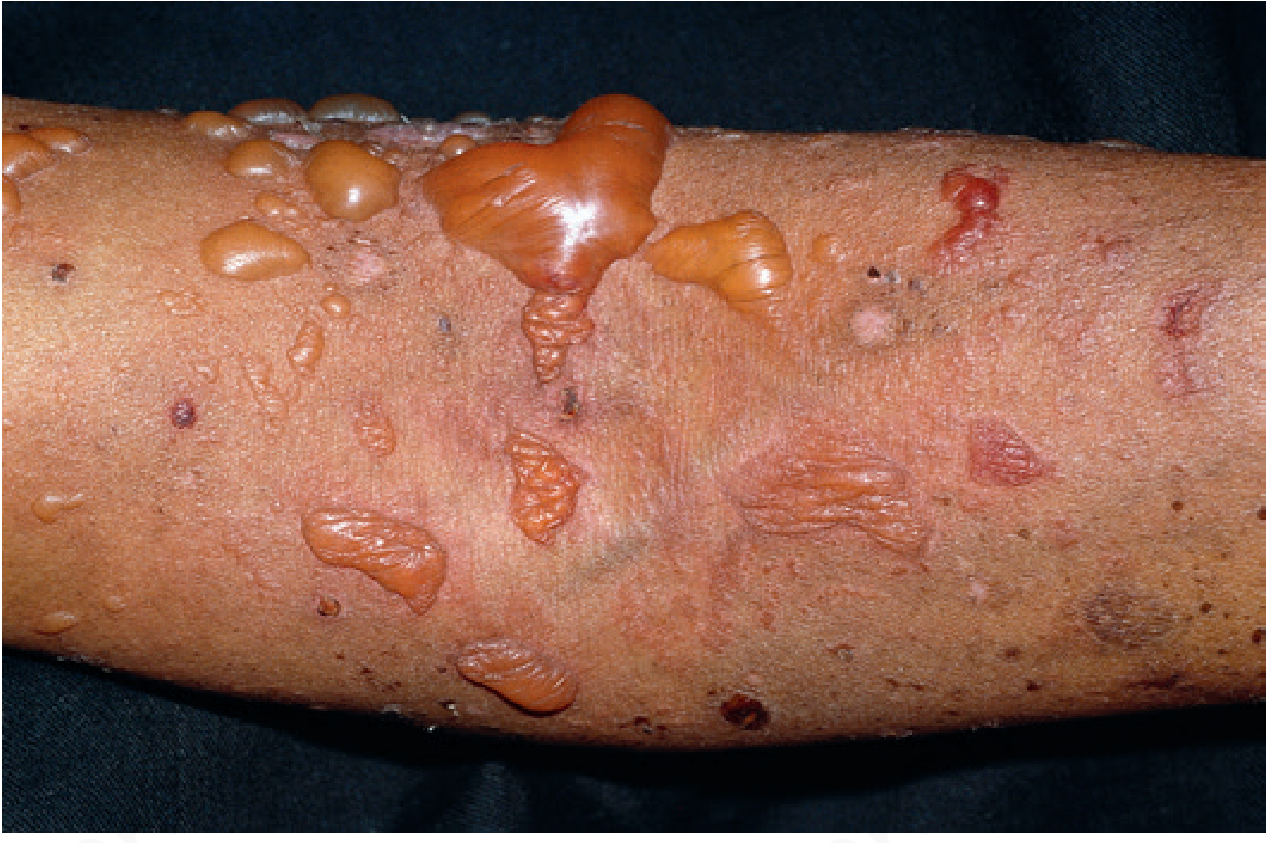
That image is actually bullous lupus lesions. Let me now compile the comprehensive SLE notes.
Systemic Lupus Erythematosus (SLE) - Complete Notes
1. Overview
SLE is a chronic, remitting and relapsing, multisystem autoimmune disease that affects predominantly women. It is considered the classic human immune complex disease.
- Incidence: ~1 in 700 women aged 20-60 years in the US; ~1 in 250 among Black women
- Sex ratio: Female:Male = 10:1
- Principal manifestations: Rashes, arthritis, glomerulonephritis; also hemolytic anemia, thrombocytopenia, neuropsychiatric disorders
Source: Cellular and Molecular Immunology, p. 1274
2. Pathogenesis
SLE is driven by a breakdown of tolerance in self-reactive B and T lymphocytes, triggered by genetic and environmental factors.
Genetic Factors
- HLA alleles: HLA-DR2 or HLA-DR3 each confer an odds ratio of 2-3; both together = ~5
- Complement deficiencies (C1q, C2, or C4) in ~5% of patients - leads to defective clearance of immune complexes and apoptotic cells, and failure of B cell tolerance
- FcγRIIB polymorphism (inhibitory Fc receptor) - may contribute to inadequate B cell activation control
- PTPN22 polymorphism and multiple other genes identified by genome-wide association studies
Environmental Triggers
- UV light - induces apoptosis of skin cells, releases nuclear antigens
Key Pathogenic Mechanisms
- IFN-α signature: Blood cells in SLE patients show striking gene expression indicating exposure to IFN-α (produced mainly by plasmacytoid DCs). Plasmacytoid DCs from SLE patients produce abnormally large amounts of IFN-α.
- TLR activation: Toll-like receptors TLR9 (DNA-recognizing) and TLR7 (RNA-recognizing) activate B cells specific for self nuclear antigens.
- Defective apoptotic clearance: Inadequate clearance of apoptotic nuclei (due to complement defects and nucleases like TREX1) leads to a large burden of nuclear antigens.
- Immune complex deposition: Autoantibodies form complexes with their antigens, which deposit in small arteries and capillaries causing glomerulonephritis, arthritis, and vasculitis.
- Direct autoantibody cytotoxicity: Anti-erythrocyte antibodies → hemolytic anemia; anti-platelet antibodies → thrombocytopenia
Source: Cellular and Molecular Immunology, p. 1274-1275
3. Classification Criteria
ACR Criteria (11 criteria - need ≥4)
| # | Criterion |
|---|---|
| 1 | Malar rash |
| 2 | Discoid rash |
| 3 | Photosensitivity |
| 4 | Oral ulcers (21%) |
| 5 | Arthritis |
| 6 | Proteinuria >0.5 g/day or casts |
| 7 | Neurologic disorders (seizures or psychosis - no other cause) |
| 8 | Pleuritis/pericarditis |
| 9 | Blood abnormalities (hemolytic anemia, leukopenia, thrombocytopenia) |
| 10 | Immunologic disorders (anti-dsDNA, anti-Sm, APLAs, false-positive VDRL) |
| 11 | Positive ANA |
SLICC revision: ≥4 criteria (at least 1 clinical + 1 immunologic), OR biopsy-proven lupus nephritis + ANA or anti-dsDNA. Provides greater sensitivity with equal specificity.
Source: Andrews' Diseases of the Skin, p. 189
2019 EULAR/ACR Weighted Scoring Criteria
Entry criterion: ANA titer ≥1:80 - if absent, do NOT classify as SLE.
Score ≥10 points = SLE. Within each domain, count only the highest-weighted criterion.
| Clinical Domain | Points | Immunologic Domain | Points |
|---|---|---|---|
| Fever | 2 | Anticardiolipin OR anti-β2GPI antibodies | 2 |
| Delirium | 2 | Low C3 OR low C4 | 3 |
| Psychosis | 3 | Low C3 AND low C4 | 4 |
| Seizure | 5 | Anti-dsDNA antibody | 6 |
| Non-scarring alopecia | 2 | ||
| Oral ulcers | 2 | ||
| Subacute cutaneous OR discoid lupus | 4 | ||
| Acute cutaneous lupus | 6 | ||
| Joint involvement | 6 | ||
| Pleural/pericardial effusion | 5 | ||
| Acute pericarditis | 6 | ||
| Leukopenia | 3 | ||
| Thrombocytopenia | 4 | ||
| Autoimmune hemolysis | 4 | ||
| Proteinuria >0.5 g/24h | 4 | ||
| Renal biopsy: class II or V LN | 8 | ||
| Renal biopsy: class III or IV LN | 10 |
Source: Goldman-Cecil Medicine, Table 245
4. Autoantibodies
| Target Antigen | Approximate Frequency (%) |
|---|---|
| Nuclear antigens (ANA) | 99 |
| Double-stranded DNA (anti-dsDNA) | 70 |
| Sm (Smith) nuclear antigen | 38 |
| RNP (U1-RNP) | 33 |
| Ro (SSA) | 49 |
| La (SSB) | 35 |
| Phospholipids (antiphospholipid) | 21 |
| Ribosomal P | 10 |
Key points:
- Anti-dsDNA and anti-Sm are highly specific for SLE
- Anti-dsDNA levels correlate with disease activity (especially nephritis)
- ANA is the most sensitive test (99% frequency)
Source: Goldman-Cecil Medicine
5. Clinical Manifestations
Cutaneous (80% of patients - 4 of 11 ACR criteria are mucocutaneous)
Acute cutaneous LE:
- Butterfly (malar) rash - erythema over malar area and bridge of nose; nasolabial folds characteristically spared (distinguishes from dermatomyositis); may have associated edema; resolves without scarring
- Histology: interface dermatitis, scant perivascular lymphoid infiltrate
Bullous lupus erythematosus:
- Single or grouped vesicles/bullae, often widespread, predilection for sun-exposed areas
- Neutrophils accumulate at DEJ and within dermal papillae
- DIF: IgG, IgM, IgA, or C3 in continuous granular pattern at BMZ
- Most patients are HLA-DR2 positive
- Dramatic response to dapsone (unlike epidermolysis bullosa acquisita)
Other cutaneous findings:
- Vascular lesions in 50% - fingertip/toe edema, erythema, telangiectasia
- Nailfold capillary loops: wandering glomeruloid loops (vs. symmetric dilation in DM/scleroderma)
- Diffuse non-scarring alopecia; "lupus hairs" - short fragile hairs in frontal region
- Discoid lupus (scarring)
- Leg ulcers - deeply punched out
- Mucous membrane lesions in 20-30%: oral erosions, shallow ulcers, gingivitis; erythema and ulceration of hard palate
Source: Andrews' Diseases of the Skin, p. 189
Musculoskeletal
- Arthritis (non-erosive, unlike RA) - one of the most common manifestations
Renal - Lupus Nephritis
Classified by biopsy (ISN/RPS classification):
| Class | Description |
|---|---|
| I | Minimal mesangial - immune complex deposits without mesangial hypercellularity |
| II | Mesangial proliferative - mesangial deposits with mesangial hypercellularity |
| III | Focal glomerulonephritis - <50% of glomeruli involved |
| IV | Diffuse glomerulonephritis - ≥50% of glomeruli involved (most severe) |
| V | Membranous lupus nephritis |
| VI | Advanced sclerotic lesions |
Additional lesions: lupus podocytopathy, collapsing glomerulopathy, thrombotic microangiopathy (often with antiphospholipid syndrome).
Neuropsychiatric (NPSLE)
- MRI: white matter lesions, cerebral infarction, venous sinus thrombosis, atrophy (seen in 19-70%)
- Manifestations: seizures (5 pts in EULAR criteria), psychosis (3 pts), delirium (2 pts)
- MR angiography and spectroscopy for cerebral blood flow and neuronal metabolism assessment
Cardiopulmonary
- Pericarditis, pleuritis, pleural/pericardial effusions
- Diffuse alveolar hemorrhage (DAH) - rare but potentially catastrophic complication
- Mortality up to 62%
- Almost invariably accompanied by other active SLE manifestations
- Glomerulonephritis present in >90% of DAH cases
- Presents with bilateral alveolar infiltrates, dyspnea, hypoxemia; hemoptysis in minority at presentation
- Histology: intra-alveolar hemorrhage and capillaritis, without macroscopic necrosis
- BAL is adequate for diagnosis in critically ill patients (avoid biopsy in severe DAH)
- Factors associated with mortality: MV, infection, prior cyclophosphamide use
Source: Fishman's Pulmonary Diseases, p. 4263-4276
Hematologic
- Leukopenia, thrombocytopenia, autoimmune hemolytic anemia
Antiphospholipid syndrome overlap
- Increased thrombosis risk; thrombotic microangiopathy
6. Treatment
Counseling & General
- Avoid UV light exposure
- Adequate rest
- Sunscreen
Mild-Moderate Disease (no major organ involvement)
- NSAIDs - for arthralgia, serositis, constitutional symptoms
- Low-dose corticosteroids - prednisone 5-30 mg/day; effective for constitutional symptoms, arthralgias, pericarditis, pleuritis, skin disease; disease may flare if maintenance doses as low as 5 mg/day are stopped
- Hydroxychloroquine (HCQ) - 200-400 mg/day (max ≤5 mg/kg actual body weight/day)
- Controls skin and arthralgias; used routinely in most lupus patients
- Associated with decreased incidence of thrombosis
- Well tolerated; most serious toxicity = retinal damage (cumulative dose-dependent)
- Ophthalmologic exam before starting, then every 12 months
Serious Organ-System Disease
- Induction therapy for proliferative lupus nephritis:
- Option A: Mycophenolate mofetil (MMF) 2-3 g/day + IV methylprednisolone 500-2500 mg over 1-3 days then oral prednisone 0.3-0.5 mg/kg/day
- Option B: Low-dose cyclophosphamide (500 mg IV every 2 weeks × 6 doses) + IV methylprednisolone then oral prednisone
- Multitarget therapy (tacrolimus 4 mg/day + MMF 1 g/day + pulse methylprednisolone then prednisone) is superior to IV CYC for induction
- Voclosporin (23.7 mg BID) + background immunosuppression: improves clinical response in LN
- Belimumab (10 mg/kg IV or 200 mg SC weekly) + background immunosuppression: approved for LN
- Target: reduce prednisone to ≤7.5 mg/day by 3-6 months
- Maintenance: MMF preferred over azathioprine
- CNS disease, severe flares: High-dose CS ± cyclophosphamide or MMF
- Pericarditis: Prednisone 20-30 mg/day
Biologics & Emerging Therapies
| Drug | Mechanism | Indication |
|---|---|---|
| Belimumab | Anti-BAFF (B-cell survival factor) | Moderate-sustained SLE activity; lupus nephritis |
| Anifrolumab | Anti-type I interferon receptor (300 mg IV q4w) | Moderate-severe SLE (FDA approved) |
| Rituximab | Anti-CD20, B-cell depleting | Refractory SLE |
| Obinutuzumab | 2nd-gen anti-CD20 | Promising for refractory disease |
| Voclosporin | Calcineurin inhibitor | Lupus nephritis |
| Deucravacitinib | JAK inhibitor | Under investigation |
| Daprolizumab pegol | Anti-CD40 ligand | Under investigation |
| Low-dose IL-2 | Promotes T-reg expansion | Under investigation |
Cyclophosphamide
- Given to ~33% of lupus patients during disease course
- Dose: 0.5-1 g/m² BSA IV monthly × 6 months, then quarterly × 2 years + oral prednisone
- Toxicities: hemorrhagic cystitis (give mesna + hydration), infertility, infection, malignancy
Source: Goldman-Cecil Medicine, p. 2817-2818
7. Monitoring
| Parameter | Frequency |
|---|---|
| Ophthalmology (for HCQ retinal toxicity) | Before starting, then annually |
| Anti-dsDNA levels | Monitor disease activity |
| Complement (C3, C4) | Low levels = active disease |
| Urinalysis, creatinine | Renal monitoring |
| CBC | Hematologic involvement |
8. Prognosis & Complications
- Chronic remitting-relapsing course
- Major causes of morbidity: nephritis, NPSLE, cardiovascular disease, infections (from immunosuppression)
- Hydroxychloroquine reduces organ damage and thrombosis over time
- DAH carries mortality up to 62%
Sources:
- Cellular and Molecular Immunology, p. 1274-1275
- Goldman-Cecil Medicine, International Edition, Table 245, p. 2817-2818
- Andrews' Diseases of the Skin: Clinical Dermatology, p. 189
- Fishman's Pulmonary Diseases and Disorders, p. 4263-4282
This is a shared conversation. Sign in to Orris to start your own chat.