
Internal capsule
It looks like this message only contains internal budget/session information and no actual question or task. Could you let me know what you'd like help with?
Internal capsule for 6marks
I still don't see an actual question or topic in your message — just budget/session metadata again. Could you tell me what topic or question you'd like a "6 marks" answer for (e.g., a specific medical, tax, or exam topic)? Once you specify the subject, I can research it properly and give you a well-structured 6-mark answer.
Neuro anatomy
"Neuroanatomy" is a huge topic - I need to know the specific structure/subject for a focused 6-mark answer.
Asking for Preferences
Which neuroanatomy topic do you want the 6-mark answer on?
White matter of cerebrum
It looks like this message only contains internal budget/session information and no actual question or task. Could you let me know what you'd like help with?I still don't see an actual question or topic in your message — just budget/session metadata again. Could you tell me what topic or question you'd like a "6 marks" answer for (e.g., a specific medical, tax, or exam topic)? Once you specify the subject, I can research it properly and give you a well-structured 6-mark answer."Neuroanatomy" is a huge topic - I need to know the specific structure/subject for a focused 6-mark answer.
Reading File
Finding Sources
Finding Sources
Finding Sources
Finding Sources
Reading File
Finding Sources
Reading File
Finding Sources
Let me read the continuation covering commissural and association fibers (lines 320-380) to complete the picture.
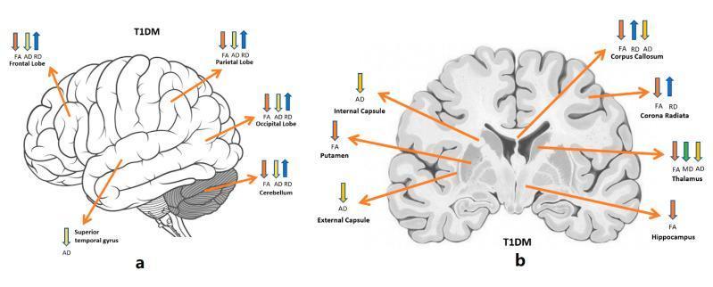
White Matter of the Cerebrum (6 Marks)
Definition
The white matter of the cerebrum lies deep to the cortical grey matter and consists of billions of myelinated axons that give it its characteristic pale color. These fibers carry information to, from, and between areas of the cerebral cortex. White matter tracts are classified into three functional groups: projection fibers, commissural fibers, and association fibers (Kaplan & Sadock's Comprehensive Textbook of Psychiatry, p. 197).
1. Projection Fibers
Connect the cerebral cortex with subcortical structures (brainstem, spinal cord, thalamus).
- Corticofugal fibers - originate in cortex, project to subcortical targets (e.g., corticospinal, corticobulbar, corticopontine, corticothalamic tracts).
- Corticopetal fibers - originate outside the telencephalon and project to the cortex (e.g., thalamocortical fibers).
- These fibers converge to pass through the internal capsule, a compact bundle bordered laterally by the lentiform nucleus (putamen + globus pallidus) and medially by the thalamus and caudate head. It has five parts:
- Anterior limb, genu (corticobulbar fibers), posterior limb (corticospinal fibers)
- Retrolenticular part - carries the optic radiation (lateral geniculate body to visual cortex)
- Sublenticular part - carries the auditory radiation (medial geniculate body to auditory cortex)
- Above the internal capsule, these fibers fan out to form the corona radiata. (Kaplan & Sadock's Comprehensive Textbook of Psychiatry, p. 196-198)
2. Commissural Fibers
Interconnect corresponding (and some non-corresponding) areas of the two cerebral hemispheres.
- Corpus callosum - the largest fiber bundle in the brain (~300 million axons); has four parts - rostrum, genu, body, and splenium. Connects mostly homotopic cortical areas, except the hand areas of the primary motor/sensory cortex and most of the primary visual cortex.
- Anterior commissure - lies in front of the fornix, connects the two temporal lobes and olfactory structures.
- Posterior commissure - connects caudal diencephalic structures.
- Hippocampal commissure - connects the two hippocampal formations. (Kaplan & Sadock's Comprehensive Textbook of Psychiatry, p. 198-200)
3. Association Fibers
Connect cortical areas within the same hemisphere.
- Short association (U) fibers - connect adjacent gyri within the same lobe.
- Long association fibers (5 major tracts):
- Superior longitudinal fasciculus - frontal, parietal, occipital cortices
- Arcuate fasciculus - frontal and temporal lobes (important in language; injury causes conduction aphasia)
- Uncinate fasciculus - orbitofrontal cortex to anterior temporal lobe
- Inferior occipitofrontal fasciculus - occipital to frontal lobe
- Cingulum - beneath the cingulate gyrus, connecting it to the parahippocampal gyrus
- Also: inferior longitudinal fasciculus (temporal-occipital), external capsule (between claustrum and putamen), extreme capsule (between claustrum and insula). (Kaplan & Sadock's Comprehensive Textbook of Psychiatry, p. 201-202)
Clinical Relevance (brief)
- Lacunar infarcts of the internal capsule/corona radiata produce pure motor hemiparesis or ataxic hemiparesis.
- Arcuate fasciculus damage causes conduction aphasia (impaired repetition with preserved comprehension and fluency).
- Corpus callosum, internal capsule, and cingulum white matter changes are implicated in schizophrenia, depression, and Alzheimer disease on DTI/MRI studies.
Diagram tip for exam: Draw a coronal section showing the corpus callosum arching over the lateral ventricles, the internal capsule between the thalamus/caudate and lentiform nucleus, and the corona radiata fanning up to the cortex - label projection (internal capsule), commissural (corpus callosum), and association (external/extreme capsule, cingulum) fibers.
Internal capsule
Finding Sources
Finding Sources
Reading File
Finding Sources
Reading File
I now have comprehensive material. Here's the 6-mark answer on the internal capsule.
Searching Images
internal capsule anterior limb posterior limb genu horizontal section basal ganglia thalamus diagram
Internal Capsule (6 Marks)
Definition
The internal capsule is a compact, curved lamina of white matter forming the largest projection fiber tract of the cerebrum. It carries afferent and efferent nerve fibers passing between the cerebral cortex and the brainstem/spinal cord. It is continuous rostrally with the corona radiata and caudally with the cerebral peduncle (Localization in Clinical Neurology, 8e, p. 30).
Location and Relations
On a horizontal (axial) section, the internal capsule is curved with its convexity facing medially (inward). It lies:
- Laterally - lentiform nucleus (putamen + globus pallidus)
- Medially - caudate nucleus (anteriorly) and thalamus (posteriorly)
Five Parts and Their Fiber Content
| Part | Location | Fibers Carried |
|---|---|---|
| Anterior limb (~2 cm) | Between caudate nucleus (medial) and lentiform nucleus (lateral) | Frontopontine fibers, thalamocortical/corticothalamic fibers, caudatolenticular fibers |
| Genu | Bend at the level of the interventricular foramen (of Monro) | Corticobulbar fibers (explains facial and lingual weakness with mild limb involvement in "capsular genu syndrome") |
| Posterior limb (3-4 cm) | Between thalamus (medial) and lentiform nucleus (lateral) | Corticospinal fibers (somatotopically arranged - arm fibers anterior, then trunk, then leg fibers posteriorly), corticorubral, corticothalamic, and thalamocortical sensory fibers |
| Retrolenticular part | Behind (posterior to) the lentiform nucleus | Optic radiation (geniculocalcarine/Gratiolet's tract) from lateral geniculate body to visual cortex; also corticotectal and corticonigral fibers |
| Sublenticular part | Below (inferior to) the lentiform nucleus | Auditory radiation from medial geniculate body to auditory cortex |
(Localization in Clinical Neurology, 8e, p. 30-31; Neuroanatomy through Clinical Cases, 3rd ed.; Gray's Anatomy for Students)
Blood Supply
- Anterior limb - recurrent artery of Heubner (branch of anterior cerebral artery)
- Genu and middle/inferior posterior limb - anterior choroidal artery (from internal carotid artery)
- Superior anterior and posterior limb - lenticulostriate arteries (branches of middle cerebral artery)
Clinical Significance
- Because motor, sensory, visual, and auditory fibers are tightly packed in a small area, even a small lacunar infarct (commonly from lenticulostriate artery occlusion) can cause dense contralateral hemiplegia with relatively little cortical deficit (pure motor stroke).
- Anterior choroidal artery syndrome - contralateral hemiplegia, hemianesthesia, and homonymous hemianopia (posterior limb + retrolenticular involvement).
- Capsular genu syndrome - facial and lingual weakness with only mild limb weakness.
- The internal capsule is a classic site of hypertensive intracerebral hemorrhage from rupture of lenticulostriate ("Charcot's artery of cerebral hemorrhage") branches.
Exam tip: Draw a horizontal section showing the caudate, thalamus, and lentiform nucleus with the capsule curving between them - label anterior limb, genu, posterior limb, retrolenticular, and sublenticular parts, and mark the corticospinal tract, optic radiation, and auditory radiation.
This is a shared conversation. Sign in to Orris to start your own chat.