I now have more than enough information for a comprehensive 30-mark PG-level answer. Let me compile it:
Carcinoma of the Cervix - A Comprehensive 30-Mark Answer
Introduction
Carcinoma of the cervix is the second most common gynecological malignancy worldwide and the most common in developing countries. It remains one of the leading causes of cancer-related mortality in women in low- and middle-income nations, including India. It is largely a preventable disease, caused predominantly by human papillomavirus (HPV) infection.
Incidence and Epidemiology
- Accounts for ~6.6% of all female cancers globally; incidence is highest in Sub-Saharan Africa and South Asia.
- Peak incidence: 45-55 years for invasive cancer; precursor lesions (CIN) peak at ~30 years.
- In India, it is the most common gynecological cancer.
Etiology and Risk Factors
Primary Cause: HPV
HPV DNA is detectable in >99% of cervical cancers. High-risk HPV types 16 and 18 account for approximately 70% of cases (Robbins & Kumar Basic Pathology).
Risk Factors (mnemonic - SHEMP)
| Factor | Detail |
|---|
| Sexual behavior | Early coitarche (<16 yr), multiple sexual partners, partner with multiple partners |
| HPV infection | Types 16, 18 (high-risk); 6, 11 (low-risk, condylomas) |
| Smoking | Acts as co-carcinogen; impairs local immunity |
| Multiparity / high parity | Repeated birth trauma to cervix |
| Oral contraceptive use | RR 1.6 at 5-9 years use; 2.2 at ≥10 years use (returns to baseline after cessation) (Berek & Novak) |
| Immunosuppression | HIV/AIDS, transplant recipients |
| Low socioeconomic status | Lack of screening access |
| STIs | Herpes simplex virus 2 as co-factor |
Pathogenesis
HPV Oncogenesis (from Robbins Pathology)
HPV is a double-stranded DNA virus. The transformation zone (squamo-columnar junction) at the cervical os is the primary target, as it contains immature metaplastic squamous cells.
Key molecular events:
- HPV infects immature squamous cells in the transformation zone via micro-abrasions.
- Viral E6 protein binds and degrades p53 (tumor suppressor) and upregulates telomerase.
- Viral E7 protein binds retinoblastoma (Rb) protein, releasing E2F transcription factors, driving cell cycle progression.
- High-risk HPV (16, 18) integrates into host genome - this disrupts the negative regulator of E6/E7, increasing their expression, and leads to genomic instability.
- Low-risk HPV (6, 11) remains episomal - E7 binds Rb with low affinity; E6 does not bind p53.
- Persistent infection + co-carcinogens --> CIN --> invasion.
Progression: CIN → Invasive Carcinoma
- LSIL (CIN I): HPV cytopathic effect (koilocytes); usually regresses
- HSIL (CIN II/III): Full-thickness dysplasia; higher risk of progression
- Invasive carcinoma: Breach of basement membrane
- SIL peaks at age 30; invasive cancer peaks at age 45 - suggesting a 10-15 year window for intervention
Pathology
Histological Types
| Type | Frequency | Key Features |
|---|
| Squamous cell carcinoma | 70-80% | Arises at transformation zone |
| Adenocarcinoma | 15-20% | Arises from endocervical glands; increasing incidence; harder to detect on Pap |
| Adenosquamous carcinoma | 5% | Mixed |
| Small cell / neuroendocrine | Rare | Most aggressive; oat cell type; immunohistochemistry needed |
Squamous Cell Carcinoma Variants (Berek & Novak)
- Large cell keratinizing: Keratin pearls; best prognosis
- Large cell non-keratinizing: Individual cell keratinization; no pearls
- Small cell anaplastic: Resembles oat cell lung cancer; worst prognosis; diffuse infiltration
Spread
- Direct: Parametrium, vagina, bladder, rectum
- Lymphatic: Paracervical → obturator → internal/external iliac → common iliac → para-aortic nodes (most important route)
- Hematogenous: Lungs, liver, bone (late, uncommon)
Clinical Features
Symptoms
- Early: Asymptomatic (detected on screening); post-coital bleeding, intermenstrual bleeding, vaginal discharge (watery, then bloody, then offensive)
- Advanced: Continuous bleeding, pelvic pain, low back pain (sciatic involvement), urinary symptoms (frequency, hematuria, fistula), rectal bleeding, leg edema (lymphatic obstruction), weight loss, cachexia
Signs
- Cervix may appear normal (microinvasive), show an ectocervical erosion, exophytic cauliflower growth, ulcer, or barrel-shaped cervix
- Parametrial involvement felt on pelvic/rectal examination
- Nodal enlargement on abdominal examination
Diagnosis and Investigations
1. Screening
- Pap smear (Papanicolaou): Mainstay of screening since mid-20th century; sensitivity ~51%, specificity ~98%
- Bethesda System classification: ASC-US, LSIL, HSIL, ASC-H, AGC, carcinoma
- HPV co-testing: Combined Pap + HPV DNA test; recommended from age 30 (ACOG)
- VIA (Visual Inspection with Acetic Acid): Low-cost alternative for low-resource settings; aceto-white lesions indicate CIN
2. Colposcopy
Indicated for: abnormal Pap smear, positive HPV, VIA positive. Allows directed biopsy.
- Aceto-white epithelium, abnormal vascular patterns (punctation, mosaic, atypical vessels)
- Satisfactory colposcopy: squamo-columnar junction fully visible
3. Biopsy
- Punch biopsy: From colposcopically directed site
- Cone biopsy (conization): For microinvasive disease - diagnostic and potentially therapeutic for stage IA1
4. Staging Investigations (FIGO 2018 allows imaging)
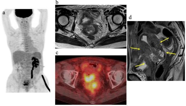
- MRI pelvis: Gold standard for local tumor extent, parametrial invasion, vaginal involvement
- CT chest/abdomen/pelvis: Lymph node metastases, systemic spread
- PET-CT: Most sensitive for lymph node staging (especially para-aortic), recurrence
- Cystoscopy + rectosigmoidoscopy: For advanced disease (stages III-IV)
- IVP/renal ultrasound: Hydronephrosis in stage IIIB
- CXR: Pulmonary metastases
- Exam under anesthesia (EUA)
- Tumor markers: SCC antigen (squamous), CA 125 (adenocarcinoma) - monitoring treatment response
Staging: FIGO 2018
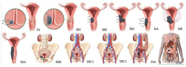
The 2018 FIGO update allowed imaging and pathology to be incorporated into staging (previously clinical only).
| Stage | Description |
|---|
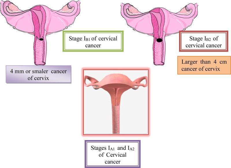
| IA | Microscopic only; depth ≤5 mm |
| IA1 | Stromal invasion ≤3 mm depth |
| IA2 | Stromal invasion >3 mm, ≤5 mm |
| IB | Clinically visible lesion or microscopic >IA |
| IB1 | ≤2 cm greatest dimension |
| IB2 | >2 cm, ≤4 cm |
| IB3 | >4 cm |
| IIA | Beyond uterus; no parametrial invasion |
| IIA1 | ≤4 cm |
| IIA2 | >4 cm |
| IIB | Parametrial invasion (no pelvic wall) |
| IIIA | Lower 1/3 vagina; no pelvic wall |
| IIIB | Pelvic wall and/or hydronephrosis |
| IIIC1 | Pelvic lymph node metastasis (r = radiological, p = pathological) |
| IIIC2 | Para-aortic lymph node metastasis |
| IVA | Bladder/rectal invasion |
| IVB | Distant metastases |
Key rule: If doubt exists about a stage, assign the lower (earlier) stage. Once treatment begins, stage must not be changed.
Incidence at diagnosis (old 2008 data): Stage I - 38%; Stage II - 32%; Stage III - 26%; Stage IV - 4%.
Treatment
Summary by Stage (Berek & Novak Table 38-4 concept)
| Stage | Treatment |
|---|
| IA1 (no LVSI) | Cone biopsy (fertility desired) or simple/extrafascial hysterectomy |
| IA1 (LVSI present) | Modified radical hysterectomy + pelvic lymphadenectomy |
| IA2 | Modified radical hysterectomy + pelvic lymphadenectomy OR radiotherapy |
| IB1-IIA1 | Radical hysterectomy (Wertheim's) + pelvic LND OR concurrent chemoradiation |
| IB2-IIA2 | Concurrent chemoradiation (preferred) ± brachytherapy; surgery if chemo-RT not feasible |
| IIB-IVA | Concurrent chemoradiation + brachytherapy (standard of care) |
| IVB | Palliative chemotherapy; cisplatin-based ± bevacizumab |
5-year survival rate for Stage I: ~85% with either radical surgery or radiation.
Surgical Treatment
Wertheim's Radical Hysterectomy (Type III)
- Includes: uterus + cervix + upper 1/3 vagina + parametrium + paracervical tissue + pelvic lymph nodes
- Indicated: Stages IA2, IB, IIA in medically fit patients
- Ovaries can be conserved (metastasis in only 0.9% of early disease)
- Advantages over radiotherapy: Preserves ovarian function, avoids radiation side effects, allows pathologic assessment of nodes
Wertheims Hysterectomy vs Radiotherapy
| Parameter | Surgery | Radiotherapy |
|---|
| Ovarian preservation | Yes | No |
| Vaginal function | Better (stretches with use) | Worse (fibrosis, atrophy) |
| Bladder/bowel morbidity | Fistula <2%; repairable | 8% chronic; difficult to repair |
| Age | Younger women | Any age, especially older |
Minimally Invasive vs Open (Critical Update)
A landmark RCT showed MIS radical hysterectomy was inferior to open surgery:
- Disease-free survival: MIS 86.0% vs Open 96.5% at 4.5 years
- Overall survival (3-year): MIS 91.2% vs Open 97.1%
- Possible cause: Uterine manipulator spreading malignant cells; CO2 insufflation
- Current recommendation: Open radical hysterectomy is preferred for cervical cancer.
Trachelectomy (Fertility-Sparing)
- Radical trachelectomy (cervical removal + pelvic nodes) with uterine preservation
- Indicated: Stage IA2 to IB1, tumor ≤2 cm, no lymph node involvement, desire for future fertility
Sentinel Lymph Node (SLN) Mapping
- Injection of blue dye/Tc-99m/ICG into cervix (3 and 9 o'clock)
- SLN most commonly at: medial to external iliac, ventral to hypogastric, superior obturator space
- NCCN recommends for tumors <2 cm
Radiation Therapy
External Beam Radiotherapy (EBRT)
- Whole pelvis radiation (45-50 Gy in 25-28 fractions)
- Can sterilize pelvic lymph nodes and primary tumor
Brachytherapy (Intracavitary/Interstitial)
- Essential component for local tumor control
- LDR (Cs-137): Inpatient; 1-3 days continuous
- HDR (Ir-192): Outpatient; multiple fractions; modern standard
- Classic 2D: Point A (2 cm superior to external os + 2 cm lateral to canal) = 7,000-8,000 cGy
- Modern 3D image-guided brachytherapy (IGA-BT): MRI/CT-planned; 8,500-9,500 cGy to HR-CTV D90; reduces toxicity by half compared to 2D (French STIC trial)
- Dose limits: Bladder <90 Gy EQD2; Rectum/Sigmoid <75 Gy EQD2
Concurrent Chemoradiation (CRT) - Standard for Locally Advanced Disease
Regimen: Cisplatin 40 mg/m² weekly + external beam RT + brachytherapy
Evidence (Berek & Novak):
- GOG Protocol 85: Cisplatin + 5-FU + RT vs hydroxyurea + RT - CRT showed significantly improved PFS and OS
- GOG Protocol 120: Weekly cisplatin arm showed superior results vs hydroxyurea; established weekly cisplatin as standard
- Intergroup trial (stages IA2-IIA with high-risk features post-surgery): Chemoradiation 4-yr survival 81% vs radiation alone 71%
- Current standard: Cisplatin-based concurrent chemoradiation for stages IIB-IVA and high-risk post-surgical patients
High-risk features post radical hysterectomy requiring adjuvant CRT:
- Positive pelvic lymph nodes
- Positive parametrial margins
- Positive vaginal margins
Intermediate-risk features requiring adjuvant RT:
- LVSI present + >1/3 stromal invasion + large tumor (Sedlis criteria)
Prognosis by Stage
| Stage | 5-Year Survival |
|---|
| IA | >95% |
| IB | 80-90% |
| IIA | 75-85% |
| IIB | 65-70% |
| IIIA-B | 40-50% |
| IVA | 15-20% |
| IVB | <5% |
Prognostic factors (adverse): Positive lymph nodes, parametrial involvement, LVSI, large tumor, poor differentiation, adenocarcinoma histology, small cell type.
Cervical Cancer in Pregnancy
- Most common gynecological cancer in pregnancy (1 in 1,000-10,000 pregnancies)
- Diagnosis: Colposcopy safe in pregnancy; biopsy if clinically suspicious
- Stage IA1: Delay treatment until delivery; cone biopsy if needed
- Stages IA2-IB1: Delay to fetal maturity (>34 weeks) acceptable; neoadjuvant cisplatin possible
- Advanced disease, early pregnancy: Immediate treatment with termination
- Mode of delivery: Caesarean section preferred to avoid seeding in birth canal; classical uterine incision
Recurrent Cervical Cancer
- Central pelvic recurrence (post-radiation): Pelvic exenteration (total - bladder + uterus + rectum with stomas; anterior - bladder only; posterior - rectum only)
- Isolated lateral recurrence: Chemoradiation if not previously radiated
- Distant recurrence: Palliative systemic chemotherapy
- Chemotherapy regimens: Cisplatin + paclitaxel ± bevacizumab (anti-VEGF monoclonal antibody; GOG 240 trial showed survival benefit)
Prevention
Primary Prevention - HPV Vaccination
| Vaccine | Types Covered | Doses |
|---|
| Cervarix (bivalent) | 16, 18 | 2 (9-14 yr); 3 (≥15 yr) |
| Gardasil (quadrivalent) | 6, 11, 16, 18 | 2-3 |
| Gardasil-9 (nonavalent) | 6, 11, 16, 18, 31, 33, 45, 52, 58 | 2-3 |
A 2025 Cochrane network meta-analysis (PMID: 41276263) confirmed HPV vaccination is effective for prevention of cervical cancer and other HPV-related diseases.
Key points:
- Best given before sexual debut (9-12 years, girls and boys)
- India's national program: Cervavac (quadrivalent, indigenously produced)
- WHO's 90-70-90 strategy: 90% girls vaccinated by 15; 70% of women screened by 35 and 45; 90% of women identified with disease treated
Secondary Prevention - Screening
- Cytology (Pap smear): Every 3 years from age 21-65
- Co-testing (Pap + HPV): Every 5 years from age 30-65
- Primary HPV testing: Emerging as preferred in many guidelines (2025 Canadian guidelines update - PMID: 41152983)
- VIA/VILI: For resource-limited settings
Summary Flowchart for Exam
Cervical Cancer
├── Etiology: HPV 16/18 (70%) → E6 degrades p53, E7 inactivates Rb
├── Precursor: CIN I → II → III → Invasion (10-15 yr window)
├── Histology: SCC 75%, Adenocarcinoma 15-20%
├── Diagnosis: Pap smear → colposcopy → biopsy → staging
├── Staging: FIGO 2018 (allows imaging + pathology)
│   ├── IA: Microscopic (≤5 mm depth)
│   ├── IB: Clinically visible, confined to cervix (IB1 ≤2cm, IB2 2-4cm, IB3 >4cm)
│   ├── II: Beyond uterus (IIA = no parametrium; IIB = parametrium)
│   ├── III: Pelvic wall/lower vagina/hydronephrosis/LN (IIIC new in 2018)
│   └── IV: Bladder/rectum (IVA); distant (IVB)
├── Treatment
│   ├── Early (IA-IIA): Surgery (Wertheim's) OR chemoradiation - equivalent 5yr survival ~85%
│   ├── Advanced (IIB-IVA): Concurrent cisplatin chemoradiation + brachytherapy
│   └── Recurrence: Pelvic exenteration (central) / palliative CRT/chemo
└── Prevention: HPV vaccine + Pap/HPV screening
Sources: Berek & Novak's Gynecology (9th ed.) | Robbins & Kumar Basic Pathology | Goldman-Cecil Medicine | PMID 41276263 (Cochrane 2025 - HPV vaccination meta-analysis) | PMID 41152983 (Canadian cervical screening guidelines 2025)