
Give the treatment of UTI in short using algorithm
UTI treatment algorithm urinary tract infection management flowchart
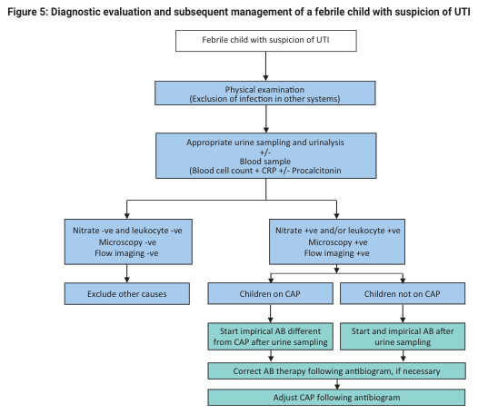
Summary : This flowchart outlines the diagnostic evaluation and management steps for a febrile child with suspected urinary tract infection (UTI), including physical examination, urine and blood testing, and subsequent treatment decisions based on test results. flowchart: # Nodes : • Febrile child with suspicion of UTI (rectangle) • Physical examination (Exclusion of infection in other systems) (rectangle) • Appropriate urine sampling and urinalysis +/- Blood sample (Blood cell count + CRP +/- Procalcitonin) (rectangle) • Nitrate -ve and leukocyte -ve, Microscopy -ve, Flow imaging -ve (rectangle) • Exclude other causes (rectangle) • Nitrate +ve and/or leukocyte +ve, Microscopy +ve, Flow imaging +ve (rectangle) • Children on CAP (rectangle) • Start empirical AB different from CAP after urine sampling (rectangle) • Correct AB therapy following antibiogram, if necessary (rectangle) • Adjust CAP following antibiogram (rectangle) • Children not on CAP (rectangle) • Start and empirical AB after urine sampling (rectangle) • Correct AB therapy following antibiogram, if necessary (rectangle) • Adjust CAP following antibiogram (rectangle) # Connectors : • Downward arrows connect each step in sequence. • After urine and blood sampling, the flow splits into two branches: – Left branch: Nitrate -ve and leukocyte -ve, Microscopy -ve, Flow imaging -ve → Exclude other causes. – Right branch: Nitrate +ve and/or leukocyte +ve, Microscopy +ve, Flow imaging +ve → further split into Children on CAP and Children not on CAP. • Children on CAP → Start empirical AB different from CAP after urine sampling → Correct AB therapy following antibiogram, if necessary → Adjust CAP following antibiogram. • Children not on CAP → Start and empirical AB after urine sampling → Correct AB therapy following antibiogram, if necessary → Adjust CAP following antibiogram. # Layout : • Vertical flow from top to bottom. • Main decision split after urine and blood sampling, with two branches for test results. • Further split in the positive test branch for children on CAP vs. not on CAP. • Each treatment branch ends with adjustment based on antibiogram. # Analysis : • The flowchart provides a clear, stepwise approach for evaluating and managing febrile children with suspected UTI. • Initial exclusion of other infections is followed by targeted urine and blood testing. • Negative test results prompt consideration of other causes, while positive results lead to empiric antibiotic therapy, tailored based on whether the child is on continuous antibiotic prophylaxis (CAP). • Final therapy is adjusted according to antibiogram results, ensuring appropriate antibiotic use.
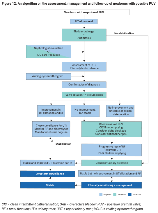
Summary : This figure presents a flowchart algorithm for the assessment, management, and follow-up of newborns with possible posterior urethral valve (PUV), detailing diagnostic steps, treatment options, and surveillance strategies based on clinical progression and response. flowchart: # Nodes : • New-born with suspicion of PUV (rectangle) • UT ultrasound (rectangle) • Bladder drainage + Antibiotics (rectangle) • Nephrologist evaluation +/- ICU care if required (parallelogram) • Assessment of RF + Electrolyte disturbance (rectangle) • Voiding cystourethrogram (rectangle) • Confirmation of diagnosis (rectangle) • Valve ablation +/- circumcision (rectangle) • Improvement in UT dilatation and RF (rectangle) • No improvement, but stable (rectangle) • No improvement and unstable or clinical deterioration (rectangle) • Check residual PUV, CIC if not emptying, Consider alpha blockade, Consider anticholinergics (rectangle) • Progressive loss of RF, Recurrent UTI, Poor bladder emptying (rectangle) • Consider Urinary diversion (rectangle) • Stable and improved UT dilatation and RF (rectangle) • Stable but no improvement in UT dilatation and RF (rectangle) • Long term surveillance (rectangle) • Stable (rectangle) • Intensify monitoring + management (rectangle) • Close surveillance for UTI, Monitor RF and electrolytes, Monitor nocturnal polyuria (rectangle) • Stabilisation (rectangle) • No stabilisation (label, right branch) # Connectors : • Arrows indicate progression from suspicion to diagnosis, treatment, and follow-up. • Branches split after "Valve ablation +/- circumcision" into three paths: improvement, no improvement but stable, and no improvement with instability/clinical deterioration. • Feedback loops from surveillance and management nodes back to stabilisation and further monitoring. • Rightmost branch for "No stabilisation" leads to more intensive interventions. # Layout : • Vertical flow from top (initial suspicion) to bottom (stable outcome or intensified management). • Horizontal branches after key decision points (e.g., post-treatment response). • Colour coding: Diagnosis (light blue), Treatment (teal), Follow-up (dark blue). • Explanatory abbreviations at the bottom: CIC, OAB, PUV, RF, UT, UUT, VCUG. # Analysis : • The algorithm is structured to escalate care based on clinical stability and response to initial treatment. • Early steps focus on diagnosis and stabilisation, followed by targeted interventions (valve ablation, antibiotics). • Surveillance and monitoring are emphasised for stable patients, while non-responders or those with deterioration are directed to more intensive management or urinary diversion. • The flowchart ensures systematic assessment and minimises missed steps in the management of newborns with suspected PUV.
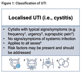
Summary : This figure presents the classification of localised urinary tract infection (UTI), specifically cystitis, including its typical signs and symptoms, absence of systemic infection, applicability to all sexes, and the importance of addressing risk factors. It also includes four schematic illustrations related to the clinical features or diagnostic considerations. flowchart/illustration: # Title & Content : • Title: "Figure 1: Classification of UTI" • Main heading: "Localised UTI (i.e., cystitis)" • Bullet points: – Cystitis with typical signs/symptoms (e.g. frequency¹, urgency², suprapubic pain³) – No signs/symptoms of systemic infection – Applies to all sexes⁴ – Risk factors may be present and should be addressed # Illustrations : • Four schematic diagrams labeled 1 to 4, each depicting anatomical or clinical features: – 1: Highlighted bladder region (suggesting frequency) – 2: Arrows indicating urgency – 3: Marked suprapubic area (suggesting pain) – 4: Overlapping male and female symbols (indicating applicability to all sexes) # Design Encodings : • Blue highlight in diagram 1 • Arrows in diagram 2 • Marked area in diagram 3 • Gender symbols in diagram 4 # Analysis : • The figure clearly distinguishes localised UTI (cystitis) by its typical symptoms and lack of systemic involvement, emphasizing that it affects all sexes and that risk factors should be considered. The illustrations visually reinforce the key symptoms and inclusivity of the classification.
Treatment of UTI - Algorithm
STEP 1: CLASSIFY THE UTI
Suspected UTI (dysuria, frequency, urgency, suprapubic pain)
              |
              ▼
    Is the patient LOW RISK?
    (Non-pregnant, non-diabetic adult woman,
     no fever, no flank pain, no structural abnormality)
              |
     YES ─────────── NO
      |                |
      ▼                ▼
 Uncomplicated    Complicated / Upper Tract
   Cystitis         UTI → see Step 4
STEP 2: UNCOMPLICATED CYSTITIS (Women, Outpatient)
| Drug | Dose | Duration | Notes |
|---|---|---|---|
| Nitrofurantoin (1st line) | 100 mg BD | 5 days | Avoid if CrCl <30 mL/min |
| TMP-SMX (1st line) | 160/800 mg BD | 3 days | Avoid if local resistance >20% |
| Fosfomycin (1st line) | 3 g single sachet | 1 day (single dose) | Useful for MDR; E. coli only |
| Ciprofloxacin (2nd line) | 250 mg BD | 3 days | Reserve; don't use 1st line |
| Levofloxacin (2nd line) | 250 mg OD | 3 days | Reserve; don't use 1st line |
Fluoroquinolones are NOT first-line for uncomplicated cystitis - increasing resistance + side-effect profiles.
STEP 3: UNCOMPLICATED CYSTITIS IN MEN
Men with lower UTI symptoms
         |
         ▼
  Treat for 7 days (not 3)
  TMP-SMX or Fluoroquinolone
  (prostate tissue penetration needed)
         |
         ▼
  Urology referral if recurrent
STEP 4: ACUTE PYELONEPHRITIS (Upper Tract UTI)
Fever + flank pain/CVA tenderness ± systemic symptoms
              |
     ─────────────────────
     |                   |
     ▼                   ▼
 OUTPATIENT           INPATIENT
(Mild-Moderate)       (Severe/Sepsis)
Able to tolerate PO   Vomiting, Sepsis signs
                      Unable to comply

OUTPATIENT:                    INPATIENT (IV first):
────────────                   ────────────────────
Ciprofloxacin 500 mg BD × 7d  Ciprofloxacin 400 mg IV q12h
 (if FQ resist <10%)          OR Ceftriaxone 1-2 g IV OD
OR TMP-SMX DS BD × 10-14d     OR Pip-Tazo 3.375 g IV q6h
OR Ceftriaxone 1g IM then      OR Gentamicin 3 mg/kg/d ± Ampicillin
   oral cephalosporin × 14d    → Switch to oral after improvement
                               Total duration: 10-14 days
Nitrofurantoin and Fosfomycin do NOT achieve adequate tissue/blood levels - do not use for pyelonephritis.
STEP 5: COMPLICATED UTI (Catheter, Obstruction, Immunocompromised, Pregnancy)
Complicated UTI
      |
      ▼
  Send urine culture first
      |
      ▼
  Empiric IV therapy:
  - Pip-Tazo / Cefepime / Ceftriaxone
  - Carbapenems if ESBL suspected
      |
      ▼
  De-escalate to oral based on culture sensitivities
  Duration: 10-14 days (21 days if sepsis)
      |
      ▼
  Remove/replace catheter (CAUTI) before starting antibiotics
STEP 6: ASYMPTOMATIC BACTERIURIA (ASB)
Bacteriuria WITHOUT symptoms
         |
   ──────────────────────
   |                    |
   ▼                    ▼
TREAT only if:       DO NOT TREAT:
• Pregnant            • Elderly
• Pre-urology         • Diabetics
  procedure           • Catheterized patients
                      • Non-pregnant adults
STEP 7: RECURRENT UTI (>2 episodes in 6 months or >3 in 12 months)
Recurrent UTI
      |
      ▼
Culture + empiric treatment (3-day regimen)
      |
      ▼
Refer to primary care / urology in 1-2 weeks
      |
      ▼
Prophylaxis options:
  • Continuous low-dose (e.g., nitrofurantoin 50 mg OD)
  • Postcoital single-dose
  • Patient-initiated self-treatment
      |
      ▼
Non-antibiotic options: Intravaginal estriol (post-menopausal),
Lactobacillus suppositories, Cranberry juice (limited evidence)
Avoid spermicide-based contraception
QUICK REFERENCE SUMMARY TABLE
| Type | Setting | Drug | Duration |
|---|---|---|---|
| Uncomplicated cystitis (F) | Outpatient | Nitrofurantoin / TMP-SMX / Fosfomycin | 5 / 3 / 1 days |
| Uncomplicated cystitis (M) | Outpatient | TMP-SMX or FQ | 7 days |
| Pyelonephritis (mild) | Outpatient | Ciprofloxacin / TMP-SMX | 7 / 10-14 days |
| Pyelonephritis (severe) | Inpatient IV | Ceftriaxone / Pip-Tazo / FQ IV | 14 days |
| Complicated UTI | Inpatient IV | Broad spectrum → narrow by culture | 10-21 days |
| CAUTI | Inpatient | Remove catheter + treat per culture | 7-14 days |