
Feto placental unit in md physiology
Reading File
Finding Sources
Finding Sources
Reading File
Reading File
Finding Sources
Reading File
Loading Image
Loading Image
Reading File
Loading Image
Loading Image
I now have all the key content and diagrams. Here is a comprehensive answer on the Feto-Placental Unit for MD Physiology:
Feto-Placental Unit (Fetoplacental Unit)
The feto-placental unit refers to the functional cooperation between the fetus and the placenta in the synthesis of steroid hormones during pregnancy. Neither the fetus nor the placenta alone can complete the full steroidogenic pathway - each lacks certain enzymes that the other supplies, making them interdependent.
Why a "Unit" is Necessary
- The placenta can convert cholesterol to pregnenolone and progesterone, but lacks the 17α-hydroxylase and C17-20 lyase enzymes needed to produce androgens (C19 steroids) from progesterone.
- The fetal adrenal gland is rich in sulfokinases and 16α-hydroxylase but cannot synthesize sufficient pregnenolone on its own.
- Together they complete the steroid synthesis pathways that neither could accomplish independently.
(Ganong's Review of Medical Physiology, 26th Ed.; Creasy & Resnik's Maternal-Fetal Medicine)
Key Pathways
1. Progesterone Synthesis
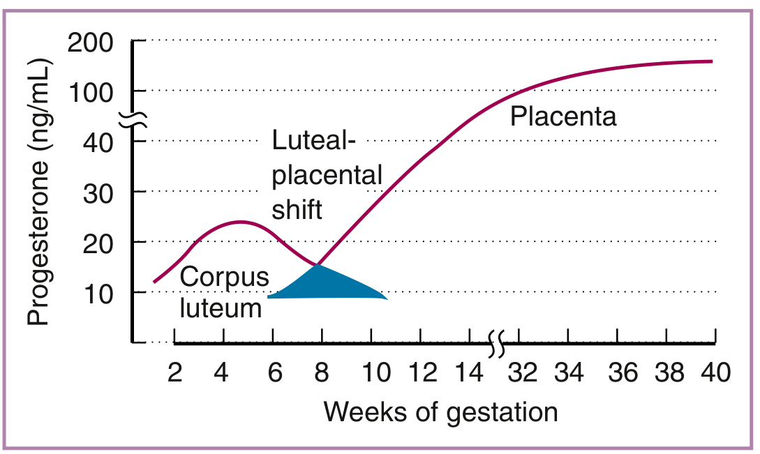
- For the first 6-10 weeks: corpus luteum is the main progesterone source.
- After that, the placenta takes over completely using maternal LDL cholesterol as the substrate.
- By term: placental progesterone production = ~250 mg/day; maternal serum levels = 130-150 ng/mL (vs. 2.5 mg/day in the follicular phase and 25 mg/day in the luteal phase).
- ~90% of placental progesterone enters the maternal compartment; the rest goes to the fetal adrenal to serve as substrate for cortisol and corticosterone synthesis.
- Metabolized to pregnanediol and excreted in urine as a glucuronide.
2. Estrogen Synthesis - The Core of the Fetoplacental Unit
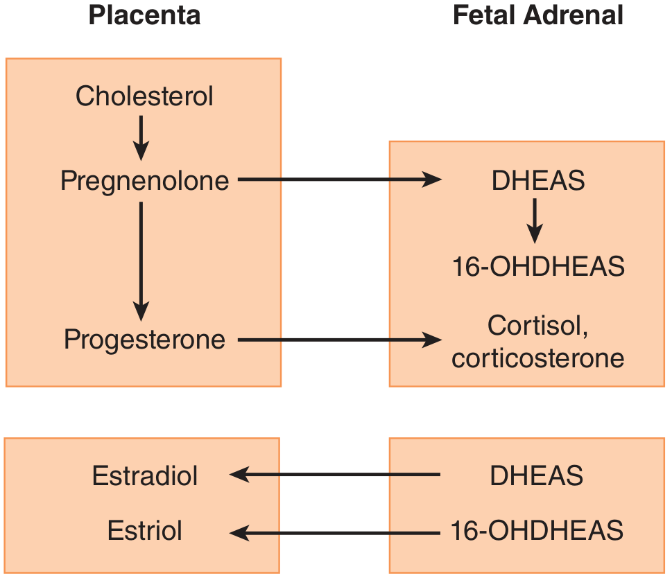
The pathway for estrogen synthesis is a 3-compartment collaborative process:
| Step | Location | Reaction |
|---|---|---|
| 1 | Placenta | Cholesterol → Pregnenolone → Progesterone |
| 2 | Fetal adrenal | Pregnenolone → DHEAS (dehydroepiandrosterone sulfate) |
| 3 | Fetal liver | DHEAS → 16-OH-DHEAS (16α-hydroxylation) |
| 4 | Placenta (via sulfatase) | 16-OH-DHEAS → 16-OH-DHEA (desulfation) |
| 5 | Placenta (via aromatase) | 16-OH-DHEA → Estriol (E3) |
Also:
- Fetal adrenal DHEAS → placenta → aromatase → Estradiol (E2) and Estrone (E1)
- ~60% of estradiol/estrone is derived from fetal androgen precursors; ~40% from maternal DHEAS.
3. Estriol - The Dominant Estrogen of Pregnancy
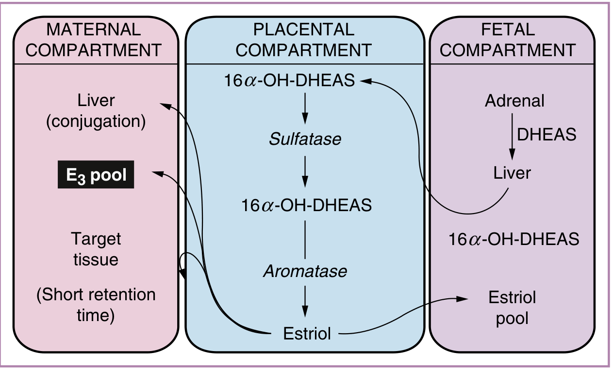
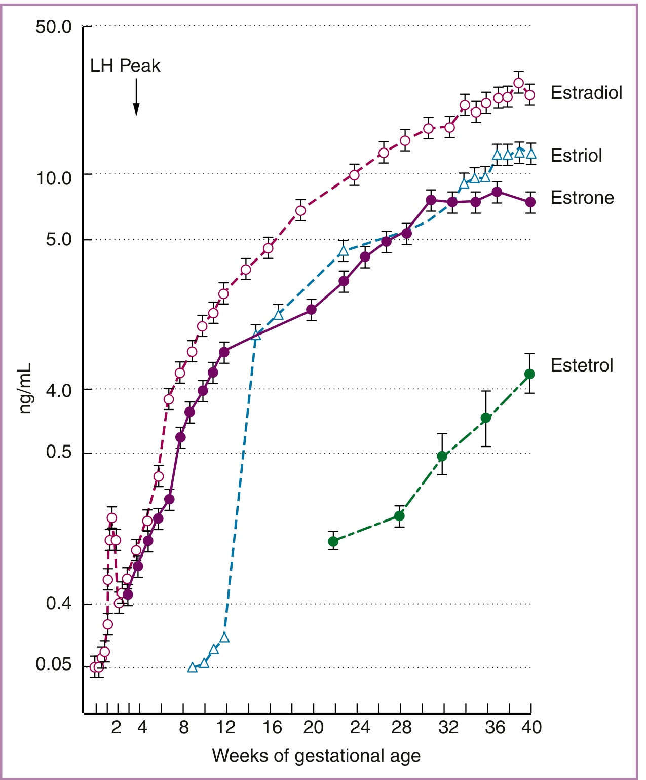
- Estriol accounts for >90% of urinary estrogen in pregnant women.
- It is not produced by the non-pregnant ovary - its presence indicates active fetoplacental function.
- Maternal serum estriol levels rise to 12-20 ng/mL by term.
- Because fetal 16-OH-DHEAS is the principal substrate, urinary estriol excretion in the mother is a clinical index of fetal well-being.
4. Cortisol / Corticosterone Synthesis (Fetal Adrenal)
- Placental progesterone is transported to the fetal circulation.
- Fetal adrenal converts progesterone → cortisol and corticosterone.
- Fetal cortisol promotes lung maturity (surfactant production) and, near term, stimulates uterine prostaglandin production contributing to labor initiation.
Clinical Significance
| Condition | Fetoplacental Implication |
|---|---|
| Low maternal urinary estriol | Suggests fetal distress or fetal death (fetal adrenal not producing DHEAS precursor) |
| Anencephaly | Hypothalamic-pituitary axis absent → low fetal DHEAS → very low estriol |
| Molar pregnancy / pseudocyesis | No fetal compartment → low estrogens |
| Placental sulfatase deficiency | X-linked; pregnancy reaches term but cervical ripening fails; male offspring develop ichthyosis |
| Placental aromatase deficiency | Pregnancy can continue but virilization of mother and fetus occurs; cervical ripening impaired |
Estetrol - Pregnancy-Unique Estrogen
- Estetrol (E4) = the 15α-hydroxy derivative of estriol.
- Derived exclusively from fetal precursors - unique to pregnancy.
- Not used clinically as it offers no advantage over estriol measurement.
Summary Concept
Maternal cholesterol
        ↓ (Placenta)
    Pregnenolone → Progesterone
        |                ↓ (Fetal adrenal)
        |           Cortisol / Corticosterone
        ↓ (Fetal adrenal)
      DHEAS
        ↓ (Fetal liver)
    16-OH-DHEAS
        ↓ (Placenta: sulfatase + aromatase)
      ESTRIOL (E3)  ← main estrogen of pregnancy
Key point for exams: The placenta has aromatase but lacks 17α-hydroxylase. The fetal adrenal has 17α-hydroxylase but depends on placental pregnenolone. This complementarity is the physiological basis of the "feto-placental unit."
Sources:
- Ganong's Review of Medical Physiology, 26th Ed. - Fig 22-21, p.418
- Creasy & Resnik's Maternal-Fetal Medicine - Figs 10.4, 10.5, 10.6, pp.223-224
What's the clinical role of estriol monitoring?
Finding Sources
Finding Sources
Searching PubMed
unconjugated estriol prenatal screening Down syndrome
Reading File
Finding Sources
Finding Sources
Reading File
Finding Sources
Finding Sources
Reading File
Finding Sources
I now have comprehensive textbook content. Here is a complete answer:
Clinical Role of Estriol Monitoring
Estriol has two distinct clinical roles: (1) prenatal aneuploidy screening via unconjugated estriol (uE3) as part of the maternal serum screen, and (2) historical fetal well-being surveillance, which is now largely obsolete. These must be distinguished clearly.
1. Prenatal Aneuploidy Screening - The Main Current Role
Unconjugated estriol (uE3) is measured in maternal serum at 16-18 weeks as one component of multi-marker screening panels.
Marker Patterns in Aneuploidy
| Condition | uE3 | AFP | hCG | Inhibin-A |
|---|---|---|---|---|
| Trisomy 21 (Down syndrome) | ↓ (0.7 MoM) | ↓ (0.75 MoM) | ↑↑ (2.3 MoM) | ↑ (1.3-2.5 MoM) |
| Trisomy 18 (Edwards syndrome) | ↓↓ | ↓ | ↓ | Normal |
| Normal | 1.0 MoM | 1.0 MoM | 1.0 MoM | 1.0 MoM |
Screening Panels
Triple screen (AFP + hCG + uE3):
- Sensitivity for Down syndrome in women <35 years: 57-67% at 5% false-positive rate
- Improved over maternal age alone (1:100 odds → 1:33 to 1:62 with positive screen)
Quad screen (AFP + hCG + uE3 + Inhibin-A):
- Detection rate for Down syndrome: ~75% at 5% false-positive rate in women <35 years
- For women >35 years: ~92% detection, but 13% false-positive rate
Integrated screening (first trimester NT + PAPP-A + second trimester quad screen combined):
- Detection rate: ~95% at 5% false-positive rate - the most sensitive analyte-based approach
- Downside: withholds risk estimate until second trimester, delaying CVS option
(Creasy & Resnik's Maternal-Fetal Medicine; Thompson & Thompson Genetics and Genomics, 9th Ed.)
2. Extremely Low uE3 - Rare Single-Gene Disorders
Very low (near-zero) unconjugated estriol should prompt evaluation for specific genetic conditions that disrupt the fetoplacental steroidogenic pathway:
| Condition | Mechanism of Low uE3 | Additional Features |
|---|---|---|
| Steroid sulfatase deficiency (X-linked ichthyosis) | Placenta cannot cleave sulfate from DHEAS - no substrate for aromatization | Male offspring develop ichthyosis after birth; X-linked recessive |
| Smith-Lemli-Opitz syndrome | Defect in cholesterol synthesis (7-DHCR enzyme) - entire steroid pathway upstream is compromised | Dysmorphic features, intellectual disability |
| Congenital adrenal hypoplasia | Fetal adrenal cannot produce DHEAS precursor | Adrenal insufficiency in neonate |
| Anencephaly | Absent fetal hypothalamic-pituitary axis → no ACTH → no fetal adrenal DHEAS | Incompatible with sustained life |
3. Historical Role: Urinary Estriol as Fetal Well-Being Index
Since fetal 16-OH-DHEAS is the principal precursor for estriol, declining maternal urinary estriol was historically used as a marker of fetoplacental compromise. The reasoning:
- Healthy fetus → active adrenal → ample DHEAS → high estriol
- Compromised fetus → reduced DHEAS output → falling estriol
Why It Was Abandoned
Several serious limitations led to its replacement by biophysical methods (NST, BPP, Doppler):
- Wide normal variation - ±1 standard deviation is very large at any gestational age, making a single value hard to interpret.
- Moment-to-moment fluctuations - a single plasma measurement is unreliable; serial measurements were needed.
- Positional effects - bed rest vs. ambulation alters uteroplacental blood flow and thus estriol levels.
- Drug interference - glucocorticoids, ampicillin, and other drugs cause spuriously low estriol.
- Poor predictive value - low estriol does not reliably predict outcome before fetal death is already imminent.
(Creasy & Resnik's Maternal-Fetal Medicine, p.224)
4. Low Estriol in Specific Obstetric Contexts
| Clinical Scenario | Estriol Level | Reason |
|---|---|---|
| Fetal death in utero | Very low / undetectable | No fetal adrenal DHEAS production |
| Anencephaly | Very low | No fetal hypothalamic-pituitary-adrenal drive |
| Molar pregnancy / pseudocyesis | Low | No fetal compartment at all |
| Aromatase deficiency | Very low | Placenta cannot aromatize DHEAS precursors; also causes maternal and fetal virilization |
| Placental sulfatase deficiency | Very low | Placenta cannot desulfate DHEAS; pregnancy still reaches term but cervical ripening fails |
Summary: Current vs. Obsolete Uses
| Use | Status |
|---|---|
| uE3 in triple/quad screen for trisomy 21 & 18 | Active - standard of care (where cfDNA not used) |
| Extremely low uE3 to flag Smith-Lemli-Opitz, steroid sulfatase deficiency | Active - clinical reflex |
| Serial urinary estriol for fetal surveillance | Obsolete - replaced by NST, BPP, umbilical Doppler |
Bottom line: The value of uE3 today is almost entirely as a prenatal screening analyte for chromosomal aneuploidies, not as a standalone marker of fetal well-being. Where cell-free fetal DNA (cfDNA/NIPT) is available, it has superseded all serum analyte screens for aneuploidy detection, with far higher sensitivity and specificity.
Sources: Creasy & Resnik's Maternal-Fetal Medicine, pp.223-224, 650; Thompson & Thompson Genetics and Genomics in Medicine, 9th Ed., Ch.18
This is a shared conversation. Sign in to Orris to start your own chat.