
ELISA microbiology
Reading File
ELISA (Enzyme-Linked Immunosorbent Assay) is a serological technique used to detect and quantify antigens or antibodies using enzyme-labeled conjugates. There are four main types: direct, indirect, sandwich, and competitive ELISA.
Searching sources...
Finding Sources
Finding Sources
Good - I found strong sources. Let me now read the most relevant sections from Sherris Medical Microbiology and Janeway's Immunobiology for ELISA principles.
Reading File
Reading File
Reading File
Excellent content. Now let me fetch the key ELISA diagrams and read more from Sherris Microbiology for the full picture.
Loading Image
Loading Image
Reading File
I now have excellent content and diagrams. Let me also fetch the direct ELISA principle diagram from Janeway's and get more detail from the Tietz lab medicine text.
Reading File
Reading File
I now have comprehensive content. Let me compile the full, detailed answer.
ELISA - Enzyme-Linked Immunosorbent Assay
Definition
ELISA is a heterogeneous enzyme immunoassay (EIA) technique used to detect and quantify antigens or antibodies. One reaction component is nonspecifically adsorbed or covalently bound to a solid phase (typically a microtiter well, magnetic particle, or plastic bead), which facilitates separation of bound and free labeled reactants.
- Tietz Textbook of Laboratory Medicine, 7th Edition
Basic Principle
The core idea is straightforward:
- An antigen or antibody is coated onto the surface of a plastic well (solid phase)
- Residual sticky sites on the plastic are blocked by adding irrelevant proteins
- The test sample is added and allowed to react
- An enzyme-linked antibody (conjugate) is added - it binds only if the target antigen/antibody is present
- Unbound antibody is washed away
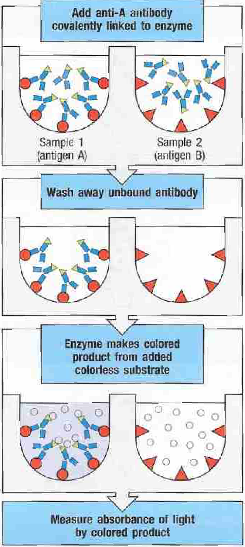
- An enzyme substrate (colorless) is added - the enzyme converts it to a colored product
- The color change is measured spectrophotometrically (optical density/absorbance)
The amount of color produced is proportional to the amount of antigen/antibody in the sample.
Types of ELISA
1. Direct ELISA
- Antigen is adsorbed directly to the plate
- Enzyme-labeled primary antibody binds directly to the antigen
- Simple but less sensitive; no signal amplification
- Detects: antigens
2. Indirect ELISA
- Antigen is coated on the plate
- Unlabeled primary antibody (from patient serum) binds the antigen
- An enzyme-linked anti-immunoglobulin (secondary) antibody then binds the primary antibody
- More sensitive than direct (signal amplified because multiple secondary antibodies bind each primary antibody)
- Detects: antibodies in patient serum (e.g., HIV antibody detection, Lyme disease serology)
3. Sandwich (Capture) ELISA
- A capture antibody is coated on the plate
- Antigen from the sample binds to the capture antibody
- A second enzyme-labeled detection antibody (recognizing a different epitope) then binds the antigen
- The antigen is "sandwiched" between two antibodies
- Most sensitive format; used for cytokine measurement, antigen detection
- Example equation: Plate-Ab → Ag → Ab-Enzyme → Substrate → Color
- Sherris & Ryan's Medical Microbiology, 8th Edition
4. Competitive (Inhibition) ELISA
- Unlabeled antigen in the patient sample competes with a labeled antigen for binding to a fixed amount of antibody
- High antigen concentration in sample = LESS color (inhibition of binding)
- Used for small molecules (drugs, hormones, haptens)
- Inverse relationship: more analyte = less signal
Enzymes Used as Labels
The enzyme label must be stable, catalytically active when conjugated, and produce a measurable product. Common enzymes:
| Enzyme | Substrate | Detection |
|---|---|---|
| Horseradish peroxidase (HRP) | TMB, OPD | Colorimetric / Chemiluminescent |
| Alkaline phosphatase (ALP) | p-Nitrophenyl phosphate | Colorimetric / Chemiluminescent |
| Glucose-6-phosphate dehydrogenase | Glucose-6-phosphate + NAD+ | Spectrophotometric |
| β-galactosidase | ONPG | Colorimetric |
- Tietz Textbook of Laboratory Medicine, 7th Edition
Key Advantages of ELISA over RIA
| Feature | ELISA | RIA |
|---|---|---|
| Radioactivity | None | Uses 125I |
| Safety | Safer | Radiation hazard |
| Cost | Lower | Higher |
| Shelf life | Longer (stable enzymes) | Short (radioactive decay) |
| Signal | Color change (spectrophotometric) | Radioactive counts |
| Sensitivity | High | Very high (slightly more sensitive) |
ELISA avoids the hazards of radioactivity while maintaining high sensitivity - making it the preferred method for most direct-binding diagnostic assays.
- Janeway's Immunobiology 10e
Clinical Applications in Microbiology
ELISA is one of the most widely used serological tools in clinical microbiology:
| Disease / Pathogen | Use of ELISA |
|---|---|
| HIV | Screening for HIV antibody (indirect ELISA); p24 antigen detection (sandwich) |
| Hepatitis B/C | HBsAg, anti-HBc, anti-HCV antibody detection |
| Lyme disease | Detection of IgM/IgG antibodies to Borrelia burgdorferi (sensitive but not specific - positive/indeterminate ELISA requires confirmatory Western blot) |
| Rabies | Detection of anti-rabies antibody in serum/CSF (antibody not detectable until late in disease) |
| Rotavirus | Rapid antigen detection in stool (sandwich ELISA) |
| RMSF | Detection of antibodies to Rickettsia rickettsii |
| Dengue | Detection of NS1 antigen and IgM/IgG antibodies |
| Toxoplasmosis, rubella, CMV, EBV | IgM/IgG serology |
| Autoimmune diseases | Detection of autoantibodies (e.g., anti-dsDNA, anti-BP180, anti-BP230 in bullous pemphigoid) |
| Cytokine measurement | Capture/sandwich ELISA for IL-6, TNF, interferon levels |
Step-by-Step Procedure (Sandwich ELISA for Antigen Detection)
- Coat: Capture antibody bound to microtiter wells overnight
- Block: Add blocking buffer (BSA or skimmed milk) to prevent nonspecific binding
- Add sample: Patient sample added; antigen binds capture antibody
- Wash: Remove unbound material
- Add detection antibody: Enzyme-conjugated antibody specific for a different epitope
- Wash: Remove unbound detection antibody
- Add substrate: Colorless substrate added (e.g., TMB for HRP)
- Stop reaction: Stop solution (e.g., H2SO4) changes color from blue to yellow
- Read: Absorbance measured at 450 nm in a plate reader (ELISA reader)
- Interpret: Compare OD to standard curve or cut-off value
Reading and Interpretation
- Results reported as optical density (OD) or absorbance units
- For quantitative assays: a standard curve (serial dilutions of known antigen concentrations) is run in parallel
- Cut-off values are established: OD > cut-off = positive; OD < cut-off = negative
- A ratio of sample OD to cut-off OD (S/CO ratio) is commonly used
Limitations
- Cross-reactivity: False positives due to non-specific antibody binding (especially in indirect formats)
- Requires washing steps and laboratory infrastructure (not point-of-care)
- Confirmatory testing needed in some cases (e.g., HIV ELISA confirmed by Western blot; Lyme ELISA confirmed by Western blot)
- Hook effect (prozone): Very high antigen concentrations can paradoxically give falsely low readings in sandwich ELISA
- Requires pure antigen or antibody as a reference standard
Related Techniques
- EMIT (Enzyme Multiplied Immunoassay Technique): Homogeneous EIA - no wash step needed; widely used for therapeutic drug monitoring
- CEDIA (Cloned Enzyme Donor Immunoassay): First genetically engineered EIA; uses fragments of β-galactosidase
- Western blot: Uses electrophoretically separated antigens - used as confirmatory test after ELISA screening
- Immunohistochemistry/Immunoperoxidase: Same principle applied to tissue sections (surgical pathology)
- dot-ELISA: Results read visually on nitrocellulose membrane (used in field settings)
Summary: ELISA is a sensitive, specific, safe (no radioactivity), and versatile immunoassay. The four formats (direct, indirect, sandwich, competitive) cover a broad range of diagnostic applications. In microbiology, it serves as the primary screening tool for viral, bacterial, and parasitic diseases, with its ability to process hundreds of samples simultaneously on 96-well microtiter plates making it ideal for large-scale screening programs.
Principle of ELISA
The principle of ELISA is based on the specific antigen-antibody reaction, where one component is bound to a solid phase and detection is achieved through an enzyme-labeled conjugate that produces a measurable color change when its substrate is added.
Searching sources...
Finding Sources
Finding Sources
Reading File
Reading File
Now let me fetch the key principle diagrams:
Loading Image
Loading Image
Loading Image
I now have everything needed for a focused, clear explanation of the principle. Here is the complete answer:
Principle of ELISA
Core Concept
ELISA is based on two fundamental biological and chemical principles working together:
"ELISA and RIA are direct binding assays for antigen or antibody; both work on the same principle, but the means of detecting specific binding is different."
- Janeway's Immunobiology, 10e
The two pillars are:
- Specific antigen-antibody binding - an antibody recognizes and binds only its complementary antigen (lock-and-key specificity)
- Enzyme amplification - an enzyme covalently linked to an antibody acts as a signal amplifier, converting a colorless substrate into a measurable colored product
The Principle - Step by Step
Step 1: Solid Phase Immobilization (the "Sorbent")
One reaction component (antigen or antibody) is nonspecifically adsorbed or covalently bound to a solid support - most commonly the wells of a 96-well polystyrene microtiter plate. This is what the "S" in ELISA stands for - Immunosorbent. The solid phase physically separates bound from free reactants.
Step 2: Blocking
Residual protein-binding sites on the plastic are blocked by adding irrelevant proteins (e.g., BSA, skimmed milk, casein). This prevents nonspecific binding of the enzyme-labeled antibody to the plate, which would cause false positives.
Step 3: Antigen-Antibody Reaction
The test sample is added. If the target antigen (or antibody) is present, it binds specifically to the immobilized partner. No antigen = no binding.
Step 4: Addition of Enzyme-Labeled Conjugate
An enzyme-linked antibody (conjugate) is added. This antibody is covalently attached to an enzyme (e.g., horseradish peroxidase or alkaline phosphatase). It binds to the captured antigen or antibody.
Step 5: Washing
Unbound enzyme-labeled antibody is washed away. Only antibody that has specifically bound remains on the plate. This is the separation step - essential to every ELISA.
Step 6: Substrate Addition (Signal Generation)
A colorless substrate is added. The enzyme converts it into a colored product. The amount of color produced is directly proportional to the amount of antigen or antibody in the sample.
Step 7: Reading
The optical density (absorbance) of the colored product is measured in a plate reader (spectrophotometer) at a specific wavelength. The result is compared to a standard curve to quantify the analyte.
Visual Summary of the Principle
Fig. A.5 - The principle of ELISA. Sample 1 contains the target antigen A - the enzyme-linked antibody binds and generates color. Sample 2 contains a different antigen - no binding, no color. Absorbance is then measured. - Janeway's Immunobiology, 10e
The Sandwich ELISA Principle (Most Common Format)
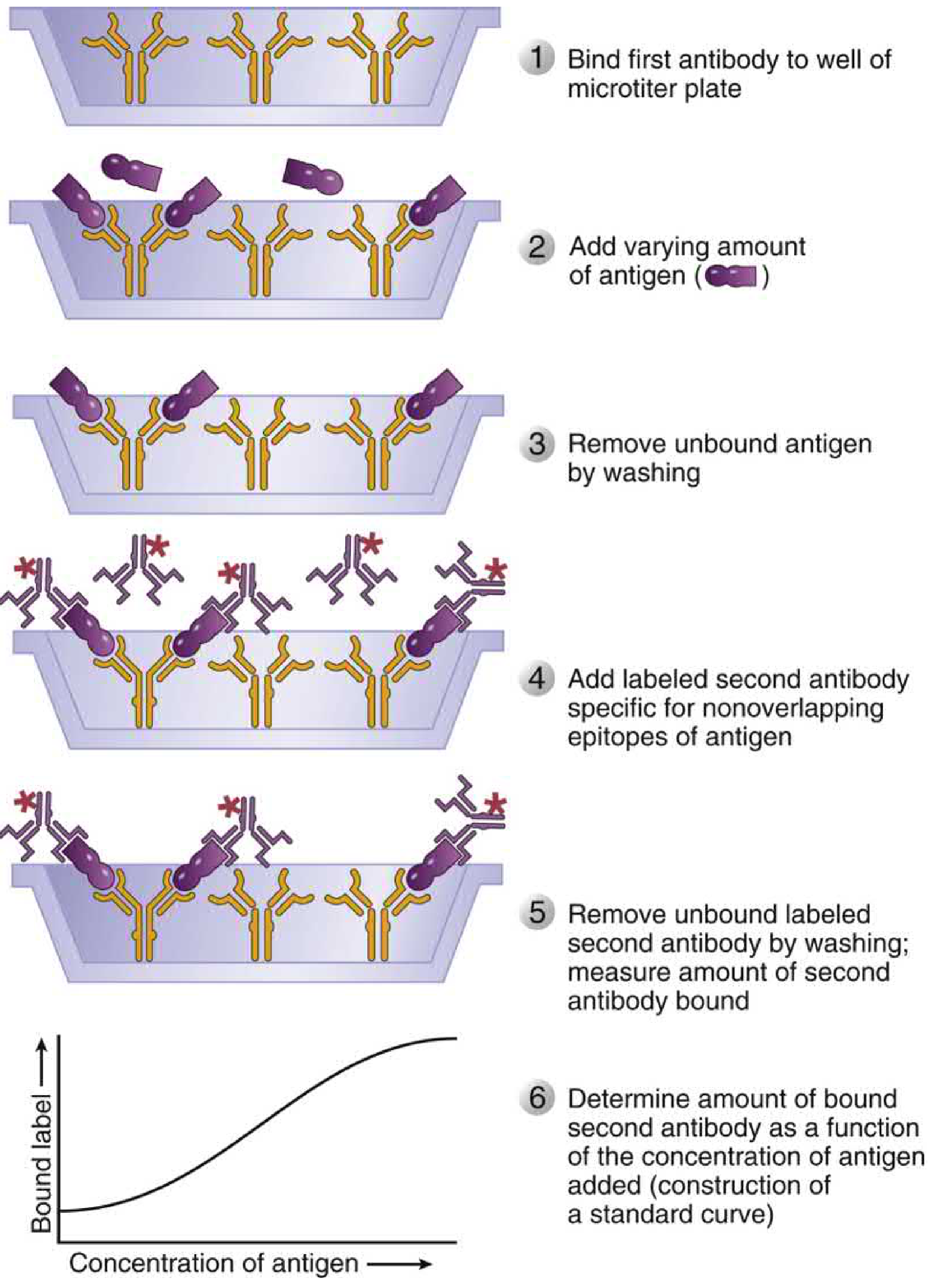
Sandwich ELISA for antigen quantification. Steps 1-6 shown. The bound label (signal) increases proportionally with antigen concentration, enabling construction of a standard curve. - Cellular and Molecular Immunology
In sandwich ELISA:
- A capture antibody (Ab1) is bound to the plate
- Antigen from the sample is "captured" and held in place
- A second detection antibody (Ab2) labeled with enzyme recognizes a different (non-overlapping) epitope on the same antigen
- The antigen is "sandwiched" between two antibodies: Plate-Ab1 → Ag → Ab2-Enzyme
The Indirect ELISA Principle (for Antibody Detection)
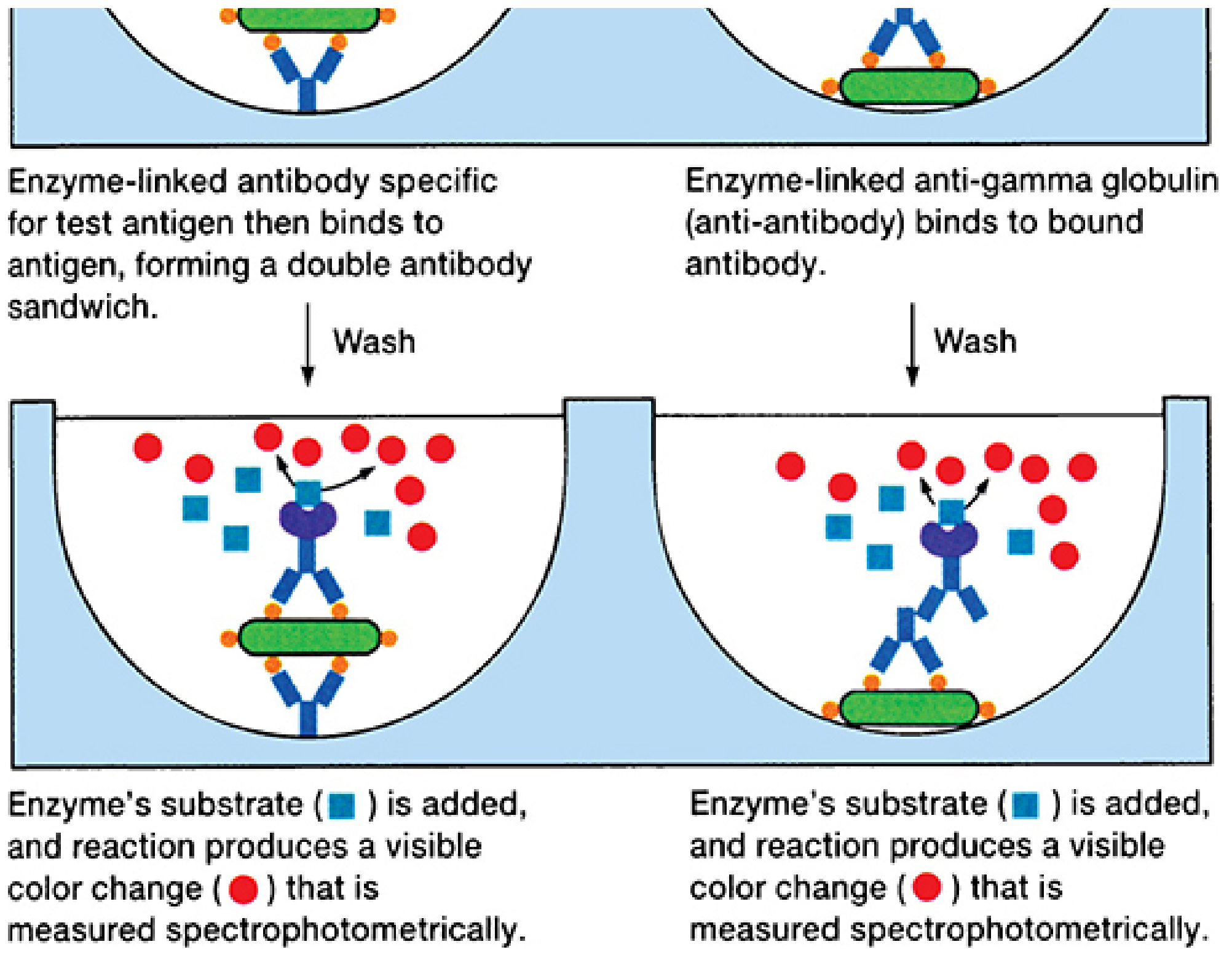
Left: Enzyme-linked antibody specific for test antigen forms a double antibody sandwich. Right: Enzyme-linked anti-gamma globulin (anti-antibody) binds to the patient's antibody that is already bound to antigen. Both produce a visible color change measured spectrophotometrically. - Sherris & Ryan's Medical Microbiology, 8th Edition
In indirect ELISA (used to detect patient antibodies e.g. anti-HIV):
- Known antigen is coated on the plate
- Patient's serum is added - if specific antibody is present, it binds the antigen
- An enzyme-linked anti-human immunoglobulin (secondary antibody) then binds the patient's antibody
- Substrate → color confirms the presence of antibody in the serum
Key Principle Summary Table
| Component | Role |
|---|---|
| Microtiter well (polystyrene) | Solid phase; immobilizes one reactant |
| Blocking protein | Prevents nonspecific binding |
| Antigen-antibody reaction | Specific binding - the recognition event |
| Enzyme-labeled conjugate | Signal molecule (HRP, ALP most common) |
| Washing step | Separates bound from free labeled reactant |
| Colorless substrate | Converted to colored product by enzyme |
| Spectrophotometer (plate reader) | Measures absorbance = quantifies reaction |
| Standard curve | Converts absorbance values to antigen/antibody concentration |
Why Enzyme Instead of Radioactivity?
ELISA replaced RIA (radioimmunoassay) because:
- No radioactive hazard - enzymes replace radioactive labels (¹²⁵I)
- Signal amplification - one enzyme molecule converts thousands of substrate molecules, giving very high sensitivity
- Longer shelf life - enzymes are stable; radioisotopes decay
- Easy to read - color change visible in the plate tray; quantified by simple spectrophotometer
- Janeway's Immunobiology, 10e
In one sentence: The principle of ELISA is that a specific antigen-antibody interaction on a solid phase is detected and quantified through an enzyme covalently linked to an antibody, which generates a colored product from a colorless substrate in direct proportion to the amount of analyte present.
This is a shared conversation. Sign in to Orris to start your own chat.