
Intestinal obstruction
"intestinal obstruction"[MeSH Terms] AND management
small bowel obstruction abdominal radiograph dilated loops air-fluid levels
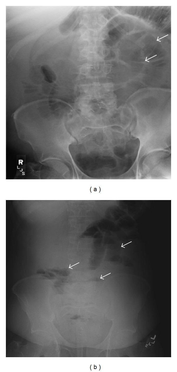
This composite diagnostic image features two abdominal radiographs (X-rays) of a single patient, illustrating classic signs of a distal small bowel obstruction. Image (a) is a supine abdominal radiograph demonstrating multiple dilated loops of small bowel in the central abdomen, characterized by thin transverse lines (valvulae conniventes). Notably, there is a paucity or near-total absence of gas within the large bowel, suggesting a complete or high-grade distal obstruction. Image (b) is an upright (erect) abdominal radiograph of the same patient, which reveals several prominent air-fluid levels (indicated by white arrows) within the dilated proximal and central small bowel loops. These tiered horizontal interfaces are a hallmark radiological finding of mechanical intestinal obstruction. The combination of dilated small bowel loops and multiple air-fluid levels, in the absence of significant colonic gas, is highly indicative of a distal small bowel obstruction, which in this clinical context was secondary to ileocolic intussusception.
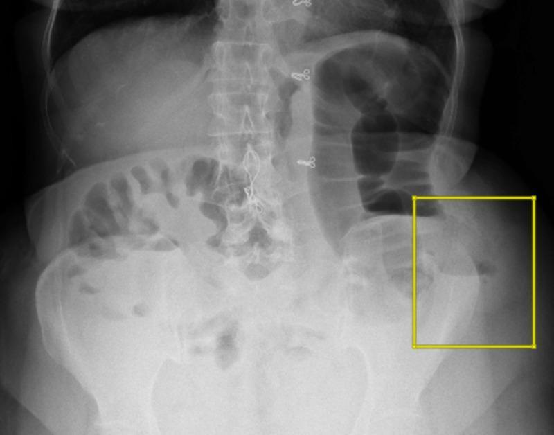
This diagnostic image is a plain erect abdominal radiograph (X-ray) demonstrating signs of intestinal obstruction. The radiograph reveals multiple significantly dilated loops of small and large bowel distributed across the abdominal cavity. Characteristic multiple air-fluid levels are visible, representing the gravitational separation of liquid and gas within the distended lumen. A yellow square highlights the left lower quadrant (left iliac region), where focal dilated bowel loops and prominent air-fluid levels are concentrated near the iliac crest. These findings are clinically significant as they indicate a mechanical bowel obstruction or a localized paralytic ileus. The image serves as a teaching tool for identifying radiological hallmarks of intestinal distension and fluid stasis in acute surgical abdominal presentations, specifically correlating with bowel herniation through an iliac crest defect.
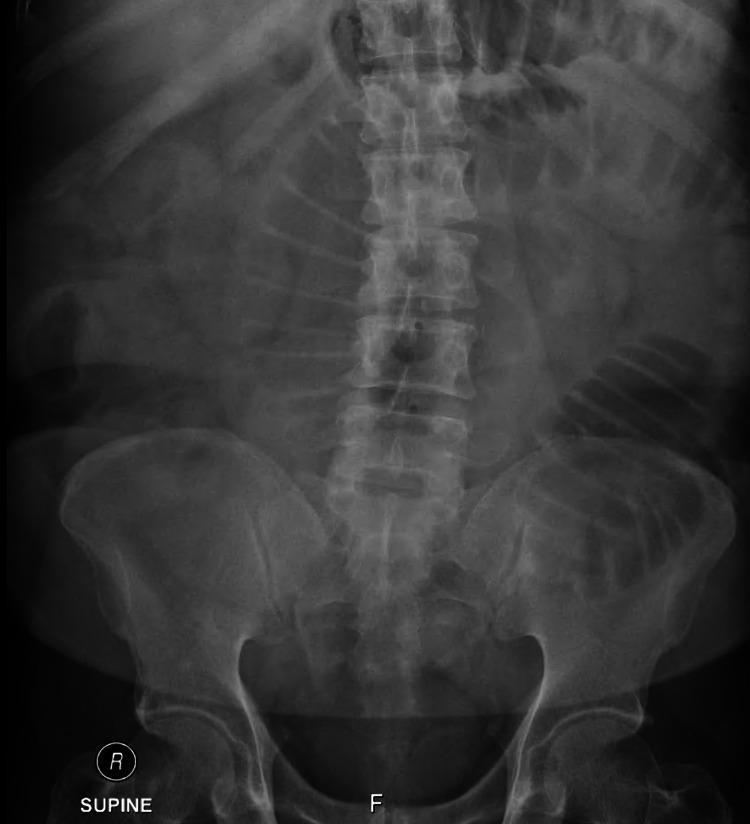
This diagnostic image is a plain abdominal radiograph (X-ray) in the supine position. The primary findings are multiple dilated loops of small bowel located centrally within the abdomen. Distinctive horizontal air-fluid levels are visible within these dilated segments, a hallmark radiological sign of mechanical bowel obstruction or ileus. The valvulae conniventes (circular folds) are evident across the full width of the dilated loops, identifying them as small intestine rather than large bowel. There is a notable absence of significant gas within the distal colon and rectum, further supporting the diagnosis of a high-grade small bowel obstruction. Bony structures, including the lumbar spine and pelvis, appear unremarkable in this view. This image serves as a classic educational example of the radiographic presentation of small bowel obstruction, demonstrating the 'stepladder' appearance often seen when multiple air-fluid levels are present in various loops.
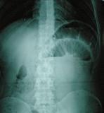
This diagnostic image is an erect abdominal plain radiograph (X-ray) demonstrating signs of a small bowel obstruction. The radiograph reveals multiple dilated loops of small bowel in the mid and upper abdomen. A classic radiographic hallmark of mechanical obstruction is present: distinct horizontal air-fluid levels, where radiolucent (dark) gas is visible superior to radio-opaque (lighter) fluid within the distended bowel lumen. The valvulae conniventes (plicae circulares) are identifiable crossing the full width of the dilated loops, confirming a small bowel rather than large bowel origin. The distribution is widespread, involving multiple quadrants, which is consistent with a generalized proximal or mid-small bowel obstructive process. This image serves as a clinical example for medical students and clinicians in identifying the 'stepladder' appearance or air-fluid level sign associated with pathologies such as intussusception or adhesive disease.
Intestinal obstruction
Classification
- Site: small bowel (SBO) or large bowel (LBO).
- Degree: partial or complete.
- Mechanism: mechanical or functional.
- Mechanical causes are intraluminal, mural or extraluminal (Sabiston, Box 95.3).
- Vascular status: Maingot's separates complete obstruction into simple, closed-loop and strangulation. In simple obstruction there is no vascular compromise and the bowel can decompress proximally (Maingot's Abdominal Operations, Mechanical Bowel Obstruction).
Causes
- Postoperative adhesions are now the most common cause. Less common causes are incarcerated hernias, metastases, radiation, Crohn's disease, intussusception and gallstone ileus (Yamada's Textbook of Gastroenterology, p. 2631).
- The classic mnemonic is adhesions, hernias, neoplasm. In Mulholland and Greenfield, simple obstruction is most often due to adhesion, groin hernia or neoplasm. A hernia can act as a tourniquet and cause closed-loop obstruction and strangulation.
- LBO is 4 to 5 times less common than SBO and affects an older population.
- Colorectal adenocarcinoma, colonic volvulus and diverticular stricture account for about 90% of cases.
- About 30% of colorectal cancers present obstructed. Most obstructing cancers lie distal to the splenic flexure, where the lumen is narrower and stool more solid.
- Volvulus is 5 to 10% of LBO in the USA. The sigmoid (about 75%) and cecum (about 22%) are the usual sites (Sleisenger and Fordtran's, Large Bowel Obstruction).
- Ileus is expected after any operation that violates the peritoneum. Small bowel tone usually recovers within 24 hours and colonic function within 3 to 5 days.
- Prolonged ileus warrants a search for peritonitis, abscess, electrolyte disturbance, sepsis, pneumonia or drugs (Tintinalli's Emergency Medicine, p. 599-600).
Clinical features
- Colicky abdominal pain, vomiting, distension and obstipation (absolute constipation) are the main symptoms.
- Level of obstruction changes the picture. High obstruction gives early, profuse vomiting with little distension. Distal obstruction gives marked distension, late vomiting and early constipation.
- Examination: distension, high-pitched or tinkling bowel sounds, and visible peristalsis. Always examine the hernial orifices and scars.
- Warning signs of strangulation: continuous pain, fever, tachycardia, localized tenderness or peritonism, and rising lactate or white cell count.
Imaging
- In SBO, the supine film shows dilated small bowel loops and a paucity of colonic gas. The upright or decubitus film shows multiple air-fluid levels.
- Small bowel is distinguished from colon by its central position and valvulae conniventes that cross the full width of the lumen (Yamada, p. 2631).
- Patients who swallow little air may show minimal gas distension but still have air-fluid levels on horizontal-beam views. A "string of pearls" sign or a gasless abdomen can occur.
- An advanced cecal carcinoma can obstruct the ileocecal valve and mimic distal SBO. An incompetent ileocecal valve can mask a distal colonic obstruction. Exclude colonic carcinoma, particularly if there is no history of surgery (Yamada, p. 2632).
- Closed-loop obstruction can cause strangulation, and CT is sensitive for detecting it (Yamada).
- CT is the key investigation. It shows the transition zone, ischemia and the likely cause.
- In malignant obstruction, CT may show a mass, adenopathy, an abrupt transition zone and irregular bowel thickening. Benign causes show mesenteric vascular changes, a smooth transition zone and smooth thickening (Harrison's 22e, Intestinal Obstruction).
- Ultrasound has a sensitivity of about 85% and avoids radiation (Tintinalli's).
- Dilute water-soluble contrast (for example 75 mL Gastrografin in 75 mL water) assesses whether contrast reaches the colon. Its osmotic effect can occasionally be therapeutic (Bailey and Love, 28th ed.).
Management
- Resuscitation: IV fluids and electrolyte correction, nasogastric decompression, nil by mouth, analgesia and antiemetics, and a urinary catheter to monitor output. This step is general surgical practice.
- Surgical review early. Once mechanical obstruction is suspected or confirmed, consult surgery (Tintinalli's).
- Nonoperative trial for adhesive SBO
- Patients with partial adhesive SBO without strangulation are candidates for water-soluble contrast and a trial of conservative care (Current Surgical Therapy 14e, Nonoperative Management).
- Contraindications to nonoperative management are suspected ischemia, closed-loop obstruction, strangulated hernia, perforation and large bowel obstruction (Maingot's; Sabiston, Postoperative Bowel Obstruction).
- Immediate surgery is needed for complete or high-grade obstruction with fever, tachycardia, worsening leukocytosis, peritonitis, perforation or strangulation (Sabiston).
- Large bowel obstruction: treatment is usually surgical or endoscopic, and the cause (cancer, volvulus) drives the approach. Cecal dilation above 12 to 14 cm is a surgical emergency because of the risk of rupture (Harrison's).
- Malignant obstruction (Harrison's)
- Management depends on tumor extent, further cancer therapy options, life expectancy and organ function.
- Surgery carries 10 to 20% mortality. Self-expanding metal stents can palliate obstruction at the gastric outlet, duodenum, proximal jejunum, colon or rectum.
- A venting gastrostomy can relieve vomiting. Antiemetics, antispasmodics, analgesics and octreotide are used for symptom control.
- Median survival after malignant obstruction is 3 to 4 months. About 25 to 30% of these patients have a non-cancer cause, such as adhesions or drug-induced ileus.
Recent evidence flag
- Optimal timing of surgery in SBO (2026) [PMID 41061742]
- Prognostic factors for resolution without operation (2024) [PMID 37938387]