Now I have strong textbook backing. Let me compile the full comprehensive explanation.
Block N Surgery: Comprehensive High-Yield Explanation
Final Year MBBS | KMU Curriculum | General Surgery + Burns + Musculoskeletal Perioperative Care
PART I: GENERAL SURGERY & FOUNDATION-III
SECTION 1: Patient Safety and Consent in the Operating Theater
WHO Surgical Safety Checklist
The WHO Surgical Safety Checklist is one of the most evidence-backed tools in modern surgical practice. Its fundamental purpose is to improve communication and teamwork - not technical skills. Most preventable surgical deaths and complications arise from failures of communication, not from surgeon incompetence.
The checklist has three phases:
| Phase | Timing | Key Actions |
|---|
| Sign In | Before anesthesia induction | Patient confirms identity, procedure, site, allergies, consent; anesthesia team reviews equipment |
| Time Out | After induction, before skin incision | Entire team pauses; surgeon, anesthetist, and nurse all confirm patient, site, procedure verbally |
| Sign Out | Before patient leaves OR | Specimen labels confirmed, instrument/swab count completed, postop plan discussed |
Exam Pointer: The Time Out is the most critical phase - it is the last chance to prevent wrong-site or wrong-patient surgery. All team members must actively participate, not just nod.
The checklist was validated in a landmark NEJM 2009 study (Haynes et al.) showing a 36% reduction in major complications and 47% reduction in mortality across 8 hospitals worldwide.
Patient Identification and the Two-Check Rule
Wrong-patient errors are a leading Never Event. The two-check rule requires verifying the patient's ID bracelet:
- When preparing the intervention (drawing medications, labeling blood)
- Immediately before performing the act
This is extended in transfusion practice - blood products require a two-person independent check at the bedside.
Shared Responsibility Model
Patient safety operates on three tiers:
- Front-line staff: follow protocols, raise concerns, report near-misses without fear
- Clinical managers: investigate incidents, enforce compliance, support front-line reporters
- Executives: build a just culture - one that distinguishes human error from reckless behavior and creates systems that make errors visible rather than punished
Just Culture is the key term here - it is neither a blame culture (punishes all errors, drives reporting underground) nor a no-blame culture (ignores accountability). It rewards error reporting while holding individuals accountable for reckless choices.
SECTION 2: Principles of Skin Incisions
Langer's Lines
Langer's lines represent the predominant orientation of collagen fibers in the dermis. They follow the direction of maximum skin tension. When an incision is made parallel to Langer's lines, the collagen fibers are separated cleanly and the wound edges do not pull apart - healing produces a narrow, flat scar.
When incisions cross Langer's lines perpendicularly, the cut collagen fibers retract and muscle forces pull the wound edges apart, resulting in a wide, raised, or hypertrophic scar.
Important nuance from Bailey and Love's (28th Ed.): The concept of Relaxed Skin Tension Lines (RSTLs) is increasingly preferred over Langer's lines for head and neck surgery. RSTLs follow the natural crease lines visible when skin is pinched and relaxed. In practice, placing incisions along natural body creases and wrinkles achieves superior cosmesis by reducing suture line tension.
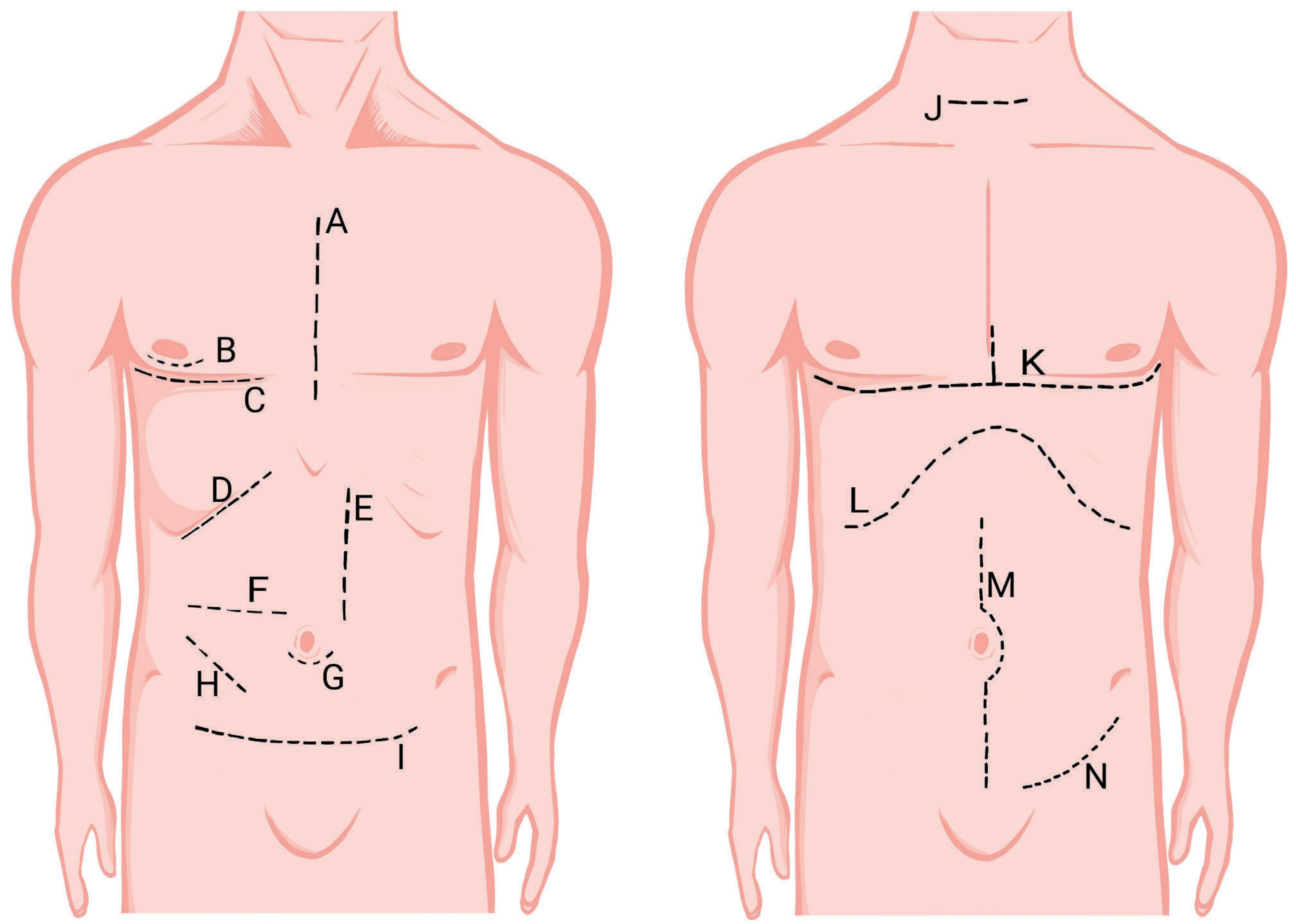
Figure: Standard skin incisions in general surgery - Bailey and Love's Short Practice of Surgery, 28th Ed.
Four Principles of Incision Planning
- Adequate exposure - the incision must allow the surgeon to see and work comfortably within the target cavity
- Extensibility - able to be lengthened if unexpected findings arise intraoperatively
- Neurovascular preservation - run incisions parallel to major nerves and vessels where possible
- Muscle-splitting over muscle-cutting - splitting muscles along fiber direction (as in the gridiron) preserves strength and reduces hernia risk
Abdominal Incision Types - Comparison Table
| Incision | Location | Technique | Uses | Key Advantage | Key Disadvantage |
|---|
| Midline | Linea alba | Cuts through avascular linea alba | Emergency laparotomy, trauma | Fastest access, fully extensible | Highest hernia risk |
| Paramedian | 2-3 cm lateral to midline | Through rectus sheath; muscle retracted | Colonic, pelvic | Lower hernia rate (muscle shutter) | Time-consuming, limits contralateral access |
| Subcostal (Kocher's) | Parallel to costal margin | Muscle-cutting | Cholecystectomy, liver, spleen | Excellent upper quadrant exposure | Painful; risk of intercostal nerve injury |
| Gridiron (McBurney's) | McBurney's point, RIF | Muscle-splitting | Open appendectomy | Very low hernia rate; preserves muscles | Limited exposure |
| Lanz | Transverse, RIF | Follows Langer's lines | Appendectomy | Better cosmetic result than gridiron | Similar exposure limitation |
| Pfannenstiel | Transverse suprapubic | Muscle-splitting through rectus | Cesarean section, hysterectomy, cystectomy | Excellent cosmesis, very strong | Poor access to upper abdomen |
McBurney's Point: Located at the junction of the lateral one-third and medial two-thirds of a line drawn from the anterior superior iliac spine (ASIS) to the umbilicus. This corresponds to the base of the appendix in most individuals.
Surgical Positioning: Rose's Position
Rose's position involves placing the patient supine with neck hyperextension - achieved by placing a sandbag or roll under the shoulders. This is the standard position for:
- Thyroidectomy
- Laryngoscopy
- Tracheostomy
- Tonsillectomy
Hyperextension stretches the anterior neck, tightens the pretracheal fascia, and brings the thyroid gland forward, maximizing surgical exposure. Pressure points must be padded, and the cervical spine must be assessed before positioning.
SECTION 3: Wound Closure, Healing, and Diathermy
Three Pathways of Wound Healing
| Type | Definition | Mechanism | Example | Scar Width |
|---|
| Primary Intention | Clean wound, edges closely apposed | Minimal granulation; epithelial bridging within 24-48h | Surgical incision, sutured laceration | Narrow |
| Secondary Intention | Large tissue loss or infected wound | Granulation tissue fills defect from base up; wound contraction by myofibroblasts | Abscess cavity, pressure ulcer | Wide |
| Tertiary (Delayed Primary) | Contaminated wound left open, closed later | Deep layers closed; skin left open 4-5 days until contamination cleared | Traumatic wounds, penetrating abdominal injuries | Variable |
Cellular timeline of healing (MCQ favorite):
- Hours 0-6: Vasoconstriction then vasodilation; platelets release PDGF, TGF-β
- 24-48 hours: Neutrophils predominate (acute inflammation, debridement)
- Day 2-3: Macrophages take over as the dominant cell - they phagocytose debris, release growth factors (VEGF, FGF, EGF), and direct angiogenesis and fibroplasia
- Day 3-5: Fibroblasts proliferate; collagen synthesis begins (Type III first, then replaced by Type I)
- Week 2 onwards: Remodeling - collagen cross-linking, scar contraction
Macrophages are the key cell for wound healing - their absence results in severely impaired healing even in the presence of neutrophils.
Factors Impeding Wound Healing
Local factors:
- Infection (most common cause of delayed healing - bacterial load >10⁵/g tissue)
- Ischemia/poor blood supply
- Foreign bodies
- Dead space
- Radiation injury
Systemic factors:
- Diabetes mellitus - impairs neutrophil function, angiogenesis, and collagen synthesis
- Malnutrition - protein and vitamin C deficiency impair collagen synthesis
- Corticosteroids - suppress inflammation and fibroplasia
- Smoking - vasoconstriction reduces tissue oxygen delivery
- Obesity - increases tension, reduces vascularity in subcutaneous fat
- Jaundice - bile salts impair wound healing
- Uraemia - impairs all phases
Suture Materials
| Suture | Type | Absorption | Mechanism | Uses |
|---|
| Vicryl (Polyglactin 910) | Synthetic absorbable | 60-90 days | Hydrolysis | Deep fascia, peritoneum, subcutaneous |
| PDS (Polydioxanone) | Synthetic absorbable | ~180 days | Hydrolysis | Mass closure, anastomoses |
| Prolene (Polypropylene) | Synthetic non-absorbable | Permanent | - | Vascular surgery, skin closure |
| Silk | Natural non-absorbable | Slow degradation | Proteolysis | Ligatures |
| Catgut | Natural absorbable | 10-14 days (plain); 21-28 days (chromic) | Enzymatic | Now largely replaced |
Vertical mattress suture is preferred for high-tension closures (laparotomy in obese patients) because it:
- Distributes tension across a wider area
- Everts wound edges (prevents inversion which impairs healing)
- Obliterates dead space
Surgical Drains
Drains serve to remove fluid accumulating in a dead space (blood, serous fluid, bile, lymph). Principles:
- Redivac (closed suction drain): removed after 24 hours in uncomplicated elective surgery when drainage is <50 mL/24h and appears serous
- Drains are NOT routinely left in clean operations
- Leaving drains unnecessarily increases infection risk
Clavien-Dindo Classification of Complications (must memorize):
| Grade | Definition |
|---|
| I | Any deviation from normal postoperative course; managed without pharmacological treatment except antiemetics/antipyretics |
| II | Drug therapy required (e.g., antibiotics, transfusion, TPN) |
| III | Requires surgical, endoscopic, or radiological intervention |
| IIIa | Intervention without general anesthesia |
| IIIb | Intervention under general anesthesia |
| IV | Life-threatening complication requiring ICU |
| IVa | Single organ dysfunction |
| IVb | Multiorgan dysfunction |
| V | Death |
Surgical Diathermy
Monopolar diathermy:
- Current flows from active electrode through the patient's body to a grounding plate
- High current density at active tip produces coagulation or cutting
- Contraindicated in pacemaker patients - can induce arrhythmias, inhibit pacing, or damage the device
- Also risky near metallic implants (prostheses, cochlear implants) due to current arcing
- Safe minimum distance from pacemaker: 15 cm
Bipolar diathermy:
- Current flows only between the two tips of the bipolar forceps
- No current traverses the patient's body
- Safe in pacemaker patients
- Used for fine hemostasis in neurosurgery, ophthalmology, and plastic surgery
SECTION 4: Laparoscopic and Robotic Surgery
Why CO₂ for Pneumoperitoneum?
From Miller's Anesthesia (10th Ed.): "Carbon dioxide (CO2) is the inert gas of choice for laparoscopy due to its high diffusion coefficient and its lower risk of gas emboli since it is easily excreted from the body through the respiratory system."
Key properties:
- Non-combustible (safe with electrosurgery)
- Highly soluble in blood (CO₂ emboli are rapidly cleared by lungs)
- Does not support combustion
- Relatively cheap and widely available
Safe insufflation pressure: ≤15 mmHg. Above 15 mmHg, the elevated intra-abdominal pressure compresses the inferior vena cava, reducing venous return and causing hemodynamic instability. It also splints the diaphragm, reducing lung compliance and increasing peak airway pressures.
Physiological Changes During Laparoscopy
- Respiratory: Diaphragm elevation reduces FRC; CO₂ absorption causes hypercapnia; increased ventilation needed
- Cardiovascular: Elevated intra-abdominal pressure reduces venous return; compensatory tachycardia
- Vagal bradycardia: Peritoneal stretch during insufflation can trigger a vasovagal response - managed by desufflation and IV atropine
- Position effects: Trendelenburg increases venous return but worsens respiratory compliance; reverse Trendelenburg (upper abdominal surgery) can cause hypotension
Advantages vs. Open Surgery
| Laparoscopic | Open |
|---|
| Smaller wounds, less pain | Better tactile feedback (haptics) |
| Faster recovery, shorter hospital stay | Faster to perform in emergency |
| Reduced wound infection rate | No learning curve |
| Better cosmesis | Unrestricted instrument movement |
| Less intraoperative blood loss | Lower equipment cost |
Absolute Contraindications to Laparoscopy
- Uncontrolled coagulopathy (absolute)
- Inability to tolerate general anesthesia
- Uncorrected hypovolemia
Relative contraindications include prior abdominal surgery (adhesions), pregnancy (second trimester and beyond), severe COPD, and massive obesity.
Conversion to Open
Must convert when:
- Uncontrollable hemorrhage
- Inability to identify anatomy safely
- Major visceral injury
- Equipment failure
Robotic Surgery
The da Vinci system provides:
- 7 degrees of freedom (vs. 4 in conventional laparoscopic instruments)
- 3D high-definition visualization with up to 10× magnification
- Tremor filtration (filters physiological hand tremor)
- Superior ergonomics for the surgeon
- Used in prostatectomy, hysterectomy, cardiac surgery, and complex colorectal procedures
PART II: MUSCULOSKELETAL-III (PERIOPERATIVE)
SECTION 9: Enhanced Recovery After Surgery (ERAS)
ERAS is a structured, evidence-based perioperative care pathway designed to attenuate the surgical stress response and optimize return to baseline function. Originally developed by Kehlet for colorectal surgery, it is now applied across general, orthopedic, urological, and gynecological surgery.
From Schwartz's Principles of Surgery (11th Ed.): "ERAS pathways are multi-modal perioperative care pathways designed to hasten recovery after elective surgery. These pathways may include preoperative education and counseling, preoperative optimization, limiting..."
Key ERAS Elements by Phase
Preoperative:
- Patient education and expectation-setting
- Fasting: Solids - stop 6 hours before; Clear fluids - allowed up to 2 hours before induction
- Carbohydrate loading: Oral carbohydrate drinks (e.g., Ensure, Fortisip) taken 2-3 hours preoperatively. This reduces:
- Preoperative thirst and hunger
- Postoperative insulin resistance
- Nitrogen catabolism
- Correction of preoperative anemia (target Hb >100 g/L)
- Optimized diabetes control (HbA1c <8.5%)
- Cessation of smoking ≥8 weeks before surgery
Intraoperative:
- Minimally invasive surgery where possible
- Goal-directed fluid therapy - guided by stroke volume variation, not fixed volume protocols
- Multimodal opioid-sparing analgesia: paracetamol + NSAIDs + regional block + low-dose opioid only as needed
- Maintenance of normothermia (active warming - Bair Hugger blankets)
- Prevention of PONV (postoperative nausea and vomiting) - ondansetron ± dexamethasone
Postoperative:
- Early oral feeding - within 6 hours of surgery where possible; not waiting for return of bowel sounds
- Early mobilization - out of bed within 24 hours; prevents DVT, ileus, pneumonia, and deconditioning
- Avoidance of routine nasogastric tubes, urinary catheters (remove early), and surgical drains
- Optimal analgesia allowing deep breathing and physiotherapy
- Avoidance of excess IV fluids - fluid overload causes gut edema, ileus, and delayed recovery
Key Outcomes with ERAS:
- Reduced length of stay by 30-50%
- Reduced complication rates
- Reduced 30-day readmission rates
- No increase in reoperation rates
SECTION 10: Perioperative Pain Management
Pathophysiology of Postoperative Pain
Surgical injury triggers a neuroendocrine stress response:
- Afferent pain signals activate the hypothalamic-pituitary-adrenal (HPA) axis
- Cortisol and catecholamines are released
- Cardiovascular effects: Hypertension, tachycardia, increased myocardial oxygen demand - risk of ischemia and MI in cardiac patients
- Respiratory effects: Splinting (guarding) prevents deep inspiration - atelectasis, retained secretions, pneumonia
- Gastrointestinal effects: Sympathetic activation inhibits gut motility - postoperative ileus
- Metabolic effects: Catabolism, hyperglycemia, nitrogen wasting - poor wound healing and infection risk
- Psychological: Anxiety, sleep deprivation, PTSD (especially in ICU patients)
Adequate pain control reduces all of these adverse effects and is a key pillar of ERAS.
WHO Analgesic Ladder
Originally designed for cancer pain, the ladder is widely applied to postoperative pain:
Step 3 - Severe Pain:   Strong opioids (Morphine, Oxycodone, Fentanyl)
                         ± adjuvants
Step 2 - Moderate Pain: Weak opioids (Tramadol, Codeine)
                         ± non-opioids ± adjuvants
Step 1 - Mild Pain:     Non-opioids (Paracetamol, NSAIDs, e.g., Ibuprofen, Ketorolac)
                         ± adjuvants
Paracetamol (Acetaminophen):
- Mechanism: Central COX inhibition; exact mechanism debated (possible endocannabinoid pathway)
- Maximum dose: 4 g/day in adults; reduce in liver disease
- Excellent safety profile; safe in renal impairment
NSAIDs (e.g., Ibuprofen, Diclofenac, Ketorolac):
- Mechanism: Inhibit cyclooxygenase (COX-1 and COX-2), reducing prostaglandin synthesis
- Reduce opioid requirements by 30-40%
- Contraindications: Renal impairment, GI ulcer, anticoagulant use, after major surgery with bleeding risk
Tramadol:
- Weak μ-opioid receptor agonist (1/6000th the potency of morphine at the receptor)
- Also inhibits serotonin and norepinephrine reuptake (mechanism of its atypical analgesia)
- Ceiling effect at ~400 mg/day
- Can cause serotonin syndrome if combined with SSRIs/SNRIs/MAOIs
- Reduces seizure threshold - caution in epilepsy
Morphine:
- Gold standard strong opioid; μ-opioid receptor agonist
- Metabolized to morphine-6-glucuronide (M6G) - active metabolite, accumulates in renal failure
- Side effects: respiratory depression, constipation, nausea, sedation, urinary retention, histamine release (avoid in asthma)
- Preferred route: IV for acute severe pain; oral for chronic/moderate
Neuropathic Pain Management
Neuropathic pain arises from nerve injury or dysfunction. Features:
- Burning, shooting, tingling, or electric shock quality
- Allodynia (pain from normally non-painful stimuli)
- Hyperalgesia (exaggerated response to painful stimuli)
- Responds poorly to standard opioids
First-line adjuvant agents:
- Pregabalin (Lyrica) and Gabapentin (Neurontin): Both are α2δ calcium channel subunit ligands - they reduce neuronal excitability and inhibit release of excitatory neurotransmitters (glutamate, substance P)
- Amitriptyline (tricyclic antidepressant): Inhibits serotonin and norepinephrine reuptake; blocks sodium channels
- Duloxetine (SNRI): Particularly used for diabetic neuropathy
Regional analgesia:
- Epidural analgesia: Gold standard for thoracic and major abdominal surgery; local anesthetic ± opioid
- Nerve blocks (e.g., femoral nerve block, TAP block): Reduce systemic opioid requirements
PART III: PLASTIC SURGERY AND BURNS
SECTION 11: Classification of Burns
Depth Classification
First-Degree (Superficial/Epidermal):
- Involves epidermis only
- Appearance: Erythema, warmth, dry, no blisters
- Sensation: Painful
- Healing: 5-7 days, no scarring
- Example: Sunburn
- NOT included in TBSA calculation for fluid resuscitation
Second-Degree (Partial-Thickness):
| Feature | Superficial Partial-Thickness | Deep Partial-Thickness |
|---|
| Depth | Epidermis + superficial dermis | Epidermis + deep dermis |
| Appearance | Blisters, moist, pink, weeping | Pale/mottled, less moist |
| Capillary refill | Present | Absent or sluggish |
| Pain | Severe (nerve endings intact) | Reduced sensation |
| Healing | 7-14 days, minimal scar | >21 days; likely needs grafting |
Third-Degree (Full-Thickness):
- Destroys all layers of skin (epidermis + dermis) - may extend to subcutaneous fat, muscle, or bone (4th degree+)
- Appearance: Dry, white, brown, or black leathery eschar; no blisters; non-blanching
- Sensation: Completely painless - nerve endings destroyed
- Healing: Cannot heal without surgical excision and skin grafting
- Requires reconstruction
Jackson's Zones of Burn Injury (from Schwartz's 11th Ed.):
- Zone of Coagulation (center): Necrotic tissue, irreversible damage, requires excision
- Zone of Stasis (middle): Ischemic but potentially viable; adequate resuscitation and wound care can prevent conversion to full thickness
- Zone of Hyperemia (periphery): Inflammatory vasodilation; will heal spontaneously
The Zone of Stasis is clinically critical - it can progress to full-thickness necrosis with inadequate resuscitation, infection, or hypotension.
SECTION 12: Assessment of Burn Patients
TBSA Calculation Methods
Wallace Rule of Nines (Adults):
| Body Region | % TBSA |
|---|
| Head and Neck | 9% |
| Anterior Trunk | 18% |
| Posterior Trunk | 18% |
| Each Upper Limb | 9% |
| Each Lower Limb | 18% |
| Perineum | 1% |
| Total | 100% |
For irregular/scattered burns: Palmar method - the patient's palm (including fingers) = approximately 1% TBSA. Useful for small scattered burns.
Lund and Browder Chart (Children - most accurate):
Adjusts for the fact that children have proportionally larger heads and smaller legs. As age increases, the head percentage decreases and leg percentage increases. This is the gold standard for pediatric burns.
Important: First-degree (superficial/epidermal) burns are NOT included in TBSA calculations for fluid resuscitation. Only second- and third-degree burns are counted.
Parkland Formula for Fluid Resuscitation
Formula: Fluid = 4 mL × body weight (kg) × % TBSA (2nd + 3rd degree burns)
Administration:
- 50% given in first 8 hours (counted from time of injury, not time of hospital arrival)
- 50% given in next 16 hours
- All as Ringer's Lactate (or Hartmann's solution)
Why Ringer's Lactate and not Normal Saline?
- Normal saline causes hyperchloremic metabolic acidosis
- Ringer's lactate is physiologically balanced; lactate is metabolized to bicarbonate
Worked example from Roberts and Hedges':
65 kg woman with 35% TBSA burns:
- Total = 4 × 65 × 35 = 9,100 mL over 24 hours
- First 8 hours: 4,550 mL (569 mL/hr)
- Next 16 hours: 4,550 mL (284 mL/hr)
Note: Add maintenance fluids to the Parkland formula. Most calculators omit this.
Monitoring Adequacy of Resuscitation
Gold standard: Hourly urine output via urinary catheter
| Patient | Target UO |
|---|
| Adult | 0.5 - 1.0 mL/kg/hr |
| Child | 1.0 - 2.0 mL/kg/hr |
| High-voltage electrical burns | 1.0 - 1.5 mL/kg/hr (to flush myoglobin) |
Oliguria = increase fluid rate. Do NOT give diuretics (they mask hypovolemia and falsely elevate urine output).
Hypervolemia signs (too much fluid): pulmonary edema, increasing oxygen requirements, worsening facial/airway edema.
SECTION 13: Initial and Long-Term Burn Management
Immediate First Aid (4 Steps)
- Remove from source - stop the burning process; safety for rescuer first
- Cool the burn - cool (not cold/ice) running water for 20 minutes continuously. Ice water causes vasoconstriction and worsens tissue injury. Hypothermia risk in children and large burns.
- Remove clothing - before swelling makes this impossible; leave adherent materials
- Cover - cling film (non-adherent) or clean sheet - prevents heat loss and contamination
Tetanus prophylaxis is mandatory for all burns breaching the skin.
ATLS Primary Survey - Burn Modifications
Standard ABCDE applies, but with burn-specific additions:
- A - Airway: First priority. Signs of inhalation injury - hoarseness, stridor, carbonaceous sputum, singed nasal hairs, facial burns, wheezing. Airway edema can develop within hours. Early intubation is mandatory - do not wait for stridor to develop.
- B - Breathing: Check for circumferential chest burns causing respiratory restriction (may need escharotomy)
- C - Circulation: IV access × 2 large-bore cannulae; begin Parkland resuscitation
- D - Disability: Check neurological status; consider carbon monoxide poisoning (give 100% O₂)
- E - Exposure: Calculate TBSA; maintain normothermia
Inhalation Injury
Inhalation injury is the single most important negative prognostic factor in burns. It increases mortality dramatically - burns of equivalent size have 2-3× higher mortality when combined with inhalation injury.
Three components:
- Upper airway thermal injury: Direct heat damage to oropharynx and larynx; edema causes obstruction
- Lower airway chemical injury: Toxic particles and gases (acrolein, hydrogen cyanide, CO) damage bronchial epithelium - inflammation, mucosal sloughing, cast formation, bronchospasm
- Systemic toxicity: Carbon monoxide (CO) binds hemoglobin with 240× affinity of O₂ → carboxyhemoglobin → tissue hypoxia despite normal PaO₂. Treat with 100% O₂ via non-rebreather mask (or intubation)
Bronchoscopy confirms inhalation injury and clears casts.
Escharotomy
Indications:
- Circumferential full-thickness burns of extremities (limb compartment syndrome)
- Circumferential chest burns causing respiratory restriction
- Signs of compartment syndrome: pain on passive stretch, tense swelling, paresthesia, pallor, pulselessness
Key facts:
- Performed by incising through the eschar (not fascia) longitudinally on medial and lateral surfaces
- No anesthesia required - full-thickness burns are insensate (nerve endings destroyed)
- Usually performed within 2-6 hours of burn injury
- Release is confirmed when compartment pressure falls below 30 mmHg
Fasciotomy is indicated when:
- Compartment pressure remains >30 mmHg after escharotomy
- High-voltage electrical burns (current passes through muscles, causing deep muscle necrosis regardless of skin appearance)
Topical Antimicrobials
| Agent | Properties | Uses | Contraindications/Side Effects |
|---|
| Silver Sulfadiazine (Flamazine) | Broad-spectrum; impedes epithelialization | Standard for partial-thickness burns | Transient leukopenia; contraindicated on face (staining), sulfa allergy, G6PD deficiency, near-term pregnancy |
| Mafenide Acetate | Penetrates eschar well | Deep wounds, electrical burns, infected burns | Very painful on application; carbonic anhydrase inhibitor → metabolic acidosis |
| Silver nitrate | Very broad spectrum | Resistant organisms | Hyponatremia, hypochloremia, electrolyte leaching; stains everything black |
| Bacitracin/Petrolatum | Minimal antimicrobial; non-adherent | Face, superficial burns | Limited depth penetration |
Long-Term Surgical Management
Timing of surgery:
- Superficial partial-thickness burns: Heal conservatively within 14 days; no surgery needed
- Deep partial-thickness burns: Failing to heal within 3 weeks → proceed to tangential excision
- Full-thickness burns: Always require surgery
Tangential excision:
- Sequential slicing of burned tissue until viable bleeding tissue is reached
- Reduces bacterial load, infection risk, and healing time
- Followed immediately by split-thickness skin graft (STSG)
Split-thickness skin graft (STSG):
- Contains epidermis + partial dermis
- Donor site heals spontaneously
- Can be meshed (expanded 1.5:1 to 6:1) to cover larger areas
- Meshed grafts leave a characteristic "fish-net" scar pattern
SECTION 14: Early and Late Burn Complications
Early Complications
| Complication | Mechanism | Management |
|---|
| Burn Shock | Massive capillary leak from inflammatory mediators (histamine, bradykinin, reactive oxygen species) → plasma loss → hypovolemia | Aggressive Ringer's Lactate resuscitation (Parkland formula) |
| Inhalation Injury | See above | Early intubation, bronchoscopy, 100% O₂ |
| Sepsis | Burn wound colonization → bacteremia; most common organisms: Pseudomonas, Staphylococcus aureus | Topical antimicrobials, early excision, systemic antibiotics only for clinical infection |
| Curling's Ulcer | Stress-induced gastric ulceration in major burns | Prophylactic proton pump inhibitors or H₂ blockers |
| Electrical Burns: Rhabdomyolysis | High-voltage (>1,000 V) causes massive skeletal muscle necrosis; myoglobin release → tubular precipitation → acute kidney injury | Aggressive IV hydration; maintain UO 1.5 mL/kg/hr; consider bicarbonate to alkalinize urine; fasciotomy |
| Carbon Monoxide Poisoning | CO binds Hb with 240× affinity of O₂; headache, confusion, cherry-red skin, coma at high levels | 100% O₂ (reduces CO half-life from 5h to 1h); hyperbaric O₂ for severe cases |
Late Complications
Hypertrophic Scarring:
- Dense, raised, erythematous scar confined to the wound boundary (vs. keloid which extends beyond)
- Risk factors: Deep partial-thickness burns healing >3-4 weeks, areas of high tension (shoulder, sternal)
- Prevention: Early excision and grafting for deep burns; pressure garments (≥24 hours/day for 12-24 months); silicone gel sheets
- Treatment: Intralesional triamcinolone, laser therapy, surgical revision
Post-Burn Contracture:
- Scar contraction across joints → flexion deformity (most common)
- Common sites: Axilla (adduction contracture), elbow, knee, neck, hand
- Prevention: Early grafting, splinting in position of function, physiotherapy
- Treatment: Z-plasty, local/free flaps, skin grafting with release of contracture
Marjolin's Ulcer:
- Malignant transformation (usually squamous cell carcinoma) in chronic burn scars or unstable scar tissue
- Characterized by rapid growth, raised rolled edges, ulceration
- Wide excision required
KEY MCQ POINTS: RAPID-FIRE SUMMARY
| Topic | Critical Fact |
|---|
| WHO Checklist - primary goal | Improve communication, not technical skills |
| Time Out - timing | After induction, before skin incision |
| Langer's lines - incision parallel | Narrow scar, minimal tension |
| Langer's lines - incision perpendicular | Wide, hypertrophic scar |
| RSTLs | Preferred for head/neck surgery |
| Most important factor for healing | Absence of infection |
| Cell dominant on Day 2 | Macrophage |
| Vicryl absorption | 60-90 days by hydrolysis |
| Monopolar diathermy CI | Pacemaker patients |
| Bipolar diathermy | Safe in pacemakers |
| Laparoscopy gas | CO₂ - non-combustible, highly soluble |
| Max insufflation pressure | 15 mmHg |
| Vagal bradycardia during laparoscopy | Desufflate + atropine |
| ERAS fasting (solids/fluids) | Solids: 6h; Clear fluids: 2h |
| ERAS carbohydrate loading | Reduces insulin resistance |
| WHO Ladder Step 2 | Tramadol (weak μ-opioid + SNRI) |
| WHO Ladder Step 3 | Morphine |
| Neuropathic pain - first line adjuvant | Pregabalin / Gabapentin |
| Burns depth: painless | Full-thickness (3rd degree) |
| Burns depth: most painful | Superficial partial-thickness (2nd degree) |
| 1st degree burns in Parkland formula | NOT included |
| Best TBSA tool for children | Lund and Browder chart |
| Fluid for burns resuscitation | Ringer's Lactate |
| Parkland formula | 4 mL × kg × %TBSA |
| Parkland: 1st 8 hours % | 50% |
| Urine output target (adult burns) | 0.5-1 mL/kg/hr |
| Oliguria management | Increase RL - NOT diuretics |
| Worst prognostic factor in burns | Inhalation injury |
| Cool water duration | 20 minutes |
| Escharotomy: anesthesia needed? | No (eschar insensate) |
| Silver sulfadiazine CI | Face, sulfa allergy, G6PD deficiency |
| Mafenide acetate mechanism of acidosis | Carbonic anhydrase inhibition |
| Deep burns: surgery timing | Failing to heal by 3 weeks |
| High-voltage injury (>1000V) | Rhabdomyolysis → AKI |
| Clavien-Dindo Grade III | Requires surgical/endoscopic/radiological intervention |
| Zone of Stasis | Can be salvaged with adequate resuscitation |
| Post-burn contracture type | Usually flexion deformity |
| Marjolin's ulcer | SCC in chronic burn scar |
Sources: Bailey and Love's Short Practice of Surgery (28th Ed.) | Schwartz's Principles of Surgery (11th Ed.) | Miller's Anesthesia (10th Ed.) | Roberts and Hedges' Clinical Procedures in Emergency Medicine | Tintinalli's Emergency Medicine | Fischer's Mastery of Surgery (8th Ed.) | Sabiston Textbook of Surgery