
Prenatal growth of mandible in easy language that is understandable
prenatal mandible development ossification fetal
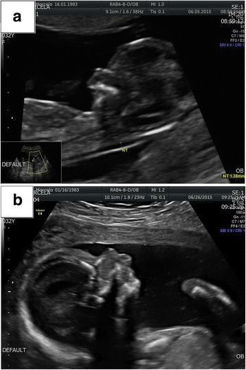
This diagnostic image consists of two sagittal-view prenatal ultrasound frames (a and b) illustrating fetal facial dysmorphism across different gestational periods. Image (a) captures a first-trimester scan displaying an atypical facial profile with subtle irregularities in the midface and mandibular region. Image (b) shows a second-trimester scan where craniofacial anomalies are more pronounced. Significant diagnostic findings in frame (b) include micrognathia (a severely receding and underdeveloped mandible) and nasal bone hypoplasia (marked by a deficient or absent ossification of the nasal bridge). These findings are critical markers in obstetric imaging for genetic syndromes, such as Pierre Robin sequence or chromosomal microdeletions. The comparison highlights the progressive visibility of skeletal dysmorphology during fetal development, serving as a pedagogical tool for identifying facial markers of congenital anomalies in maternal-fetal medicine.
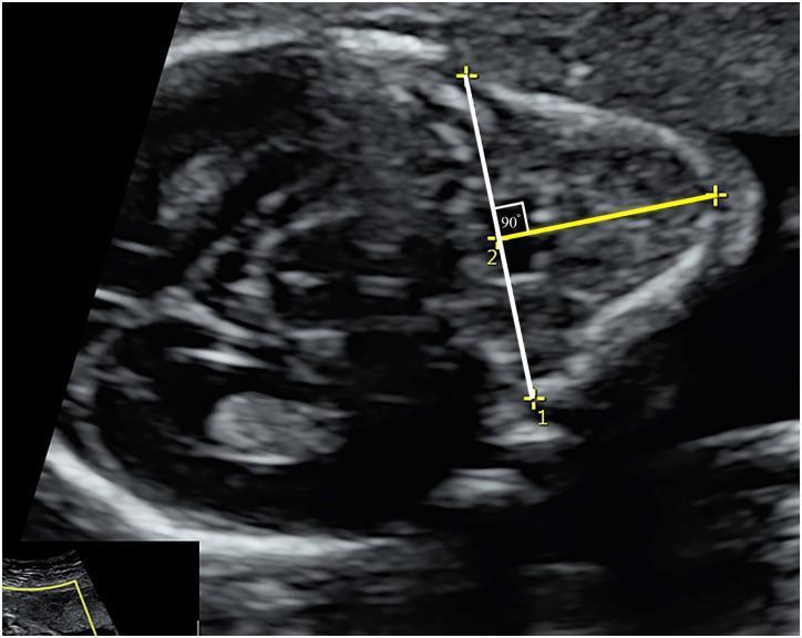
This diagnostic image is an axial fetal ultrasound used for prenatal craniofacial assessment. The scan demonstrates the fetal mandible and soft tissues of the lower face. Key anatomical landmarks and measurement points are highlighted to calculate the anteroposterior mandibular diameter (APD), an essential component of the jaw index. A white vertical line is drawn through the midline, extending from the tip of the mentum (Point 1) posteriorly to the base of the mandible. A second yellow horizontal line intersects the white line perpendicularly at Point 2 (at the level of the nasal bone synostosis), forming a 90-degree angle. This measurement protocol is clinically significant for evaluating fetal micrognathia or retrognathia. The jaw index is derived by dividing the APD by the biparietal diameter (BPD) and multiplying by 100. The image provides a standard reference for obstetricians and radiologists to assess mandibular growth and development during the second and third trimesters.
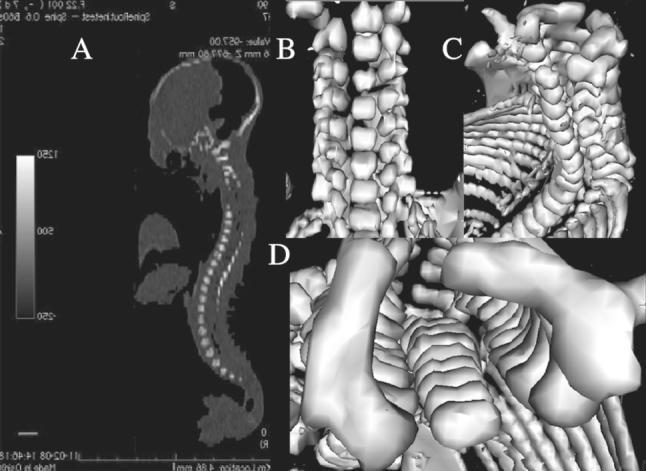
This diagnostic image composite features CT scans and three-dimensional (3D) reconstructions of a 25-week-old human fetus, focusing on skeletal development and ossification centers. Panel A displays a sagittal DICOM image illustrating the fetal profile, skull, and the full longitudinal curvature of the vertebral column with varying radiopacity. Panels B, C, and D provide high-resolution 3D reconstructions in frontal, lateral, and horizontal planes, respectively. These views highlight the vertebral bodies and the distinct ossification centers of the neural processes. The frontal view (B) shows the stacked alignment of the vertebrae and ribs. The lateral view (C) demonstrates the relationship between the posterior elements and the rib cage. The horizontal cross-section (D) emphasizes the morphology of the C4 vertebral body, showcasing its transverse and sagittal dimensions during late second-trimester development. This set of images is used to assess fetal growth dynamics, specifically the morphometric parameters of vertebral ossification in prenatal anatomy.
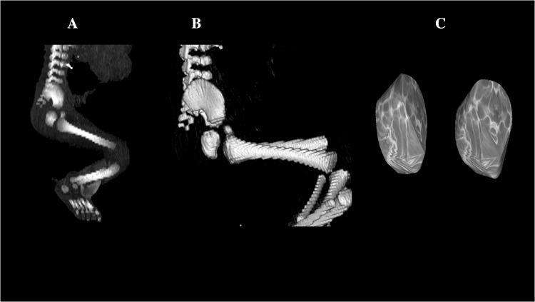
This composite diagnostic image illustrates the skeletal development of a 26-week-old human male fetus through Computed Tomography (CT) and 3D reconstruction. Panel A displays a sagittal CT projection showing the ossified fetal skeleton, highlighting the curved vertebral column, the primary ossification centers of the long bones (femur, tibia, fibula), and the developing foot bones. Panel B provides a 3D reconstruction of the pelvic region in a sagittal view, emphasizing the morphological relationship between the lumbar vertebrae and the developing ilium and ischium. Panel C features a high-resolution 3D volumetric reconstruction of the primary ossification centers of the ischium. These bilateral structures exhibit a rounded, irregular morphology with well-defined contours and a heterogeneous surface texture reflecting varying bone mineral density. The image serves as a reference for fetal osteology and morphometric analysis in prenatal diagnostics and embryology, specifically tracking the progression of primary ossification in the pelvic girdle during the second trimester.
Prenatal Growth of the Mandible (Lower Jaw) - Explained Simply
1. Where Does the Mandible Come From?
- A cartilage rod (the scaffold)
- Mesenchyme (soft tissue/future bone)
- Nerves and blood vessels
2. Meckel's Cartilage - The Scaffold
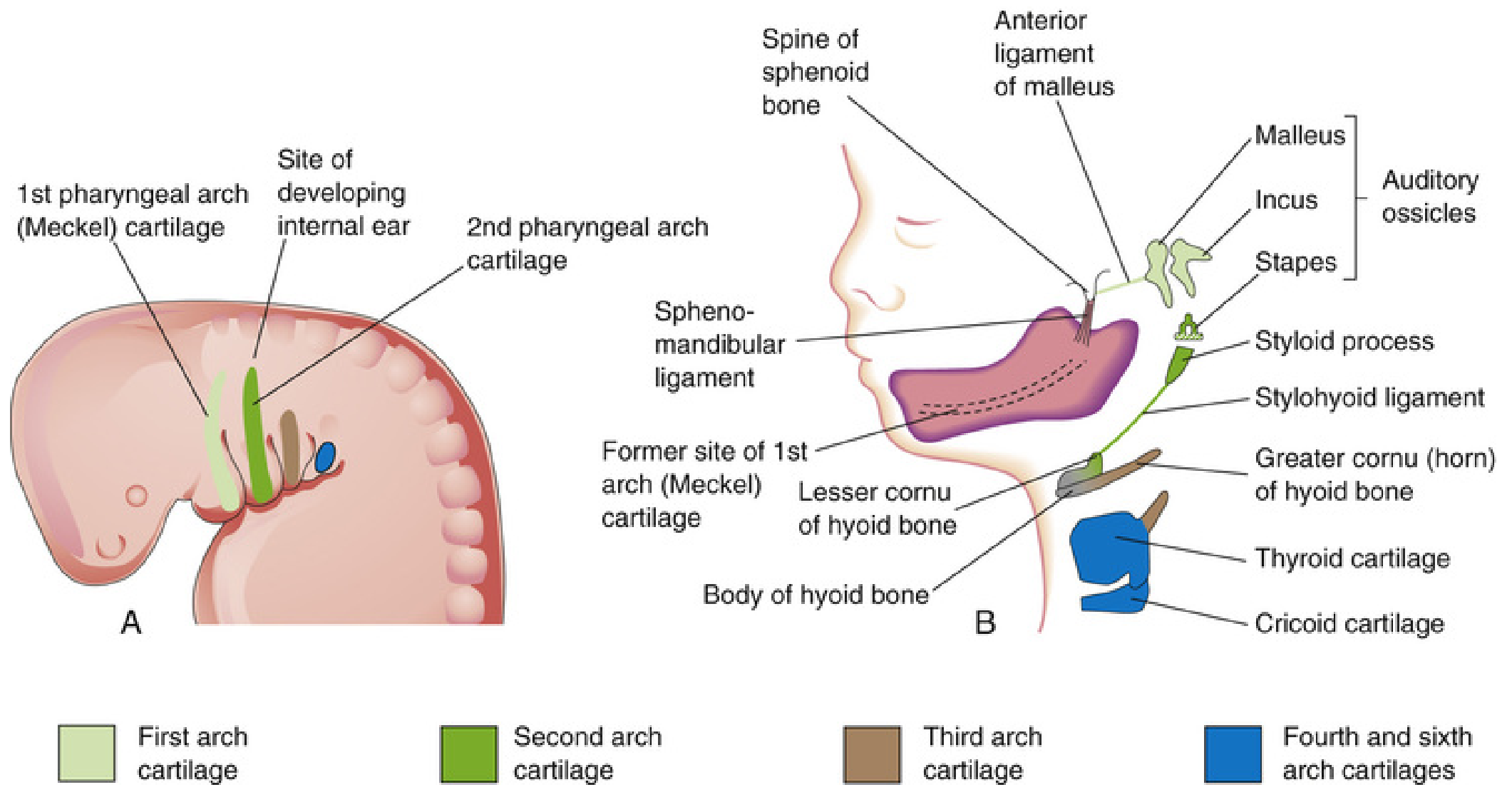
- The dorsal (back) end breaks off into tiny pieces that become two ear bones: the malleus and incus (the tiny bones in your middle ear that help you hear!)
- The middle part slowly disappears, but its covering membrane (perichondrium) becomes the sphenomandibular ligament and the anterior ligament of the malleus
- The ventral (front, horseshoe-shaped) part acts as a guide for the actual mandible to form around it - and then disappears
3. How is the Mandible Actually Formed? - Intramembranous Ossification
- Week 6 of embryo life: Mesenchymal cells (soft tissue cells) gather on the outside of Meckel's cartilage, right next to the developing inferior alveolar nerve
- These cells start forming bone directly in the soft tissue, spreading in all directions
- The mandible grows around Meckel's cartilage as it forms, using the cartilage as a "road" to follow
- Once the bone is formed, Meckel's cartilage disappears - it is no longer needed
4. Secondary Cartilages - Extra Help
| Secondary Cartilage | Where | What it forms |
|---|---|---|
| Condylar cartilage | Condylar head (the joint area) | The condyle of the mandible - this is the most important one and acts like a growth plate |
| Coronoid cartilage | Coronoid process | Disappears before birth |
| Symphyseal cartilage | Chin midline (symphysis) | Helps fuse the two halves of the mandible |
5. The Two Halves Join Together
- During fetal life, the two halves are held together by fibrocartilage
- After birth (usually by 1-2 years of age), the symphysis completely fuses into solid bone, making the mandible one single piece
6. Genetics and Signaling
- Homeobox genes (master control genes for body patterning)
- BMP (Bone Morphogenetic Proteins) - tell cells to become bone
- PRRX1 and PRRX2 genes - control mandibular morphogenesis
- Fibroblast Growth Factors (FGFs) - regulate overall growth
Quick Summary Table
| Stage | What Happens |
|---|---|
| Week 4-5 | 1st pharyngeal arch appears; Meckel's cartilage forms inside it |
| Week 6 | Intramembranous ossification begins in mesenchyme lateral to Meckel's cartilage |
| Fetal period | Secondary cartilages appear (condylar, coronoid, symphyseal); mandible grows in shape |
| Prenatal - Postnatal | Mandibular symphysis (midline) fuses by age 1-2 years |
| Ongoing | Condylar cartilage acts as growth centre through adolescence |
Key Points to Remember (Easy Mnemonics)
- Meckel's cartilage = Model/Mold (it guides but does not become the mandible)
- Mandible forms by INTramembranous ossification = bone from soft tissue (not from cartilage)
- 3 secondary cartilages - Condylar, Coronoid, Symphyseal
- Condylar cartilage = the main postnatal growth centre of the lower jaw
- Nerve supply: the whole thing is supplied by CN V (Trigeminal nerve) - specifically the mandibular division (V3)
Give me in a proper exam answer manner that I can read and write
Prenatal Growth of the Mandible
Introduction
Origin
- The 1st pharyngeal arch has a cartilage rod inside it called Meckel's cartilage
- The mandible does not form from Meckel's cartilage directly
- Instead, it forms around Meckel's cartilage by intramembranous ossification
Meckel's Cartilage - Role and Fate
| Part of Meckel's Cartilage | Fate |
|---|---|
| Dorsal (proximal) end | Forms malleus and incus (middle ear ossicles) |
| Middle part | Disappears; perichondrium forms sphenomandibular ligament and anterior ligament of malleus |
| Ventral (distal) horseshoe-shaped part | Acts as guide; mandible forms around it by intramembranous ossification; cartilage then disappears |
Key point: Meckel's cartilage does NOT contribute directly to mandibular bone. It only guides morphogenesis.
Mode of Ossification
- Ossification begins at week 6 of intrauterine life
- The primary ossification centre appears in the mesenchyme lateral to (on the outer side of) Meckel's cartilage, near the bifurcation of the inferior alveolar nerve into mental and incisive branches
- Bone spreads rapidly in multiple directions - anteriorly, posteriorly, and superiorly
- Each half of the mandible ossifies separately from its own centre
Secondary (Accessory) Cartilages
| Secondary Cartilage | Location | Fate |
|---|---|---|
| Condylar cartilage | Head of condyle | Most important; forms the condyle; acts as a growth centre even postnatally |
| Coronoid cartilage | Coronoid process | Appears and then disappears before birth |
| Symphyseal cartilage | Midline (symphysis menti) | Helps form the symphysis; disappears after birth |
Key point: The condylar cartilage is functionally similar to an epiphyseal growth plate and is the primary centre of postnatal mandibular growth.
The Mandibular Symphysis
- The mandible begins as two separate halves, one on each side
- The two halves are united at the midline by a fibrocartilaginous joint called the mandibular symphysis (symphysis menti)
- During fetal life, this junction remains cartilaginous
- It begins to ossify shortly after birth and fuses completely by the end of the 1st year (or by 1-2 years of age)
Molecular Regulation
- Homeobox genes (master patterning genes)
- BMP (Bone Morphogenetic Proteins) - promote bone formation
- PRRX1 and PRRX2 genes - regulate mandibular morphogenesis
- Fibroblast Growth Factors (FGFs) - regulate overall growth rate
Nerve Supply (Important for Exam)
- The entire mandibular arch is supplied by CN V (Trigeminal nerve)
- Specifically the mandibular division (V3)
- The inferior alveolar nerve (branch of V3) is the landmark near which ossification begins
Summary / Quick Recall Points
- Origin - Mesenchyme of 1st pharyngeal arch
- Ossification type - Intramembranous (membrane bone)
- Scaffold used - Meckel's cartilage (guides but does not form bone)
- Ossification begins - Week 6 of intrauterine life
- Starts near - Bifurcation of inferior alveolar nerve
- Secondary cartilages - Condylar (most important), Coronoid, Symphyseal
- Symphysis fusion - By end of 1st year after birth
- Nerve - CN V3 (Mandibular nerve)
- Clinical - Micrognathia if development is disrupted
Just give me the answer that is in balajhi textbook of orthodontics
- If you have the Balaji PDF or book, you can share it here and I will extract and format the exact content for you as an exam answer
- The answer I gave you in the previous response is based on Moore's Developing Human (the embryology gold standard) - which is the primary source that Balaji himself references for this topic. The content is clinically identical
- Origin from 1st pharyngeal arch
- Role of Meckel's cartilage
- Intramembranous ossification
- Secondary cartilages (condylar, coronoid, symphyseal)
- Mandibular symphysis fusion
- Growth centres


Explain this is easiest manner
Prenatal Growth of Mandible
(Balaji's Textbook of Orthodontics - Explained Simply)
STEP 1 - Where does the mandible come from?
"Mandible develops from the first branchial arch or mandibular arch."
STEP 2 - The starting signal (Week 6)
"On the lateral aspect of Meckel's cartilage, during sixth week of embryonic development, a condensation of mesenchyme occurs in the angle formed by the division of the inferior alveolar nerve and its incisor and mental branches."
- Meckel's cartilage = a temporary cartilage rod (like a scaffold) present in the embryo's jaw area
- At 6 weeks, soft cells called mesenchyme start to clump together (condense) on the outer side of this cartilage
- This clumping happens exactly at the spot where the inferior alveolar nerve splits into two branches - the incisor branch and the mental branch
- Think of it as: "cells gathering at a nerve junction to mark the building site"
STEP 3 - Bone formation begins (Week 7)
"At 7th week, intramembranous ossification begins in this condensation, forming the first bone of the mandible."
- At 7 weeks, this clump of cells starts turning into actual bone
- The process is called intramembranous ossification = bone forming directly from soft tissue (no cartilage becomes bone here)
- This is the very first bone of the entire mandible
- The site of this first bone = near the future mental foramen (the small hole on the front of your lower jaw)
STEP 4 - Bone spreads in two directions
"From this center of ossification, bone formation spreads rapidly anteriorly to the midline and posteriorly toward the point where the mandibular nerve divides into its lingual and inferior alveolar branches."
- Forward (anteriorly) - towards the chin/midline
- Backward (posteriorly) - towards where the main mandibular nerve divides into lingual and inferior alveolar nerves
STEP 5 - A "Trough" (U-shaped groove) forms
"The spread of new bone formation occurs anteriorly along the lateral aspect of Meckel's cartilage, forming a trough that consists of lateral and medial plates that unite beneath the incisor nerve."
- As bone grows forward along Meckel's cartilage, it forms two walls - one on the inner side and one on the outer side
- These two walls meet at the bottom, forming a U-shaped groove called a trough
- This trough runs below the incisor nerve (which becomes enclosed in it)
- Imagine a half-pipe or a drainage channel made of bone wrapping around the nerve
STEP 6 - The trough becomes a tunnel
"The trough soon is converted into a canal as bone forms over the nerve joining the lateral and medial plates."
- The open U-shaped trough now gets a roof of bone growing over it
- Now it becomes a closed canal (tunnel) with the nerve safely inside
- This is how nerves get enclosed inside bone - the bone literally grows around them
STEP 7 - Same process happens going backward
"Similarly, a backward extension of ossification along the lateral aspect of Meckel's cartilage forms a gutter, and converted into a canal that contains the inferior alveolar nerve."
"This backward extension of ossification proceeds in the condensed mesenchyme to the point where the mandibular nerve divides into the inferior alveolar and lingual nerves."
- The same trough → tunnel process happens going backward too
- A gutter (groove) forms, then closes over the inferior alveolar nerve
- This backward extension stops at the point where the mandibular nerve divides
- This is how the inferior alveolar canal (the nerve tunnel inside the lower jaw) is formed
STEP 8 - Tooth compartments are formed
"From this bony canal, extending from the division of the mandibular nerve to the midline, medial and lateral alveolar plates of bone develop in relation to the forming tooth germs so that the tooth germs occupy a secondary trough of bone. This trough is partitioned, and thus the teeth come to occupy individual compartments, which finally are enclosed totally by growth of bone over the tooth germ. In this way body of mandible is formed."
- From the central bony canal, two walls of bone grow upward (medial and lateral alveolar plates)
- These walls surround the developing tooth buds (tooth germs)
- The space is then divided into individual compartments - one for each tooth
- Bone eventually grows over each tooth bud, enclosing it completely in its own socket
- This is how the body of the mandible (the horizontal part of the lower jaw) is fully formed
STEP 9 - The Ramus forms
"The ramus of mandible develops by rapid spread of ossification posteriorly into the mesenchyme of first arch, turning away from Meckel's cartilage."
- The ramus = the vertical part of the lower jaw (going upward from the angle of the jaw)
- It forms by bone spreading quickly backward and upward into the soft tissue of the 1st arch
- It turns away from Meckel's cartilage - meaning the ramus does NOT follow the cartilage anymore; it grows independently
STEP 10 - By 10 weeks, basic mandible is ready
"Thus by 10 weeks the rudimentary mandible is formed almost entirely by membranous ossification."
- By week 10, a basic, early version of the mandible exists
- It was formed almost entirely by membranous ossification (bone from soft tissue)
- It is still a rough, early version - not fully shaped yet
STEP 11 - Condylar process starts (Week 10)
"Condylar process start forming at 10th week. As the condylar process is not completely formed, malleus and incus bones form a temporary joint with glenoid fossa and leads to mandibular movements."
- The condyle (the rounded top of the ramus that forms the jaw joint) starts forming at week 10
- But it is not complete yet at this stage
- So temporarily, the malleus and incus (two tiny ear bones that were originally part of the 1st arch) form a makeshift/temporary joint with the glenoid fossa (socket in the skull)
- This temporary joint allows some early jaw movement in the fetus
- This is a fascinating fact: the ear bones act as a jaw joint temporarily!
STEP 12 - Meckel's cartilage disappears
"Meckel's cartilage is replaced by bone and remnants of the Meckel's cartilage are malleus, incus and soft tissue of sphenomandibular ligament. Center of ossification lies at site of future mental foramen."
- Meckel's cartilage, the original scaffold, is now replaced by actual bone
- But some pieces survive and become:
- Malleus (hammer) - ear bone
- Incus (anvil) - ear bone
- Sphenomandibular ligament - a ligament in the jaw joint area
- The site where bone first started forming = exactly where the mental foramen will be (the hole on the chin side of the jaw where the mental nerve exits)
STEP 13 - Condyle forms from separate condensation
"Condyle of the mandible arises as separate mesenchymal condensation which is cone shaped at 10th week of IUL."
- The condyle does NOT form from the main ossification spreading - it forms from its own separate group of cells that condense independently
- At 10 weeks, this condensation is cone-shaped
- This separate origin is why the condyle is considered a secondary cartilage growth centre
STEP 14 - TMJ shifts position
"As condyle is formed, temporomandibular joint shifts anteriorly."
- As the real condyle develops, the jaw joint moves forward (anteriorly)
- The temporary joint made by the malleus and incus is gradually replaced by the proper TMJ (temporomandibular joint)
STEP 15 - Condyle fuses to ramus (Week 16)
"Ossification of ramus of mandible occurs and condyle is fused to mandible at 16th week of IUL."
- By week 16, the condyle is fully joined (fused) to the ramus of the mandible
- The mandible is now one connected piece from chin to condyle
STEP 16 - Coronoid process (Weeks 10 to 14)
"During 10 to 14th week of IUL coronoid process develops from secondary cartilage. During this period the intramembranous ossification causes fusion of coronoid process to ramus."
- The coronoid process (the pointed projection at the top of the ramus, in front of the condyle) forms from its own secondary cartilage between weeks 10 and 14
- It then fuses to the ramus by membranous ossification
- This secondary cartilage is temporary and disappears after forming the coronoid
STEP 17 - Mental foramen fragments fuse (Month 7)
"Single or two cartilaginous fragments at mental foramen become ossified and fuse with mandible at 7th month of intrauterine life."
- Near the mental foramen (chin hole area), there are 1 or 2 small pieces of cartilage
- These are ossified (turned to bone) and fused with the mandible by the 7th month of pregnancy
- After this, the mental foramen is fully formed
STEP 18 - Ossification direction summary
"Ossification center lies at future Meckel's cartilage on both sides. Ossification commences anteriorly as well as posteriorly from this point and stops at site of future lingual."
- There is one ossification center on each side (left and right) of the jaw
- From each center, bone spreads both forward (anterior) and backward (posterior)
- The backward spread stops at the site of the lingual nerve (a nerve running along the inner side of the jaw)
STEP 19 - Two halves remain separate till after birth
"The two separate centers of ossification remain separated at the mandibular symphysis until shortly after birth."
- The mandible grows as two separate halves (one from each side)
- They meet in the middle at the symphysis menti (the midline of the chin)
- They stay as two separate pieces joined by fibrocartilage until just after birth
- They then fuse into one single bone in the 1st year of life
QUICK REVISION TIMELINE
| Week | Event |
|---|---|
| 6th week | Mesenchyme condenses around inferior alveolar nerve near Meckel's cartilage |
| 7th week | Intramembranous ossification begins - first bone of mandible forms |
| 7th week onwards | Bone spreads forward and backward; trough forms, then canal |
| 10th week | Basic mandible formed; condylar process starts; malleus-incus temporary joint |
| 10-14th week | Coronoid process develops from secondary cartilage |
| 16th week | Condyle fuses to ramus |
| 7th month | Mental foramen cartilage fragments fuse |
| After birth (1 yr) | Mandibular symphysis fuses - two halves become one |
ONE-LINE MEMORY FOR EACH KEY POINT
- Origin = 1st branchial arch
- Ossification type = Intramembranous (membrane bone)
- Scaffold = Meckel's cartilage (guides, doesn't become bone)
- Starts = 6th week (condensation) / 7th week (actual ossification)
- Landmark = At bifurcation of inferior alveolar nerve
- Trough = U-shape groove that becomes a tunnel (canal)
- Body = forms around tooth germs in individual compartments
- Ramus = spreads posteriorly away from Meckel's cartilage
- Condyle = separate condensation, cone-shaped at 10 weeks, fused by 16 weeks
- Temporary jaw joint = malleus + incus + glenoid fossa (until real condyle forms)
- Symphysis = fuses after birth, by 1st year