
SLE In derma write a short note about it
Reading File
Now let me search the dermatology textbooks for SLE content in parallel:
Finding Sources
Finding Sources
The Andrews' Diseases of the Skin (clinical dermatology textbook) has a dedicated section. Let me read it:
Reading File
Finding Sources
Excellent content coming from Andrews' Dermatology. Let me read more of this section and also fetch from Harrison's:
Reading File
Reading File
I have very rich content. Let me fetch one key image to include and read the treatment section:
Reading File
Finding Sources
Finding Sources
Reading File
Reading File
I now have comprehensive content. Let me also fetch the key malar rash image:
Finding Sources
Finding Sources
I have enough comprehensive content from Andrews' Dermatology and Harrison's to write an excellent short note. Let me now compose the answer:
Systemic Lupus Erythematosus (SLE) - Short Note (Dermatology)
Definition and Epidemiology
SLE is a chronic, multisystem autoimmune disease characterised by widespread immune complex-mediated tissue damage. It predominantly affects young to middle-aged women (female-to-male ratio ~9:1). Skin involvement occurs in ~80% of cases, making dermatological findings central to its diagnosis - four of the eleven ACR diagnostic criteria are mucocutaneous.
ACR Classification Criteria (11 criteria; ≥4 needed for diagnosis)
| # | Criterion |
|---|---|
| 1 | Malar rash |
| 2 | Discoid rash |
| 3 | Photosensitivity |
| 4 | Oral ulcers |
| 5 | Arthritis |
| 6 | Proteinuria >0.5 g/day or casts |
| 7 | Neurologic disorders (seizures/psychosis) |
| 8 | Pleuritis/pericarditis |
| 9 | Haematologic disorders (haemolytic anaemia, leukopenia, thrombocytopenia) |
| 10 | Immunologic disorders (anti-dsDNA, anti-Sm, APLAs) |
| 11 | Positive ANA |
The SLICC criteria (revision of ACR) require at least 1 clinical + 1 immunological criterion, or biopsy-proven lupus nephritis with positive ANA/anti-dsDNA. This improves sensitivity.
Cutaneous Manifestations
Classified into SLE-specific (LE-specific) and non-specific lesions.
A. Acute Cutaneous LE (ACLE)
- Malar (butterfly) rash - the hallmark. Erythema over both cheeks and the nasal bridge with striking sparing of the nasolabial folds (unlike dermatomyositis which involves the folds). May be flat or slightly raised, with associated oedema.
- Resolves without scarring.
- May also manifest as a generalised maculopapular photosensitive rash on sun-exposed areas.
- Bullous lupus erythematosus (BLE): vesicles/bullae on sun-exposed areas; histology shows neutrophilic accumulation at the DEJ and subepidermal bullae; responds dramatically to dapsone; associated with HLA-DR2.
- TEN-like lesions: rare, due to intense interface inflammation.
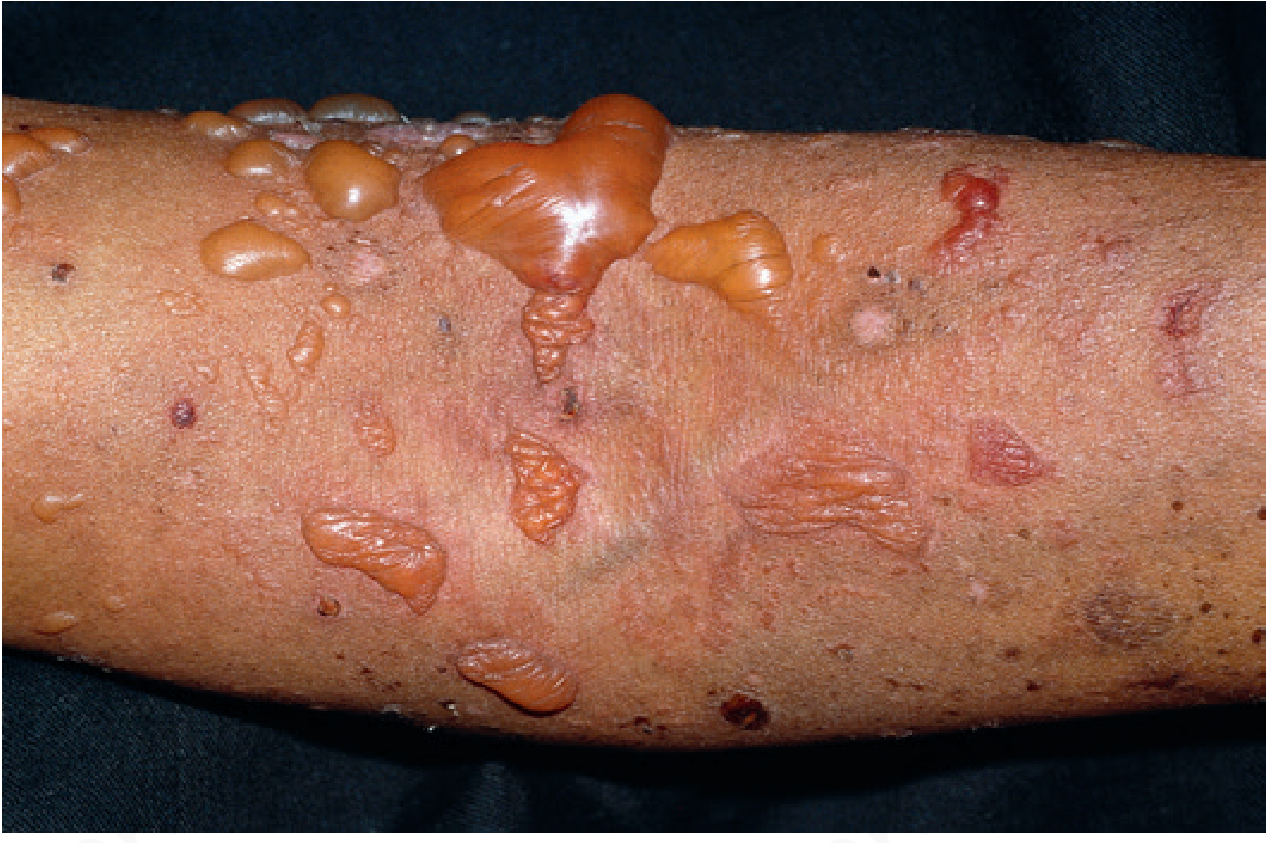
Fig. Bullous lupus erythematosus - Andrews' Diseases of the Skin
B. Subacute Cutaneous LE (SCLE)
- Photosensitive rash in sun-exposed areas (especially upper torso).
- Two morphological forms:
- Annular/polycyclic (red-rimmed rings)
- Papulosquamous/psoriasiform
- Strongly associated with anti-Ro (anti-SSA) antibodies.
- Heals without scarring but may cause dyspigmentation.
C. Chronic Cutaneous LE (CCLE) - Discoid LE
- Rough, slightly raised, circular lesions with erythematous rims, central atrophy and depigmentation, with dyspigmented or hyperpigmented periphery.
- Scarring is a hallmark - can cause permanent scarring alopecia.
- Other chronic forms: hypertrophic/verrucous lupus, lupus panniculitis (lupus profundus), tumid lupus, chilblain lupus, DLE/lichen planus overlap.
D. Vascular Lesions (~50% of patients)
- Periungual telangiectasia, red or spotted lunulae.
- Raynaud's phenomenon (~1/3 of patients): triphasic colour change (white - blue - red) on cold/stress exposure.
- Erythema multiforme-like lesions (Rowell syndrome).
- Livedo reticularis - associated with antiphospholipid antibodies (APLAs).
- Leg ulcers, thrombophlebitis.
- Nailfold capillary loops show wandering glomeruloid loops (unlike DM/scleroderma which show symmetric dilation/dropout).
E. Hair and Nails
- Diffuse non-scarring alopecia (telogen effluvium).
- "Lupus hairs" - short, broken hairs in the frontal region from increased hair fragility.
- Periungual telangiectasia, red/spotted lunulae.
F. Mucous Membrane Lesions (20-30%)
- Oral erosions, shallow angular ulcerations with surrounding erythema (Fig. 8.11).
- Petechiae and ulcerations on the hard palate.
- Conjunctivitis, episcleritis, nasal and vaginal ulcerations.
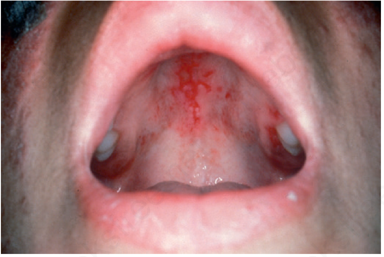
Fig. Oral lupus erythematosus - Andrews' Diseases of the Skin
Histopathology
- Interface dermatitis with vacuolar degeneration of basal layer.
- Scant perivascular lymphoid infiltrate.
- Thickening of basement membrane zone (BMZ).
- Lupus band test (LBT) - Direct immunofluorescence (DIF): granular deposits of IgG, IgM, IgA and C3 at the DEJ (BMZ). Positive in both lesional and non-lesional sun-exposed skin in SLE (unlike DLE, where positivity is only in lesional skin).
Investigations
| Test | Significance |
|---|---|
| ANA | Screening test; highly sensitive but not specific |
| Anti-dsDNA | Highly specific for SLE; titres correlate with disease activity (especially nephritis) |
| Anti-Sm | Highly specific for SLE |
| Anti-Ro/SSA | Associated with SCLE, neonatal lupus, photosensitivity |
| Anti-La/SSB | Associated with secondary Sjogren features |
| APLAs (anticardiolipin, lupus anticoagulant) | Thrombosis, recurrent miscarriages, antiphospholipid syndrome |
| C3, C4 | Decreased during active disease |
| Skin biopsy + DIF | Interface dermatitis; LBT positive |
| CBC | Haemolytic anaemia, leukopenia, lymphopenia, thrombocytopenia |
| Urinalysis | Proteinuria, casts (renal involvement) |
Etiology and Pathogenesis
- Multifactorial: genetic (>100 loci, including HLA-DR3, C4 null alleles, DNASE1L3, TREX1 mutations), hormonal (female predominance, oestrogen role), and environmental (UV radiation, infections, smoking, oral contraceptives).
- Pathomechanism: Abnormal apoptosis/reduced clearance of apoptotic cells → exposure of nucleosome antigens → anti-nuclear antibody production → immune complex formation → complement activation → tissue damage.
- UV radiation (both UVB and UVA) upregulates antigen expression and pro-inflammatory cytokines (IL-1, TNF-α), triggering photosensitivity and flares.
- Reduced T-suppressor cell function; overproduction of gamma-globulins by B cells.
Differential Diagnosis
- Dermatomyositis (involves nasolabial folds, heliotrope rash, Gottron papules)
- Rosacea (involves nasolabial folds, telangiectasia, no scarring)
- Discoid lupus erythematosus (DLE)
- Erythema multiforme
- Pemphigus erythematosus (Senear-Usher syndrome)
- Polymorphous light eruption
- Drug eruptions
- Sjogren syndrome
Treatment
General Measures (for ALL patients)
- Strict sun protection: broad-spectrum sunscreen (high SPF) daily, sun avoidance - even asymptomatic patients have photosensitivity.
- Avoid cold exposure, heat, and trauma (biopsies/scar revision can provoke flares).
- Smoking cessation.
- Calcium + Vitamin D supplementation (increased osteoporosis risk independent of steroids).
- Monitor bone density; bisphosphonates if on corticosteroids.
Local/Topical Treatment
- Potent/superpotent topical corticosteroids (first-line for skin lesions; occlusion may enhance effect).
- Intralesional triamcinolone acetonide (2.5-10 mg/mL, every 4-6 weeks; max 40 mg at once) - single most effective local treatment.
- Topical calcineurin inhibitors (tacrolimus, pimecrolimus) - second-line.
- Topical retinoids for hypertrophic lesions.
- PDL (585-595 nm) laser for vascular lesions.
Systemic Treatment
| Drug | Role |
|---|---|
| Hydroxychloroquine (HCQ) | Mainstay for all forms of LE; reduces flares, prevents organ damage, safe in pregnancy |
| NSAIDs | Arthralgia, mild systemic symptoms |
| Systemic corticosteroids | Moderate-severe disease, flares |
| Methotrexate | Skin and joint disease |
| Azathioprine | Maintenance; safer in pregnancy |
| Mycophenolate mofetil | Lupus nephritis; skin disease |
| Cyclophosphamide | Severe nephritis, CNS lupus |
| Belimumab | Anti-BAFF/BLyS biologic; approved for active SLE |
| Anifrolumab | Anti-IFN-α receptor; approved for active SLE |
| Dapsone | Specifically for bullous LE |
Special Considerations
- Neonatal lupus: caused by passive transfer of maternal anti-Ro/SSA antibodies; presents with transient cutaneous lesions (annular erythema) and, importantly, congenital heart block in the neonate.
- Drug-induced lupus: caused by hydralazine, procainamide, isoniazid, minocycline, anti-TNF agents; ANA positive, but anti-dsDNA usually negative; resolves on drug withdrawal.
- Pregnancy: increased risk of miscarriage (especially with APLAs); SLE may flare post-partum; hydroxychloroquine is safe and should be continued.
- Prognosis: Renal involvement is the most important predictor of poor prognosis. Thromboses are the most common cause of death after the first 5 years of disease.
Sources: Andrews' Diseases of the Skin: Clinical Dermatology, p. 189-190 | Harrison's Principles of Internal Medicine 22E, Ch. 368
This is a shared conversation. Sign in to Orris to start your own chat.