
Diabetes mellitus for mbbs pathology long answer
diabetic nephropathy glomerulosclerosis Kimmelstiel Wilson nodule histology
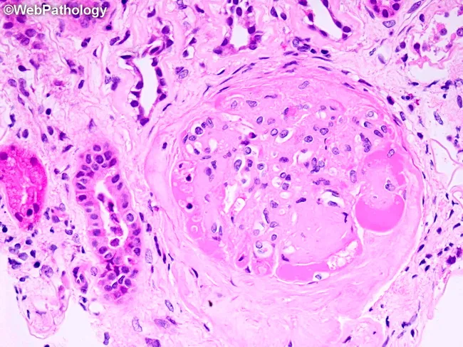
Comprehensive Description: The image depicts a renal glomerulus studied by light microscopy on a hematoxylin and eosin stained section. The cortical glomerulus shows marked mesangial expansion with sclerosis, a hallmark of diabetic nephropathy. A classical Kimmelstiel-Wilson nodular lesion is evident at approximately the 5–6 o’clock position within the tuft, appearing as a rounded, PAS-positive eosinophilic nodule composed of matrix and mesangial material encroaching on capillary loops. Surrounding capillary lumina are variably narrowed by eosinophilic hyaline caps, consistent with nodular glomerulosclerosis and hyaline arteriolosclerosis of diabetic microangiopathy. The glomerular basement membranes are thickened, and there is proliferation of mesangial matrix with increased cellularity in some areas. The periglomerular stroma shows interstitial inflammation and edema. Overall, these features reveal chronic hyperglycemia-induced glomerular damage with progressive loss of filtration surface area, correlating with proteinuria and progressive chronic kidney disease risk. The image demonstrates pathognomonic nodular sclerosis in long-standing diabetes mellitus, requiring correlation with clinical history, duration of diabetes, blood pressure control, and renal function tests. This slide exemplifies diabetic nodular glomerulosclerosis (KW disease) and is a classic teaching example for renal pathology and medical education in nephrology. Correlation with clinical data strengthens educational utility for nephropathology training.
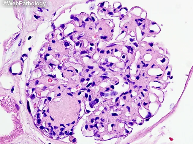
This is a renal biopsy histology slide examined under brightfield light microscopy. The primary subject is diabetic nephropathy with nodular glomerulosclerosis (Kimmelstiel-Wilson nodules) and diffuse mesangial sclerosis within the glomerular tuft of a renal cortex specimen. The glomerulus shows nodular mesangial expansion forming rounded, eosinophilic nodules that disrupt capillary loops, together with diffuse increased mesangial matrix. Capillary lumina are variably compressed, and arterioles frequently exhibit hyaline thickening consistent with diabetic microangiopathy. The cellular morphology is characterized by expanded mesangial cells with increased extracellular matrix and relatively sparse inflammatory infiltrate. The basement membrane appears thickened in cross-sectional elements. In this image, Masson’s trichrome is not visible; a subsequent stain would color collagen in blue, highlighting fibrotic remodeling. Clinically, these histologic features correlate with progressive proteinuria and reduced glomerular filtration rate in long-standing diabetes. Diagnostic significance rests on the combination of nodular sclerosis (Kimmelstiel-Wilson nodules) and diffuse mesangial sclerosis as hallmarks of diabetic nephropathy. Differential considerations include focal segmental glomerulosclerosis and hypertensive nephrosclerosis, but the nodular pattern is characteristic. This image is valuable for pathology education, nephrology training, and research into diabetic kidney disease progression and glycemic control effects. Representative for teaching clinics, this image supports correlating histology with clinical nephrology.
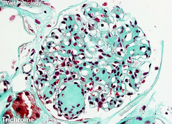
Masson's trichrome-stained renal cortex histology imaged at light microscopy reveals nodular glomerulosclerosis characteristic of diabetic nephropathy. The glomerulus shows circumscribed Kimmelstiel-Wilson nodules-eosinophilic mesangial nodules that expand the tuft and encroach on capillary loops. Diffuse mesangial sclerosis is evident as widespread mesangial matrix expansion, with increased basophilic mesangial tissue and accentuated blue-green collagen deposition in the extracellular matrix on Masson stain. The stain differentiates collagen (blue/green) from cytoplasm and nuclei (red/purple), highlighting the fibrotic nodules and mesangial expansion. Within the mesangium, there is progressive accumulation of matrix with variable cellularity; some capillary lumina appear narrowed due to nodular compression. The surrounding tubulointerstitial tissue may show mild chronic changes, consistent with chronic diabetic nephropathy. The overall pattern is typical of long-standing diabetes mellitus with nephropathy and correlates with progressive proteinuria, reduced glomerular filtration rate, and eventual renal insufficiency. Clinically, this finding supports diagnosis of diabetic kidney disease (diabetic nephropathy) and is essential for grading disease stage and guiding management. Differential diagnoses include nodular glomerulosclerosis due to amyloidosis or hypertensive nephrosclerosis, but the classic KW nodules and mesangial expansion favor diabetes. This image is valuable for medical education, pathology training, and research into diabetic renal pathology.
Diabetes Mellitus - MBBS Pathology Long Answer
Definition
Epidemiology
- Affects >30 million people in the USA (>11% of the population)
- ~422 million worldwide (WHO)
- Leading cause of end-stage renal disease, adult-onset blindness, and non-traumatic lower limb amputation
- 7th leading cause of death in the USA
- About 1/4 of affected individuals are unaware they have hyperglycemia
Diagnostic Criteria (ADA/WHO)
| Criterion | Value |
|---|---|
| Fasting plasma glucose | ≥126 mg/dL |
| Random plasma glucose (with symptoms) | ≥200 mg/dL |
| 2-hour OGTT (75 g glucose) | ≥200 mg/dL |
| HbA1c | ≥6.5% |
- Fasting glucose 100-125 mg/dL (impaired fasting glucose)
- 2-hour OGTT glucose 140-199 mg/dL (impaired glucose tolerance)
- HbA1c 5.7-6.4%
Classification
Type 1 Diabetes (T1D) - ~5-10% of cases
- Autoimmune destruction of pancreatic β cells
- Absolute insulin deficiency
- Most common in patients <20 years
Type 2 Diabetes (T2D) - ~90-95% of cases
- Combination of peripheral insulin resistance + inadequate β-cell secretory response ("relative insulin deficiency")
- Strongly linked to obesity, especially central/visceral obesity
- Classically adult-onset, but rising in adolescents
Other/Monogenic Types
- MODY (Maturity-Onset Diabetes of the Young) - single gene defects
- Gestational diabetes mellitus (GDM)
- Secondary diabetes (pancreatitis, Cushing's, acromegaly, drugs)
Normal Glucose Homeostasis
- Hepatic glucose production - regulated by glucagon (stimulates) and insulin (suppresses)
- Peripheral glucose utilization - primarily skeletal muscle (insulin-dependent via GLUT4)
- Pancreatic hormone secretion - insulin and glucagon have opposing effects
- Glucose enters β cells via GLUT2 transporter
- Glucose metabolism increases intracellular ATP:ADP ratio
- ATP-sensitive K+ channels (K-ATP channels) close → membrane depolarization
- Voltage-gated Ca²+ channels open → Ca²+ influx → exocytosis of insulin granules
Pathogenesis
Type 1 Diabetes
- Strongly linked to HLA-DR3 and HLA-DR4 (on chromosome 6p)
- Risk is ~50% in monozygotic twins (lower than T2D, suggesting environmental triggers)
- Polymorphisms in CTLA4 and PTPN22 also contribute
- Failure of self-tolerance in T cells
- Th1 CD4+ cells secrete IFN-γ and TNF, causing β-cell injury
- CD8+ cytotoxic T lymphocytes (CTLs) directly kill β cells
- Circulating autoantibodies against islet antigens (insulin, glutamic acid decarboxylase/GAD, IA-2) - found in most patients even at presymptomatic stages
- Insulitis: lymphocytic infiltration of islets
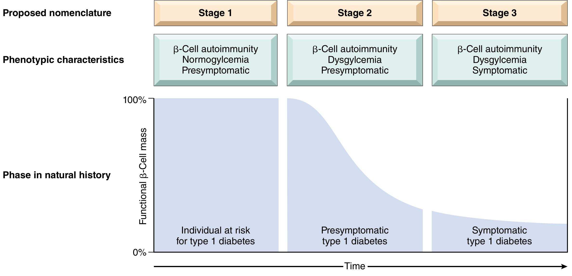
- Stage 1: Autoimmunity present; islet autoantibodies positive; normoglycemia; asymptomatic
- Stage 2: Autoimmunity + dysglycemia (impaired glucose tolerance/fasting glucose); asymptomatic
- Stage 3: Onset of clinical disease with symptoms
Type 2 Diabetes
- Concordance rate >90% in monozygotic twins (higher than T1D)
- First-degree relatives have 5-10x higher risk
-
30 loci identified by GWAS; many involve adipose tissue function and β-cell function
- Obesity (especially visceral/central) is the most important acquired risk factor - >80% of T2D patients are obese
- Sedentary lifestyle
- Sleep disorders (obstructive sleep apnea) and circadian disruption
- Metabolic syndrome: combination of obesity + hyperglycemia + dyslipidemia + hypertension
- Excess free fatty acids (FFAs): Ectopic fat deposition in skeletal muscle and liver; activate serine kinases (IRS-1 and IRS-2 serine phosphorylation) that impair downstream insulin signaling
- Adipocytokines: Adiponectin (insulin-sensitizing) is decreased; TNF, IL-6, leptin, resistin are increased - promoting inflammation and insulin resistance
- Inflammation: Infiltrating macrophages in adipose tissue release IL-1β and TNF that impair insulin signaling
- This leads to reduced GLUT4 translocation to the plasma membrane in muscle, reducing glucose uptake
- Initially, β cells compensate for insulin resistance by hypersecretion
- Over time, β cells become exhausted, worsened by:
- Glucotoxicity (excess glucose is toxic to β cells)
- Lipotoxicity (FFAs cause β-cell apoptosis)
- Amyloid deposition in islets (from islet amyloid polypeptide/IAPP secreted with insulin)
- The end result is progressive failure to secrete enough insulin to overcome resistance
Morphology (Pathological Findings)
Pancreatic Lesions
| Feature | Type 1 DM | Type 2 DM |
|---|---|---|
| Islet size reduction | Marked | Subtle |
| Insulitis (lymphocytic infiltration) | Present | Absent |
| Amyloid deposition in islets | Absent | Present (late stages) |
| Fibrosis of islets | Absent | May occur |
- Insulitis: Islets infiltrated predominantly by T lymphocytes - also seen in animal models of autoimmune diabetes
- Amyloid in T2D: Deposition begins in and around capillaries; at advanced stages, islets may be virtually obliterated
Long-Term Complications: The Big Picture
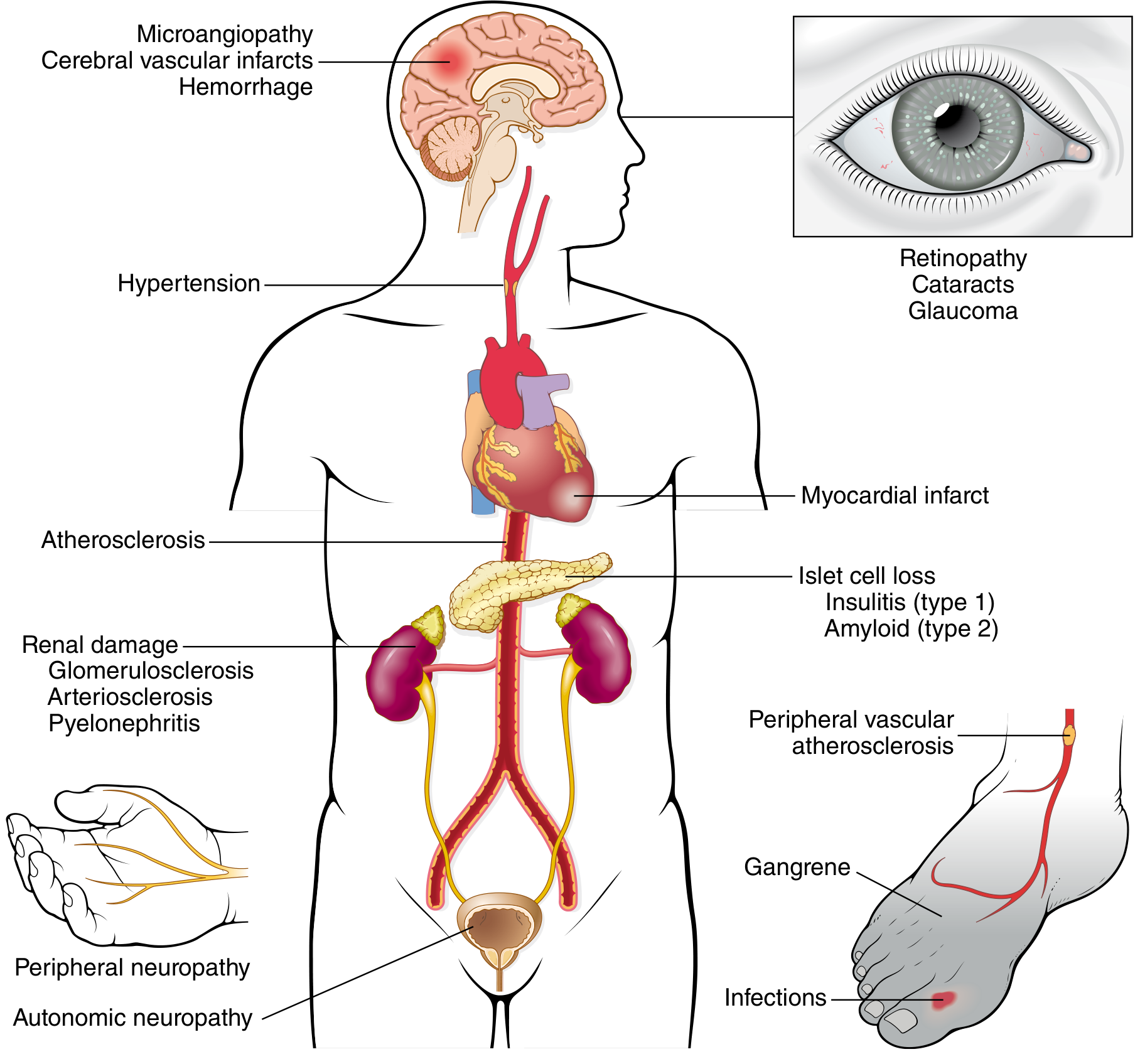
Diabetic Vascular Disease
1. Macrovascular Disease (Macroangiopathy)
- Accelerated atherosclerosis of the aorta, coronary arteries, and large vessels - morphologically identical to non-diabetic atherosclerosis but more severe and earlier in onset
- Myocardial infarction = most common cause of death in DM
- Gangrene of lower extremities - 100x more common in DM than general population
- Severe atherosclerosis of renal arteries
2. Microvascular Disease (Microangiopathy)
- The hallmark is diffuse thickening of basement membranes - most evident in capillaries of skin, skeletal muscle, retina, renal glomeruli, and renal medulla
- Despite thickened BM, capillaries are paradoxically leakier to plasma proteins
- Underlies diabetic nephropathy, retinopathy, and neuropathy
Diabetic Nephropathy
- Seen in virtually ALL cases of diabetic nephropathy
- Begins as early as 2 years after onset of T1D
- Best seen on electron microscopy
- Accompanied by mesangial widening
- Diffuse increase in PAS-positive mesangial matrix
- Correlates well with worsening proteinuria
- Progressive mesangial expansion → reduced renal function
- Pathognomonic of diabetic nephropathy
- Ovoid/spherical, laminated, PAS-positive nodules of matrix in the periphery of the glomerulus (within mesangial core)
- Often surrounded by patent peripheral capillary loops
- Accompanied by: fibrin caps (hyaline material in capillary loops) and capsular drops (hyaline adherent to Bowman's capsule)
- Both afferent AND efferent arterioles show hyalinosis (efferent arteriolar hyalinosis is nearly unique to DM)
- Occurs in ~15-30% of long-term diabetics; usually associated with renal failure
- Pyelonephritis - more common and severe in DM; especially papillary necrosis
- Renal atherosclerosis and arteriolar sclerosis
- Tubular atrophy and interstitial fibrosis (from ischemia)
- Kidney becomes contracted and granular (nephrosclerosis)
Diabetic Retinopathy
- Microaneurysms
- Flame-shaped and blot hemorrhages
- Hard exudates (lipid deposits)
- Cotton-wool spots (nerve fiber layer infarcts)
- Venous beading
- New vessel formation (neovascularization) in response to retinal ischemia
- Risk of vitreous hemorrhage, retinal detachment, and blindness
- Driven by VEGF (vascular endothelial growth factor)
- Cataracts - from sorbitol accumulation via polyol pathway (hyperglycemia → sorbitol → osmotic lens damage)
- Glaucoma - increased intraocular pressure
Diabetic Neuropathy
- Peripheral neuropathy present in up to 50% of long-term diabetics
- Types: distal symmetric sensorimotor polyneuropathy (most common), autonomic neuropathy, mononeuropathy
- Mechanism: microangiopathy of vasa nervorum + direct Schwann cell/axon damage from hyperglycemia
- Autonomic neuropathy → gastroparesis, orthostatic hypotension, erectile dysfunction, neurogenic bladder
- Contributes to diabetic foot via loss of protective sensation
Molecular Mechanisms of Hyperglycemic Damage
| Mechanism | Details |
|---|---|
| Formation of Advanced Glycation End-products (AGEs) | Non-enzymatic glycation of intracellular and extracellular proteins; AGEs cross-link collagen, trap plasma proteins, bind RAGE receptors activating inflammation |
| Activation of Protein Kinase C (PKC) | Increased intracellular diacylglycerol (DAG) from excess glucose activates PKC; impairs endothelial function, increases VEGF (retinopathy), increases permeability |
| Polyol Pathway | Excess glucose → sorbitol (via aldose reductase) → fructose; depletes NADPH and glutathione (antioxidants); osmotic damage to lens (cataracts) and Schwann cells |
| Hexosamine Pathway | Excess glucose metabolized via hexosamine pathway → altered gene expression (e.g., increased TGF-β causing fibrosis) |
Clinical Features
- Acute onset; younger patients; lean body habitus
- Classic "3 Ps": Polyuria, polydipsia, polyphagia
- Weight loss (catabolism of muscle and fat)
- Ketoacidosis (DKA) - major acute complication; due to absolute insulin deficiency → unrestrained lipolysis → ketone body formation
- Insidious onset; older (though increasingly younger)
- Often asymptomatic initially; discovered incidentally
- Associated with obesity
- Hyperosmolar hyperglycemic state (HHS) rather than DKA in acute crises
- Fatigue, blurred vision, recurrent infections (particularly UTI, skin infections)
- Increased susceptibility to infections (impaired leukocyte function)
- Poor wound healing
- Features of end-organ damage (nephropathy, retinopathy, neuropathy, vasculopathy)
Summary Box (Exam Points)
- Pathognomonic renal lesion: Kimmelstiel-Wilson nodular glomerulosclerosis (PAS-positive mesangial nodules)
- Insulitis: Lymphocytic infiltration of islets - seen in T1D
- Islet amyloid: T2D - from IAPP (amylin)
- Most common cause of death in DM: Myocardial infarction
- Second most common cause of death: Renal failure
- Efferent arteriolar hyalinosis: Nearly pathognomonic of DM
- Leading cause of non-traumatic amputation, adult-onset blindness, ESRD - all DM
- Concordance in MZ twins: T1D ~50% | T2D >90%
- HLA associations in T1D: HLA-DR3, HLA-DR4
- Key autoantibodies in T1D: Anti-insulin, anti-GAD (glutamic acid decarboxylase), anti-IA-2
- 4 mechanisms of hyperglycemic damage: AGEs, PKC activation, polyol pathway, hexosamine pathway