
Anatomy of Neck complete guide with image
Reading File
Finding Sources
Finding Sources
Finding Sources
Finding Sources
Now let me read the key sections from Gray's Anatomy for Students and Fischer's Mastery of Surgery in parallel:
Reading File
Reading File
Reading File
Finding Sources
Reading File
Finding Sources
Reading File
Finding Sources
Now let me fetch the key images and search for more content:
Loading Image
Loading Image
Loading Image
Loading Image
Loading Image
Loading Image
I now have excellent content and images. Let me read a bit more from the Fischer book to get the carotid triangle and anterior triangle details, then compile the full guide:
Reading File
Loading Image
Reading File
I now have comprehensive content and high-quality images. Let me compile the complete guide.# Complete Guide to Neck Anatomy
Sources: Fischer's Mastery of Surgery 8e, Gray's Anatomy for Students
1. Overview and Boundaries
The neck extends from the base of the skull (superior nuchal line and mastoid process) to the superior thoracic aperture (manubrium of sternum, clavicles, and first ribs) inferiorly. It is divided into anterior and posterior triangles on each side by the sternocleidomastoid (SCM) and trapezius muscles.
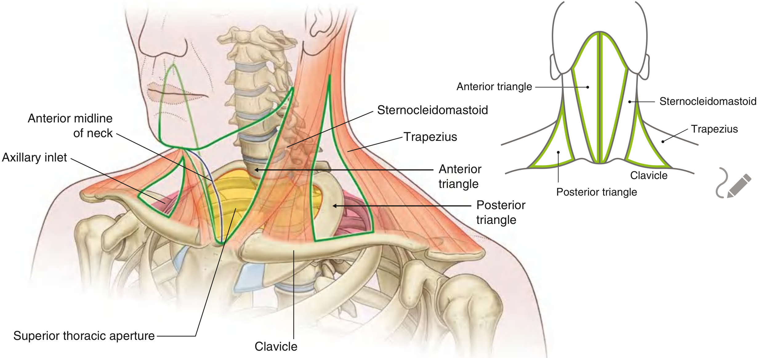
Fig. 1 - Anterior and posterior triangles of the neck with key bony landmarks (Gray's Anatomy for Students)
2. Fascial Layers of the Neck
Understanding the fascia is critical for surgery, infection spread, and regional anesthesia.
Superficial Fascia
- Loose areolar connective tissue containing the platysma (muscle of facial expression, innervated by the cervical branch of the facial nerve)
- Superficial veins (external and anterior jugular), superficial cervical plexus branches, and lymph nodes lie deep to the platysma but superficial to the investing layer of deep fascia
Deep Cervical Fascia - Three Layers
| Layer | Contents / Significance |
|---|---|
| Investing layer | Surrounds the entire neck; splits to envelope SCM and trapezius; forms the roof of both anterior and posterior triangles |
| Pretracheal layer | Muscular part surrounds infrahyoid muscles; visceral part surrounds thyroid, trachea, and esophagus; continuous with buccopharyngeal fascia |
| Prevertebral layer | Surrounds vertebral column and its musculature; contains the cervical sympathetic trunk |
| Carotid sheath | Tubular fascial sheet from skull base to root of neck; contains common and internal carotid arteries, internal jugular vein, vagus nerve (CN X), and sympathetic fibers |
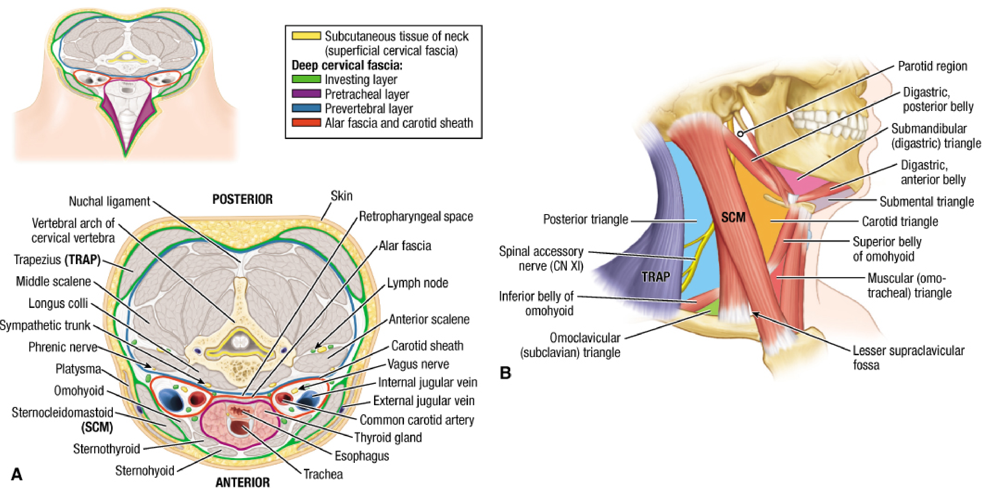
Fig. 2 - A: Cross-section of neck showing all fascial layers, carotid sheath, and key structures. B: Lateral view identifying all triangles (Fischer's Mastery of Surgery)
3. Triangles of the Neck
ANTERIOR TRIANGLE
Boundaries:
- Medially: midline of neck
- Superiorly: inferior border of mandible
- Posteriorly: anterior border of SCM
The anterior triangle is further subdivided by the digastric muscle (superiorly) and the superior belly of omohyoid (inferiorly) into four triangles:
| Sub-triangle | Boundaries | Key Contents |
|---|---|---|
| Submental (unpaired) | Between the two anterior bellies of digastric, hyoid bone | Submental lymph nodes, beginning of anterior jugular vein |
| Submandibular (Digastric) | Mandible above, anterior and posterior bellies of digastric | Submandibular gland, facial artery & vein, hypoglossal nerve (CN XII), mylohyoid nerve |
| Carotid | Posterior belly of digastric (sup.), anterior belly of omohyoid (inf.), SCM (post.) | Common/internal/external carotid arteries, internal jugular vein, CN X, CN XI, CN XII, ansa cervicalis, superior laryngeal nerve |
| Muscular (Omotracheal) | Midline (med.), superior belly of omohyoid (sup./lat.), SCM (post.) | Infrahyoid (strap) muscles, thyroid, trachea, esophagus |
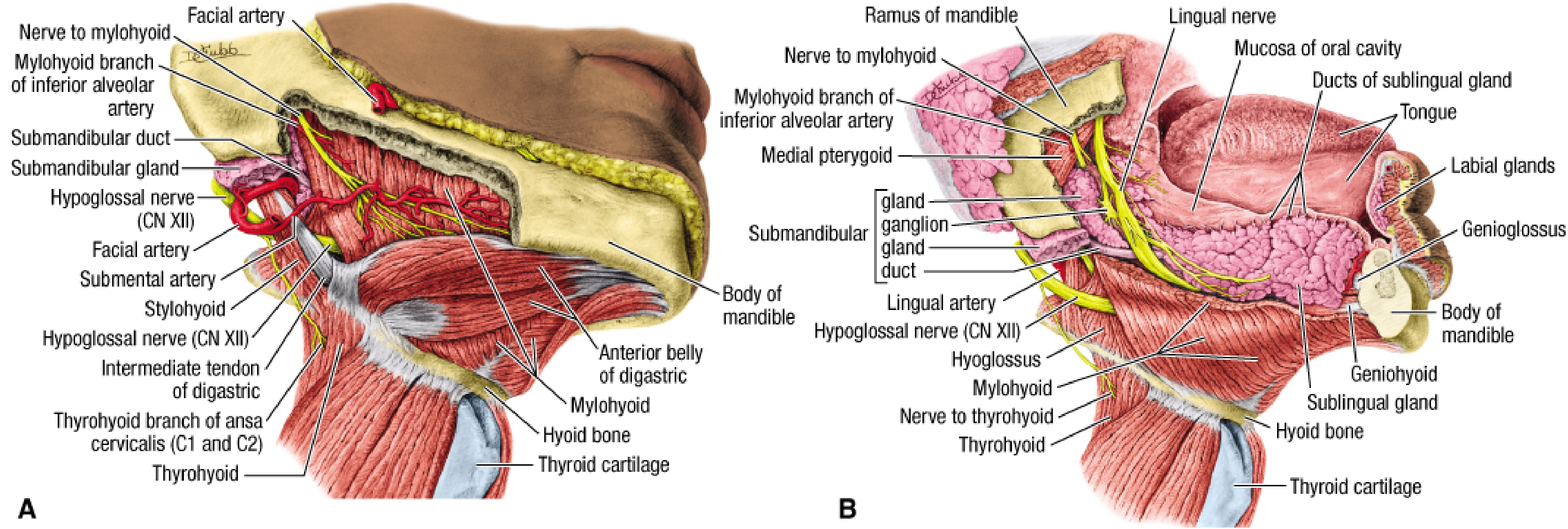
Fig. 3 - Submandibular triangle: A: Superficial dissection showing facial artery, digastric, stylohyoid, hypoglossal nerve. B: Deep dissection showing submandibular ganglion, lingual nerve, sublingual gland (Fischer's Mastery of Surgery)
POSTERIOR TRIANGLE
Boundaries:
- Anteriorly: posterior border of SCM
- Posteriorly: anterior border of trapezius
- Inferiorly: middle one-third of clavicle
The inferior belly of omohyoid divides it into:
- Occipital triangle (larger, superior)
- Omoclavicular / Subclavian triangle (smaller, inferior)
All three layers of deep cervical fascia are present; the investing layer forms the roof.
4. Key Muscles of the Neck
Sternocleidomastoid (SCM)
- Origin: Sternal head from manubrium; clavicular head from medial third of clavicle
- Insertion: Mastoid process and superior nuchal line
- Innervation: Spinal accessory nerve (CN XI) for motor; C2-C3 for proprioception
- Action: Unilateral - tilts head to same side, rotates chin to opposite side; bilateral - flexes neck, assists in forced inspiration
Platysma
- Thin sheet in superficial fascia; innervated by cervical branch of facial nerve
- Clinically important as the marginal mandibular branch of CN VII can lie up to 2.5 cm below the mandibular ramus - at risk in neck dissection
Infrahyoid (Strap) Muscles
| Muscle | Origin | Insertion | Nerve |
|---|---|---|---|
| Sternohyoid | Manubrium + medial clavicle | Hyoid body | Ansa cervicalis (C1-C3) |
| Sternothyroid | Manubrium | Thyroid cartilage | Ansa cervicalis (C1-C3) |
| Thyrohyoid | Thyroid cartilage | Hyoid body | C1 via hypoglossal nerve |
| Omohyoid | Superior scapular border | Hyoid body | Ansa cervicalis (C1-C3) |
Suprahyoid Muscles
| Muscle | Function |
|---|---|
| Mylohyoid | Forms floor of mouth; elevates hyoid/tongue |
| Digastric (2 bellies) | Depresses mandible, elevates hyoid |
| Stylohyoid | Elevates and retracts hyoid |
| Geniohyoid | Elevates and protrudes hyoid |
Prevertebral / Scalene Muscles
| Muscle | Action / Significance |
|---|---|
| Anterior scalene | Raises first rib; phrenic nerve lies on its anterior surface deep to prevertebral fascia |
| Middle scalene | Raises first rib; brachial plexus roots emerge between anterior and middle scalene |
| Posterior scalene | Raises second rib |
| Longus colli | Flexes and rotates cervical spine |
5. Arteries of the Neck
Common Carotid Artery
- Right: branch of brachiocephalic trunk
- Left: direct branch of aortic arch
- Both ascend within the carotid sheath; bifurcate at the level of the upper border of thyroid cartilage (C3-C4) into internal and external carotid arteries
- Carotid sinus (baroreceptor) and carotid body (chemoreceptor) are located at the bifurcation
External Carotid Artery (ECA) - Branches
Anterior branches:
- Superior thyroid artery
- Lingual artery
- Facial artery
Posterior branches:
4. Sternocleidomastoid artery
5. Occipital artery
6. Posterior auricular artery
Medial branch:
7. Ascending pharyngeal artery
Terminal branches (above parotid):
8. Superficial temporal artery
9. Maxillary artery
Subclavian Artery Branches at Root of Neck
- Vertebral artery (ascends through transverse foramina C6 to C1)
- Internal thoracic artery
- Thyrocervical trunk: gives suprascapular, transverse cervical, and inferior thyroid arteries
- Costocervical trunk: gives supreme intercostal and deep cervical arteries
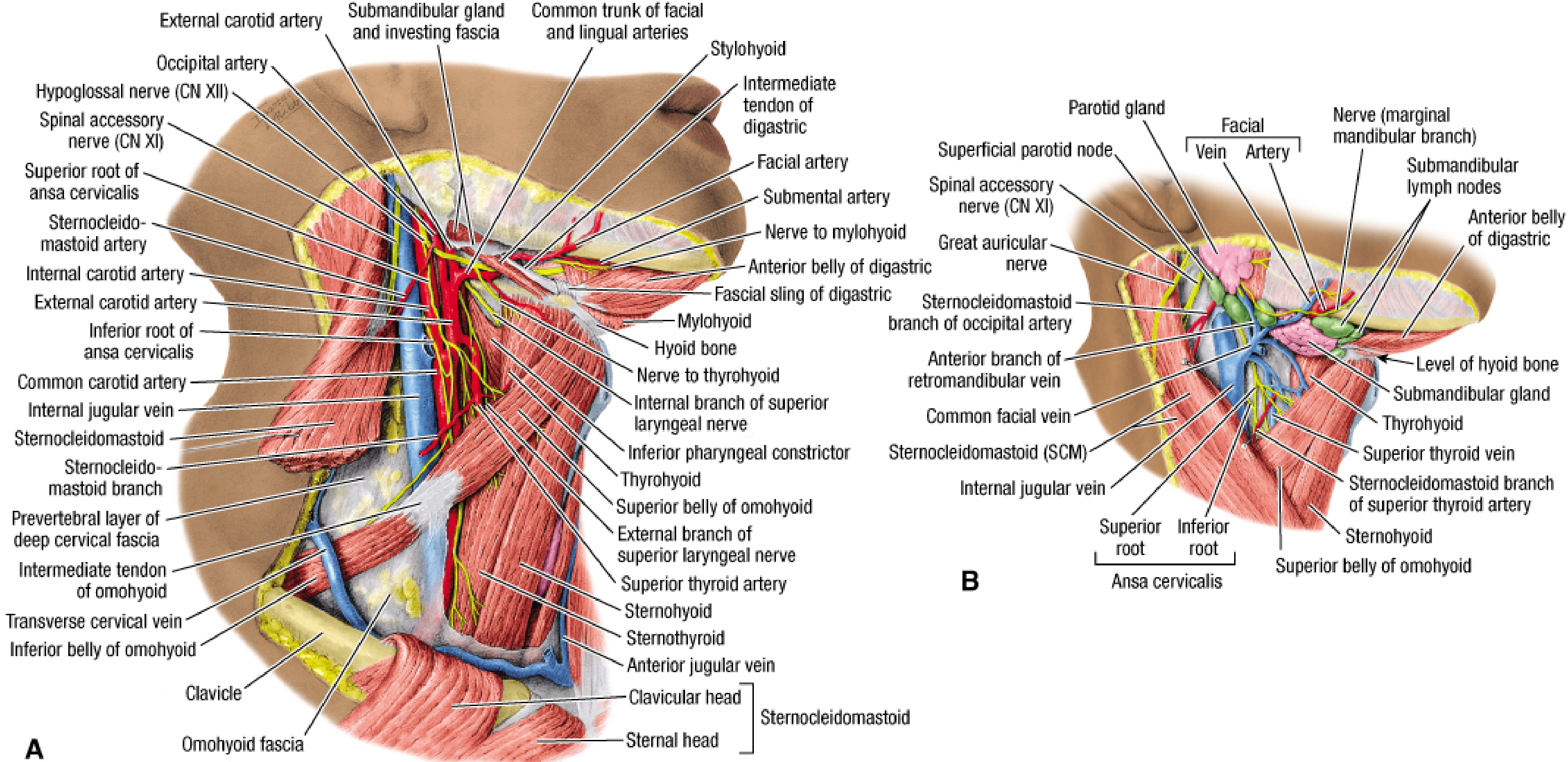
Fig. 4 - A: Deep dissection of carotid triangle showing common/internal/external carotid arteries, ansa cervicalis, jugular vein, and cranial nerves. B: Superficial dissection with submandibular gland reflected (Fischer's Mastery of Surgery)
6. Veins of the Neck
Internal Jugular Vein (IJV)
- Begins at the jugular foramen (base of skull) as a continuation of the sigmoid sinus
- Courses inferiorly in the carotid sheath, anterolateral to the carotid arteries
- Tributaries: facial, lingual, superior thyroid veins (superior triangle); middle thyroid vein (inferior)
- Joins the subclavian vein to form the brachiocephalic vein at the root of the neck
- Thoracic duct (left) and right lymphatic duct drain into the venous angle at this junction
External Jugular Vein (EJV)
- Formed in the parotid gland by the union of the posterior auricular and retromandibular veins
- Crosses superficially over the SCM from the parotid gland toward the clavicle
- Pierces the deep fascia in the subclavian triangle and enters the subclavian vein
Anterior Jugular Vein
- Begins near the hyoid, descends in midline, communicates with the EJV via the "Vein of Kocher"
7. Nerves of the Neck
Cervical Plexus (C1-C4)
Superficial (sensory) branches - all emerge at the "nerve point" behind the middle of SCM:
| Nerve | Spinal Level | Distribution |
|---|---|---|
| Lesser occipital nerve | C2 | Skin behind the ear and occipital scalp |
| Great auricular nerve | C2-C3 | Skin over parotid, ear, and mastoid |
| Transverse cervical nerve | C2-C3 | Anterior skin of neck |
| Supraclavicular nerves (medial, intermediate, lateral) | C3-C4 | Skin over clavicle and upper chest |
Deep (motor) branches:
| Nerve | Level | Supply |
|---|---|---|
| Ansa cervicalis (superior root C1-C2 + inferior root C2-C4) | C1-C4 | All infrahyoid muscles (except thyrohyoid) |
| Phrenic nerve | C3, C4, C5 | Sole motor supply to the diaphragm; also sensory and sympathetic fibers; lies on anterior scalene deep to prevertebral fascia |
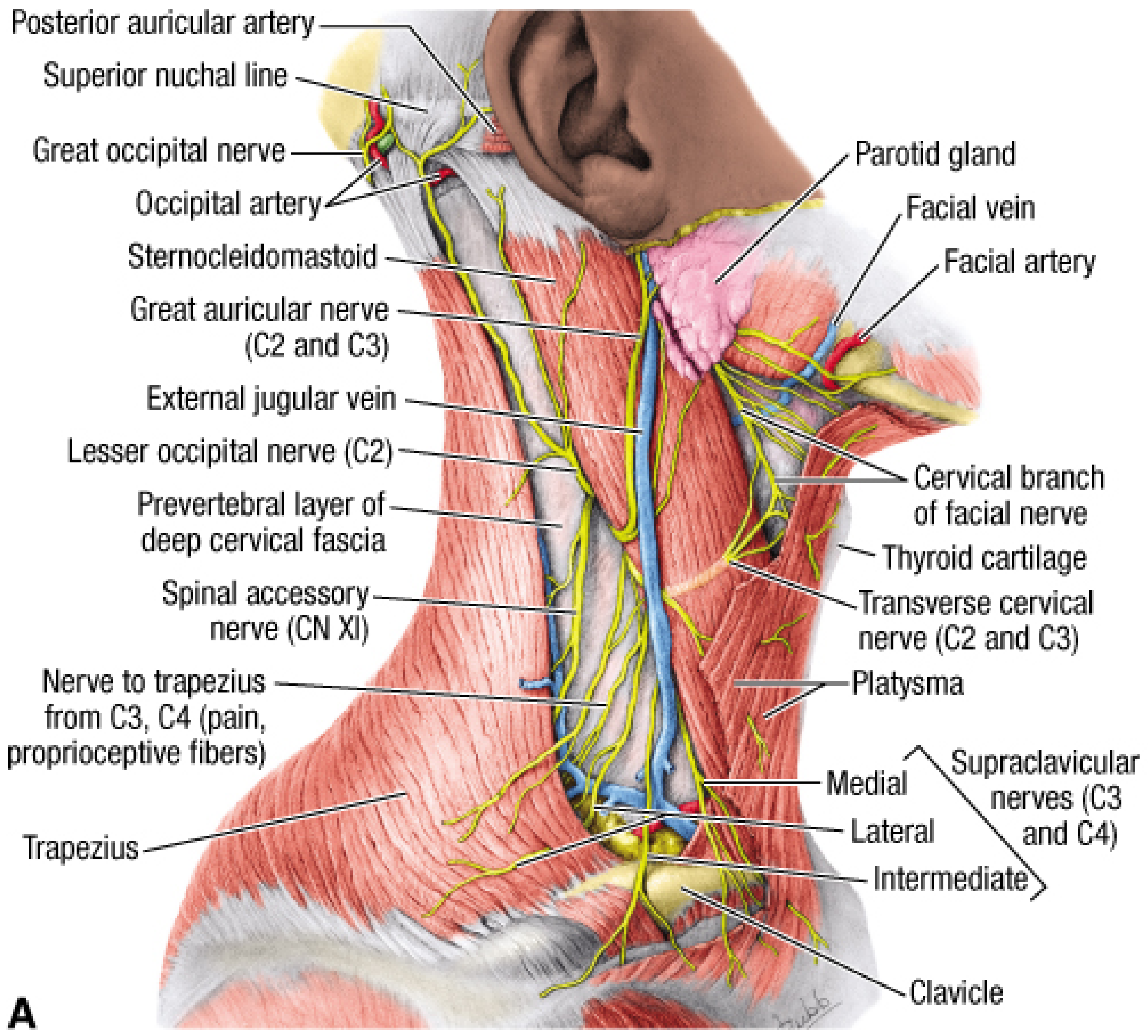
Fig. 5 - Posterior triangle superficial dissection: great auricular nerve, lesser occipital nerve, transverse cervical nerve, supraclavicular nerves, spinal accessory nerve (CN XI), and external jugular vein (Fischer's Mastery of Surgery)
Brachial Plexus (C5-T1)
Roots emerge between the anterior and middle scalene muscles. Trunks (upper C5-C6, middle C7, lower C8-T1) pass through the lower posterior triangle (omoclavicular triangle) to reach the axilla.
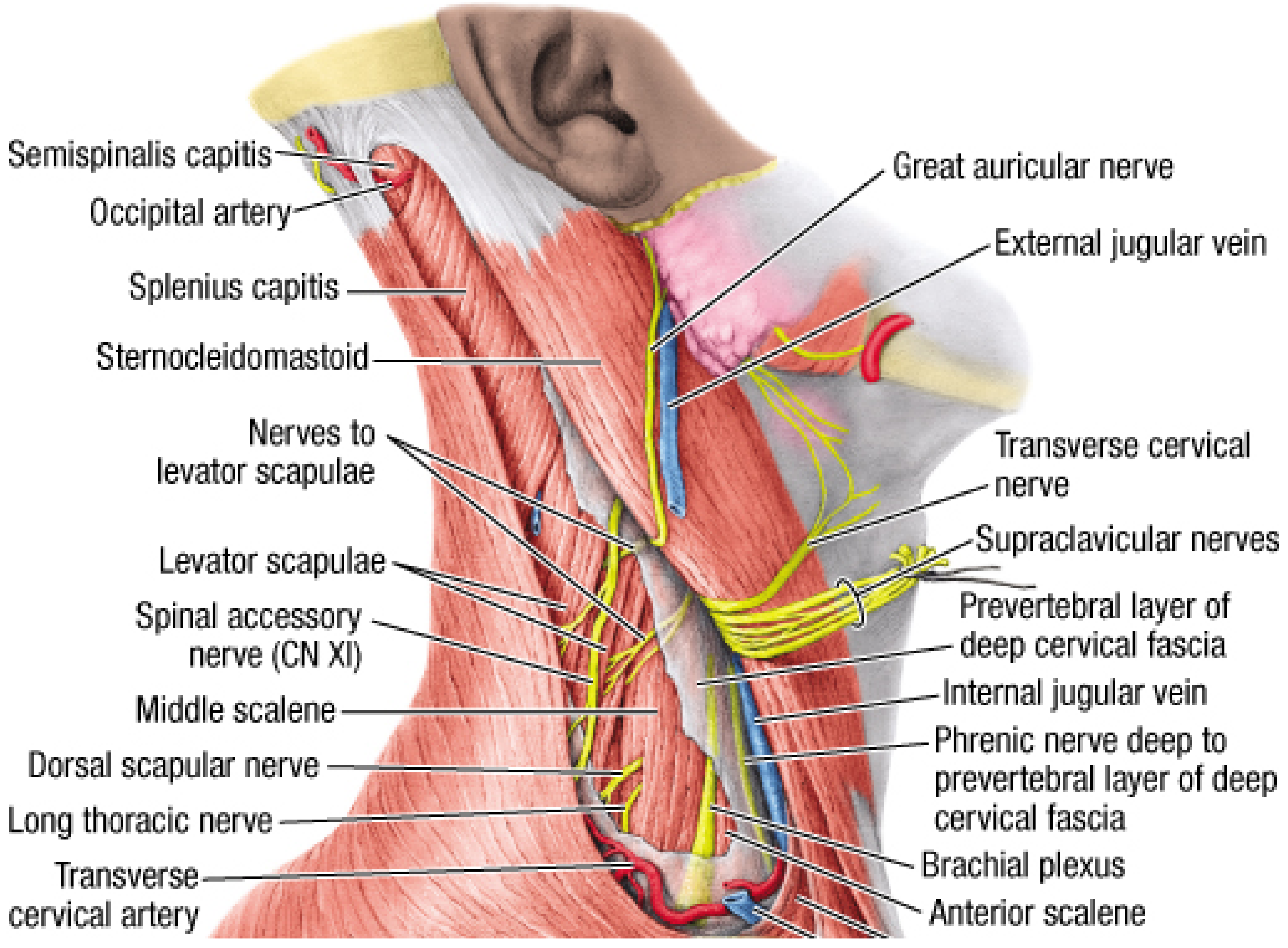
Fig. 6 - Deep posterior triangle and root of neck: brachial plexus roots C5-C8, anterior/middle/posterior scalenes, phrenic nerve, subclavian artery and vein, dorsal scapular nerve, long thoracic nerve (Fischer's Mastery of Surgery)
Cranial Nerves in the Neck
| Nerve | Location | Function in Neck |
|---|---|---|
| CN X - Vagus | Carotid sheath (posterior between artery and vein) | Gives superior laryngeal nerve (external branch: cricothyroid motor; internal branch: laryngeal sensation above cords); gives recurrent laryngeal nerve in thorax |
| CN XI - Spinal Accessory | Crosses carotid triangle, enters SCM, then crosses posterior triangle on levator scapulae | Motor to SCM and trapezius |
| CN XII - Hypoglossal | Hooks around occipital artery; passes between IJV and ICA into submandibular triangle | Motor to all intrinsic and most extrinsic tongue muscles; C1 fibers hitchhike on CN XII to form ansa cervicalis |
Cervical Sympathetic Trunk
- Lies on the prevertebral fascia, posterior to the carotid sheath
- Contains superior, middle, and inferior (stellate) ganglia
- Injury causes Horner's syndrome (ptosis, miosis, anhidrosis, enophthalmos)
Superficial Sensory Nerves (Posterior Triangle)
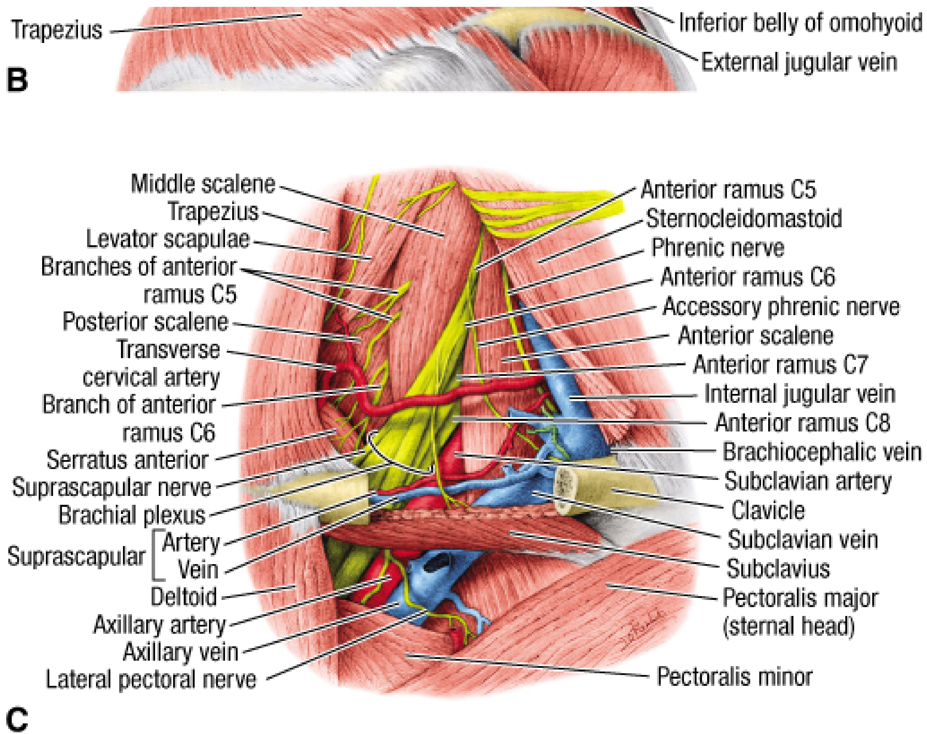
Fig. 7 - Root of neck showing brachial plexus (C5-C8 roots), anterior scalene with phrenic nerve, subclavian artery, brachiocephalic vein, and axillary vessels (Fischer's Mastery of Surgery)
8. Lymphatics of the Neck
Cervical lymph nodes are organized into six levels (used clinically for cancer staging):
| Level | Location | Drainage |
|---|---|---|
| I | Submental (Ia) and submandibular (Ib) | Floor of mouth, lip, anterior oral cavity |
| II | Upper deep cervical (around upper IJV, above hyoid) | Pharynx, oral cavity, parotid, face |
| III | Middle deep cervical (IJV, between hyoid and omohyoid) | Oral cavity, oropharynx, hypopharynx |
| IV | Lower deep cervical (IJV, below omohyoid to clavicle) | Hypopharynx, larynx, thyroid, esophagus |
| V | Posterior triangle nodes | Nasopharynx, oropharynx, skin of scalp/neck |
| VI | Central compartment (pretracheal, paratracheal, prelaryngeal) | Thyroid, larynx, trachea, esophagus |
Lymphatic drainage from the head and neck ultimately converges into the:
- Thoracic duct (left side) - drains into the left venous angle (IJV + subclavian)
- Right lymphatic duct (right side) - drains into the right venous angle
9. Root of the Neck - Key Relationships
The root of the neck contains critical structures packed into a small space:
| Structure | Position |
|---|---|
| Trachea and esophagus | Midline (esophagus slightly left) |
| Common carotid arteries | Lateral to trachea/esophagus within carotid sheath |
| Internal jugular veins | Anterolateral to carotid arteries in sheath |
| Vagus nerve | Posterior between artery and vein in sheath |
| Sympathetic trunk | On prevertebral fascia, posterior to carotid sheath |
| Subclavian artery | Arches laterally; vertebral artery leaves from first part |
| Phrenic nerve (C3,4,5) | On anterior scalene, deep to prevertebral fascia |
| Thoracic duct (left) | Arches from behind the IJV to drain at venous angle |
10. Viscera of the Neck
Thyroid Gland
- Two lateral lobes connected by an isthmus at the 2nd-4th tracheal rings
- Enclosed in the visceral (pretracheal) layer of deep fascia
- Blood supply: superior thyroid artery (from ECA) and inferior thyroid artery (from thyrocervical trunk)
- The recurrent laryngeal nerve runs in the tracheoesophageal groove - at risk during thyroid surgery
Larynx (C3-C6)
- Formed by thyroid, cricoid, and arytenoid cartilages
- Key landmark: the cricothyroid membrane is the site of emergency cricothyrotomy
- Innervation: internal laryngeal nerve (sensation above cords), recurrent laryngeal nerve (motor + sensation below cords)
Pharynx
- Lies posterior to the nasal cavity, oral cavity, and larynx
- Constrictor muscles (superior, middle, inferior) innervated by pharyngeal plexus (CN IX, X)
Quick Reference: "Nerve Point" of the Neck
The nerve point (Erb's point) is where all four superficial cervical plexus branches emerge at the posterior border of the SCM, approximately at its midpoint. It is the optimal site for superficial cervical plexus blocks and a critical landmark in neck dissection.
Clinical Highlights
| Situation | Relevant Anatomy |
|---|---|
| Carotid endarterectomy | SCM retracted; IJV, vagus nerve, hypoglossal nerve, ansa cervicalis must be preserved |
| Neck dissection | CN XI at risk as it crosses the posterior triangle; marginal mandibular branch of CN VII risks mouth asymmetry |
| Central venous access (IJV) | Target is IJV anterolateral to CCA in the carotid triangle |
| Phrenic nerve injury | Damage at anterior scalene causes ipsilateral hemidiaphragm paralysis |
| Horner's syndrome | Damage to cervical sympathetic trunk (trauma, tumor, thyroid surgery) |
| Tracheostomy | Performed through muscular (omotracheal) triangle between strap muscles |
Sources:
- Fischer's Mastery of Surgery 8th Edition, Chapter 38 (Anatomy of Neck) - pp. 1106-1116
- Gray's Anatomy for Students, Chapter 8 (Head and Neck) - Triangles of the Neck, p. 991
This is a shared conversation. Sign in to Orris to start your own chat.