
Describe anatomy of middle ear cleft and its surgical importance for 20 marks long question answer in university exam in detail and in simplified manner
middle ear anatomy cleft tympanic cavity ossicles
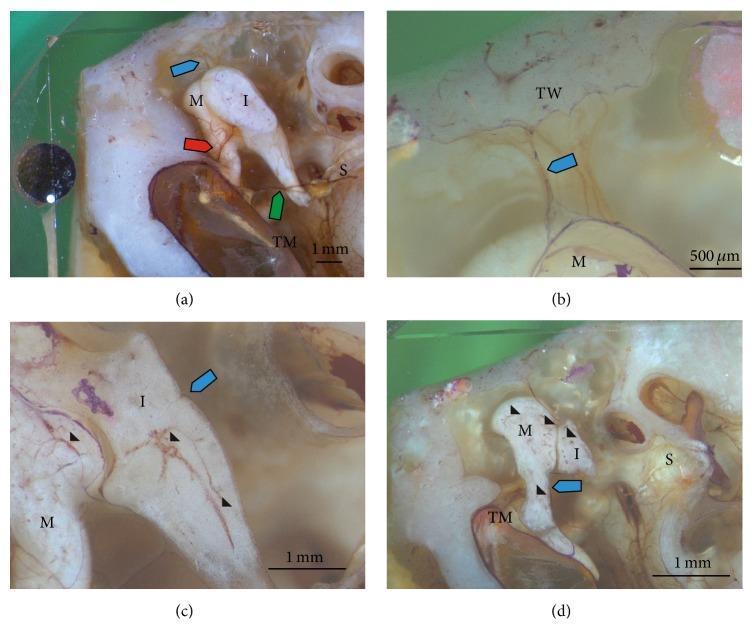
Reflected light microscopic images of the human middle ear auditory ossicles, displaying the anatomy and microvasculature of the malleus (M), incus (I), and stapes (S). The images highlight the structural relationships within the tympanic cavity, including the tympanic membrane (TM) and tympanic wall (TW). Colored arrows identify key arterial branches: the blue and red arrows mark the anterior tympanic artery pathways through the superior malleolar ligament and anterior malleolar plication, while the green arrow indicates the posterior tympanic artery crossing the incudostapedial joint. High-magnification views (c and d) utilize arrowheads to demonstrate the intricate intraosseous blood vessel system penetrating the ossicles via nutrient foramina. These sections differentiate between vascularized bone and avascular cartilage regions (indicated by asterisks). This educational visual illustrates middle ear micro-anatomy and the primary blood supply sources—the anterior and posterior tympanic arteries—essential for understanding ossicular physiology and surgical pathology in otolaryngology.
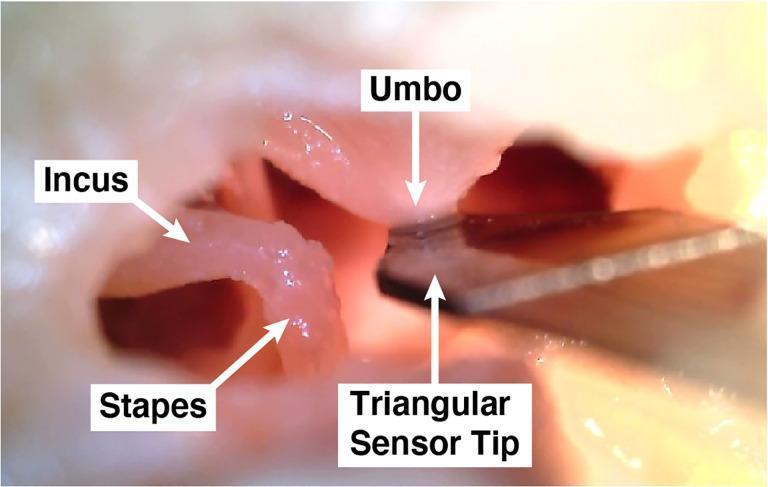
This clinical photograph shows a close-up, internal view of the human middle ear cavity from a cadaveric specimen, illustrating the anatomical components of the ossicular chain and an experimental medical device. Visible anatomical structures include the incus, the stapes, and the umbo—the conical point on the internal surface of the tympanic membrane where the manubrium of the malleus attaches. The soft tissues and ossicles appear pinkish-white with a moist texture. A dark, metallic, triangular sensor tip (part of the 'UmboMic' piezoelectric apparatus) is positioned in direct contact with the umbo. This setup is used to detect the mechanical vibrations and displacement of the tympanic membrane for research into middle ear microphone technology. The image highlights the spatial relationship between the ossicles and the positioning requirements for intra-tympanic sensing devices. The educational focus is on otologic anatomy and the integration of implantable hearing technology within the middle ear space.
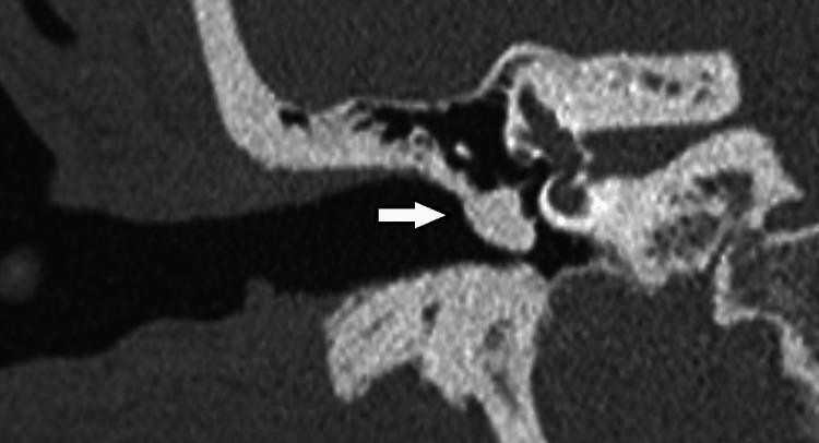
This diagnostic image is a high-resolution computerized tomography (CT) scan of the right temporal bone in a coronal view, specifically focused on the middle ear anatomy. The scan clearly depicts the auditory ossicles, including the handle of the malleus, the long process of the incus, and the head of the stapes. A significant finding is highlighted by a white arrow, pointing to a soft-tissue density opacification within the tympanic cavity. This abnormal density is shown enveloping the ossicular chain, particularly around the malleus and incus, replacing the normal air-filled space of the middle ear. The surrounding bony structures, including the external acoustic canal, the tegmen tympani, and elements of the inner ear (such as the cochlea and vestibular system), are visible for anatomical orientation. This image serves as an educational tool for identifying middle ear pathology, such as cholesteatoma, tympanosclerosis, or chronic otitis media, by demonstrating how soft-tissue masses can obscure normal air spaces and interact with the ossicular chain.
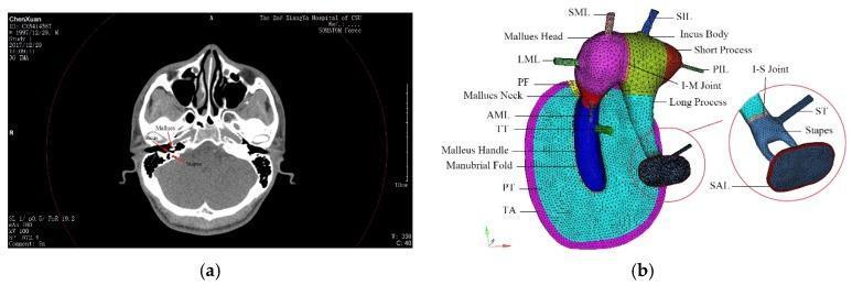
This figure presents a comparative overview of human middle ear anatomy through diagnostic imaging and biomechanical modeling. Image (a) is a clinical axial CT scan of the head at the level of the temporal bone, highlighting the cranium, nasal cavity, and middle ear structures. Specific labels identify the malleus and stapes within the tympanic cavity. Image (b) shows a high-fidelity finite element model of the human middle ear, providing a detailed anatomical diagram of the ossicular chain, ligaments, and tympanic membrane. The model uses color-coding to differentiate structures: the tympanic membrane is shown with its pars tensa (PT) and pars flaccida (PF), bordered by the tympanic annulus (TA). The ossicles are segmented into the malleus head (pink), incus body (green), and stapes (light blue). Key supporting structures are annotated, including the superior, lateral, and anterior mallear ligaments (SML, LML, AML), the superior and posterior incudal ligaments (SIL, PIL), the tensor tympani (TT), and the stapedial annular ligament (SAL). A magnified inset details the incudostapedial (I-S) joint and stapedial tendon (ST).
mastoid antrum aditus surgical anatomy
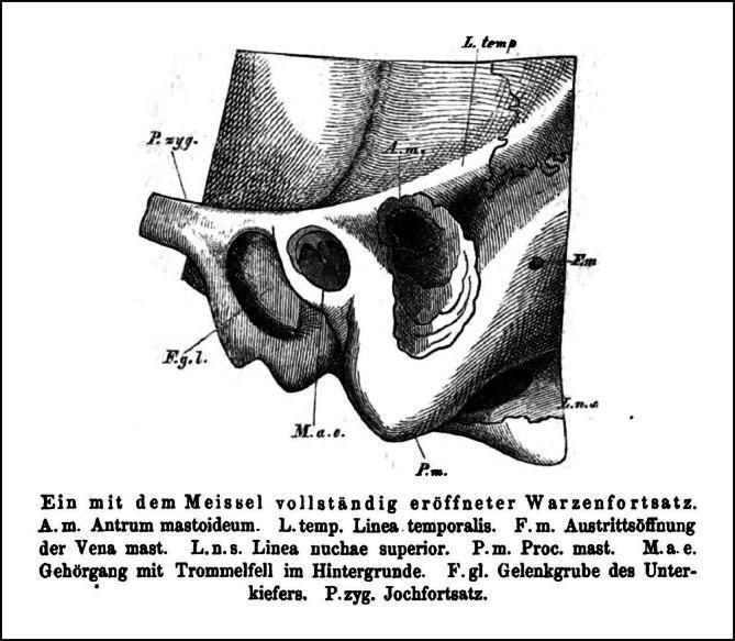
This historical anatomical illustration from an 1873 otology textbook depicts a surgical view of the lateral skull, specifically a mastoid process that has been opened using a chisel (Schwartze-type mastoidectomy). The drawing illustrates the surgical landmarks and spatial orientation required for safe access to the mastoid antrum (A.m.), which appears as a dark, funnel-shaped cavity within the bone. Key anatomical structures include: 1) The linea temporalis (L. temp.), marking the superior surgical limit; 2) The external auditory meatus (M.a.e.) with the tympanic membrane visible, situated anterior to the mastoid opening; 3) The zygomatic process (P.zyg.) and glenoid fossa (F.g.l.) located anterior-inferiorly; 4) The mastoid process (P.m.) forming the inferior border; and 5) The foramen of the mastoid vein (F.m.) and linea nuchae superior (L.n.s.) positioned posteriorly. This image serves as an educational reference for historical otological surgery and the fundamental anatomy of the temporal bone, highlighting the relationship between external surface landmarks and deeper middle ear structures.
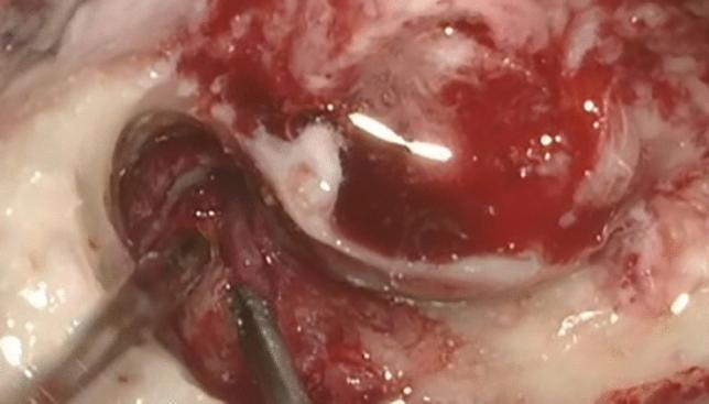
This intraoperative clinical photograph captures the surgical field during an otologic procedure, specifically targeting the middle ear and mastoid spaces. The image demonstrates extensive, hypervascularized granulation tissue located within the aditus ad antrum and extending toward the region of the facial nerve. The pathological tissue is characterized by a deep reddish hue, irregular surface texture, and friable appearance, consistent with active chronic inflammation. Adjacent anatomical structures show signs of inflammatory changes, including pale, edematous-appearing mucosa and areas of whitish exudate or mucosal thickening. Surgical instruments are visible in the lower-left quadrant, performing micro-dissection of the granulation tissue. This finding is clinically significant in the context of Granulomatosis with Polyangiitis (GPA), where such inflammatory masses in the pneumatic spaces can cause facial nerve palsy and hearing loss, necessitating surgical exploration and biopsy for histopathological confirmation and exclusion of malignancy.
tympanic cavity walls medial wall promontory oval window facial nerve
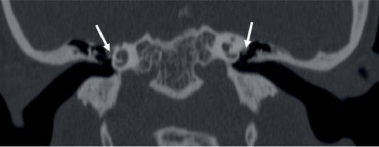
This coronal high-resolution CT (HRCT) image of the temporal bone demonstrates bilateral congenital anomalies of the facial nerve. White arrows highlight the dehiscent tympanic (horizontal) segments of the facial nerve bilaterally. The nerve exhibits an anomalous inferio-medial course, resulting in it abutting the lateral wall of the bony labyrinth. Due to this displacement and the lack of a normal bony canal covering (dehiscence), the facial nerve is positioned in close proximity to the oval window niche. The image also captures the middle ear cavity and the labyrinthine structures of the inner ear. This radiological finding is clinically significant for surgical planning, particularly in cases of conductive hearing loss or planned middle ear surgery (e.g., stapedotomy), as the dehiscent and displaced nerve is at increased risk of accidental injury or may obscure the surgical approach to the oval window.
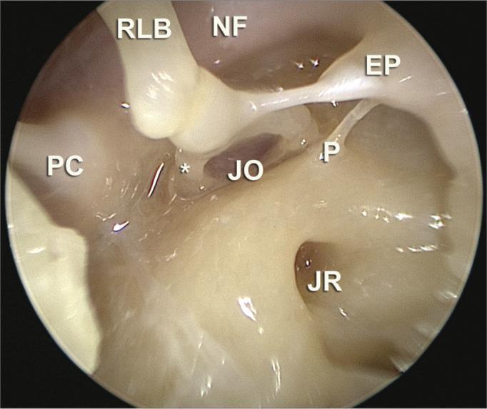
This endoscopic clinical photograph displays the detailed surgical anatomy of the middle ear (tympanic cavity) as viewed through a 30-degree scope during a stapes surgery anatomical dissection. The image provides a high-resolution view of the medial and posterior walls of the middle ear. Key visible landmarks include the long process of the incus (RLB) articulating with the stapes at the incudostapedial joint. The oval window niche (JO) and round window niche (JR) are clearly identified. Superior to the oval window niche, the facial nerve canal (NF) is visible as a smooth bony prominence. The pyramidal eminence (EP) is shown with the stapedius tendon extending toward the stapes. Additional anatomical features include the ponticulus (P), which is a bony ridge separating the oval window niche from the sinus tympani, and the cochleariform process (PC) on the anterior-superior aspect. This visual resource is essential for medical education regarding endoscopic ear surgery (EES) and middle ear microsurgery, demonstrating the spatial relationships required for procedures like stapedotomy or tympanoplasty.
Anatomy of the Middle Ear Cleft and Its Surgical Importance
DEFINITION AND OVERVIEW
- Tympanic cavity (with its sub-compartments)
- Eustachian (auditory) tube - the anterior extension
- Mastoid antrum and mastoid air cell system - the posterior extension
PART 1: THE TYMPANIC CAVITY
Subdivisions of the Tympanic Cavity
| Compartment | Location | Contents |
|---|---|---|
| Epitympanum (Attic) | Above the level of the malleolar folds | Head of malleus, body and short process of incus |
| Mesotympanum | Middle part, visible through the EAC on otoscopy | Ossicular chain, tympanic plexus, stapedius tendon |
| Hypotympanum | Below the inferior tympanic sulcus | Floor of tympanic cavity, close to jugular bulb |
- Retrotympanum - posterior to the promontory (contains sinus tympani, facial recess, oval and round window niches)
- Protympanum - anterior, contiguous with the Eustachian tube opening
WALLS OF THE TYMPANIC CAVITY (6 Walls)
1. LATERAL WALL (Membranous Wall)
- Formed by the tympanic membrane centrally
- Bony lateral wall of epitympanum (scutum) superiorly
- Bony hypotympanic wall inferiorly
- Oval, ~9-10 mm in vertical and 8-9 mm in horizontal diameter
- Set at ~55° to the floor of the ear canal
- Divided into:
- Pars tensa - the larger lower portion, with a fibrocartilaginous annulus seated in the tympanic sulcus
- Pars flaccida (Shrapnell membrane) - small triangular upper portion above the malleolar folds, within the notch of Rivinus; lacks a tympanic annulus; its lamina propria is lax with randomly oriented collagen fibres
- Both portions have 3 layers: outer epidermis (continuous with EAC skin), middle lamina propria (fibrous), inner mucosal layer (continuous with middle ear mucosa)
- The scutum (outer attic wall) is the sharp wedge-shaped inferior portion of the lateral epitympanic wall - it is thin and easily eroded by cholesteatoma, which is the classic CT finding in attic cholesteatoma
- Petrotympanic fissure on the medial surface transmits the anterior malleolar ligament and anterior tympanic branch of the maxillary artery; the chorda tympani exits anteriorly through this fissure
- Pars flaccida is the site where attic/epitympanic cholesteatoma originates due to retraction
- The thin scutum bone is used as a target for drilling in atticotomy
- Petrotympanic fissure transmits chorda tympani - damage during myringoplasty causes taste disturbance
2. MEDIAL WALL (Labyrinthine Wall)
- Promontory - a rounded eminence formed by the first turn of the cochlea. The tympanic plexus (Jacobson's nerve, branch of CN IX, carrying parasympathetics to parotid) runs over its surface.
- Oval window (Fenestra Vestibuli) - above and behind the promontory; 3.25 mm long, 1.75 mm wide; closed by the stapes footplate and its annular ligament; connects tympanic cavity to the vestibule
- Round window (Fenestra Cochleae) - below and behind the promontory, separated from the oval window by the subiculum (bony ridge); the round window membrane (2.3 × 1.9 mm) faces the scala tympani; partially obscured by overhanging bony niche and mucosal folds
- Facial nerve canal (Fallopian canal) - runs horizontally above the promontory and oval window; may have microdehiscences (in up to 25-55% of cases) making the facial nerve vulnerable during surgery; the two or three straight blood vessels visible along this line are a reliable intraoperative marker
- Processus cochleariformis - a bony shelf at the anterior end of the medial wall where the tensor tympani tendon turns 90° laterally to insert on the malleus handle; used as a key surgical landmark for the facial nerve (geniculate ganglion lies just anterosuperior to it)
- Horizontal (lateral) semicircular canal - lies above the facial nerve; its prominence forms an important intraoperative landmark in mastoid surgery
- Ponticulus - bony ridge from promontory to pyramidal eminence, separating the oval window niche from sinus tympani
- Sinus tympani - a deep recess posterior and medial to the pyramidal eminence; notoriously difficult to visualize microscopically and is a common site for residual cholesteatoma
- The facial nerve dehiscence over the oval window is the major cause of iatrogenic facial palsy in stapedectomy and tympanoplasty
- The cochleariform process is used to locate the geniculate ganglion during middle fossa approaches
- Sinus tympani is the most common site of residual disease after cholesteatoma surgery
3. POSTERIOR WALL (Mastoid Wall)
- Aditus ad antrum - the opening in the upper part of the posterior wall connecting the epitympanum to the mastoid antrum; this is the pathway through which epitympanic cholesteatoma invades the mastoid
- Pyramidal eminence (Pyramid) - hollow bony projection through which the tendon of stapedius muscle emerges; the facial nerve runs directly behind this structure
- Facial recess (posterior tympanotomy) - a recess bounded medially by the facial nerve, superiorly by the fossa incudis (which holds the short process of the incus), and laterally by the chorda tympani; access to the mesotympanum via this recess is the basis of posterior tympanotomy used in cochlear implant surgery
- Fossa incudis - a small hollow in the posterosuperior part holding the short process of the incus; loss of this bony support contributes to incus dislocation in trauma
- The facial recess is the surgical corridor for cochlear implant electrode insertion without opening the ear canal
- Aditus ad antrum is skeletonized in modified radical mastoidectomy
- The descending facial nerve immediately medial to the pyramid is at risk during posterior canal wall-down mastoidectomy
4. ANTERIOR WALL (Carotid Wall)
- Contains the opening of the Eustachian tube (lower part) and the canal for tensor tympani muscle (upper part, above the Eustachian tube)
- The internal carotid artery ascends in close relation to the anteromedial wall; the bony partition may be thin or dehiscent, making it vulnerable to injury during anterior tympanotomy or temporal bone surgery
- Petrotympanic fissure transmits chorda tympani anteriorly
5. ROOF (Tegmental Wall - Tegmen Tympani)
- A thin plate of bone separating the middle ear from the middle cranial fossa (dura mater)
- May contain dehiscences or be eroded by cholesteatoma, allowing intracranial spread
- The arcuate eminence (prominence of the superior semicircular canal) is an adjacent landmark on the middle fossa floor
- The temporal line on the mastoid surface is ~5 mm below the tegmen level
- Tegmen erosion by cholesteatoma permits extradural abscess, meningitis, or temporal lobe abscess
- The tegmen is removed during the middle cranial fossa approach to the internal auditory canal
6. FLOOR (Jugular Wall)
- Thin plate of bone separating the tympanic cavity from the jugular bulb
- May be absent or dehiscent - a high riding jugular bulb can bulge into the inferior tympanic cavity, mimicking a vascular mass (pulsatile tinnitus)
- Tympanic canaliculus in the floor transmits Jacobson's nerve (tympanic branch of CN IX), which crosses the promontory and contributes to the tympanic plexus
PART 2: THE AUDITORY OSSICLES
Malleus
- Largest ossicle; consists of head, neck, manubrium (handle), lateral process, anterior process
- The manubrium is embedded in the fibrous layer of the tympanic membrane
- Head lies in the epitympanum; articulates with the incus via the incudomalleolar joint
- Attached by the anterior malleolar ligament (through petrotympanic fissure), lateral malleolar ligament, and superior malleolar ligament
Incus
- Middle ossicle; consists of body, short process, long process, and lenticular process
- Body articulates with the head of malleus in the epitympanum
- Long process runs parallel to the manubrium and ends in the lenticular process, which articulates with the stapes
- The long process of the incus is the most vulnerable ossicle to avascular necrosis and erosion in chronic otitis media - it has a single nutrient vessel with no collateral circulation; this is why incus erosion is the most common ossicular problem in COM
- Short process projects posteriorly and rests in the fossa incudis
Stapes
- Smallest ossicle (and smallest bone in the body); consists of head, neck, two crura (anterior and posterior), and footplate
- The footplate (3 mm × 1.4 mm) sits in the oval window secured by the annular ligament
- The stapedius tendon inserts into the neck/posterior crus of the stapes
- In otosclerosis, the annular ligament becomes fixed by abnormal bone → conductive hearing loss → stapedectomy/stapedotomy is the surgical treatment
PART 3: MIDDLE EAR MUSCLES
Stapedius Muscle
- Arises from within the pyramidal eminence (cavity within the pyramid)
- Tendon emerges from the apex of the pyramid and inserts into the stapes
- Supplied by a branch of the facial nerve (CN VII)
- Contracts reflexly (stapedial reflex) in response to loud sounds, stiffening the ossicular chain and protecting the inner ear
- Facial nerve palsy distal to pyramidal eminence causes hyperacusis (loss of stapedial reflex)
Tensor Tympani
- Arises from walls of bony canal above the Eustachian tube and from the cartilaginous Eustachian tube and greater wing of sphenoid
- Runs posteriorly, turns 90° at the cochleariform process, and inserts into the medial surface of the malleus handle
- Supplied by the medial pterygoid nerve (branch of the mandibular nerve, CN V3)
PART 4: IMPORTANT NERVES IN THE TYMPANIC CAVITY
Facial Nerve (CN VII)
- The tympanic (horizontal) segment runs from the geniculate ganglion to the second genu, above the promontory and oval window
- Dehiscence of the facial canal is common (25-55%); facial nerve may prolapse into the oval window niche
- The descending (mastoid) segment runs vertically from the second genu to the stylomastoid foramen
Chorda Tympani
- Enters the tympanic cavity via the posterior canaliculus at the junction of the lateral and posterior walls
- Runs medial to the upper part of the malleus handle, between the mucosal and fibrous layers of the tympanic membrane
- Exits anteriorly via the anterior canaliculus into the petrotympanic fissure
- Carries taste from the anterior 2/3 of the tongue and preganglionic parasympathetics to the submandibular/sublingual glands
- Injury during ear surgery causes ipsilateral metallic taste and taste disturbance
Tympanic Plexus (Jacobson's nerve)
- Formed on the promontory by Jacobson's nerve (branch of CN IX), sympathetic fibres from the carotid plexus, and the caroticotympanic nerves
- Provides sensory supply to the mucosa of the middle ear, Eustachian tube, and mastoid
- Continues as the lesser petrosal nerve → parotid gland parasympathetics
- Glomus tympanicum arises from paraganglionic cells along this nerve - the most common middle ear tumour
PART 5: THE EUSTACHIAN TUBE
- Connects the anterior wall of the tympanic cavity (at the protympanum) to the nasopharynx
- Length approximately 35-36 mm; angled ~45° downward from middle ear to nasopharynx
- Bony (tympanic) third - lateral, opens into the protympanum; always open
- Fibrocartilaginous two-thirds - medial; normally closed at rest, opens during swallowing and yawning
- The tube is shaped like an inverted-J in cross section; collapsed laterally by a fibrous membrane
- Tensor veli palatini (innervated by CN V3) opens the tube on contraction
- Levator veli palatini supports the tube
- Internal carotid artery is closely related to the medial wall of the bony Eustachian tube - at risk during petrous apex surgery
- Bony-cartilaginous junction (isthmus) is the narrowest point; site where secretions accumulate
- Eustachian tube dysfunction → negative middle ear pressure → retraction pockets → cholesteatoma formation
- Failure of Eustachian tube function is the fundamental pathogenesis of secretory otitis media (glue ear); treatment is ventilation tubes (grommets) which bypass the tube's pressure equalization function
- Cleft palate patients have Eustachian tube dysfunction due to abnormal tensor veli palatini insertion → chronic otitis media with effusion
- Balloon Eustachian tuboplasty is a minimally invasive procedure for chronic Eustachian tube dysfunction
PART 6: THE MASTOID ANTRUM AND MASTOID AIR CELL SYSTEM
Mastoid Antrum
- The mastoid antrum is the largest and most constant air cell in the mastoid, always present regardless of mastoid pneumatization pattern
- Located posterosuperior to the epitympanum, communicating with it via the aditus ad antrum
- The antrum lies approximately 15 mm deep to the surface of the mastoid cortex in adults (less in children)
- Related to: tegmen (roof - middle fossa dura above), sigmoid sinus (posteriorly), posterior semicircular canal and lateral semicircular canal (medially), facial nerve (anteromedially)
- A surgical surface landmark for locating the mastoid antrum
- Bounded by the posterior root of the zygoma (temporal line) above, posterosuperior canal wall anteriorly, and a tangent line from these two points posteriorly
- The antrum lies ~1.5 cm deep to the center of this triangle
Mastoid Pneumatization
- The degree of pneumatization varies widely:
- Pneumatic type: well-aerated, with multiple large air cells extending to the tip, zygomatic root, and perisinus regions
- Diploic type: small cells, diploic bone predominates
- Sclerotic type: dense bone, minimal or no air cells (associated with chronic otitis media in childhood)
- Primary regions of pneumatization: mastoid, perilabyrinthine, petrous apex, accessory (zygomatic, squamous, occipital, styloid)
- Air cell tracts serve as pathways for spread of infection to intracranial, infralabyrinthine, and petrosal compartments
Surgical landmarks on the mastoid surface:
- Spine of Henle (suprameatal spine) - small bony projection just above and behind the posterior wall of the EAC; the antrum lies just medial and slightly superior to this
- Temporal line (linea temporalis) - marks the approximate level of the tegmen; ~5 mm below the middle fossa floor
- Sigmoid sinus - the posterior limit of mastoid dissection; its anterior wall (Trautmann's triangle area) is exposed to access the posterior fossa
- Digastric ridge - inferior landmark for the mastoid tip; the stylomastoid foramen (where the facial nerve exits) is at its anterior limit
- Sinodural angle - the angle between the tegmen and sigmoid sinus; Citelli's angle; important landmark in mastoid surgery
PART 7: SURGICAL IMPORTANCE - COMPREHENSIVE SUMMARY
1. Cholesteatoma Surgery
- Cholesteatoma typically originates from pars flaccida retraction (attic cholesteatoma) or pars tensa retraction (posterosuperior retraction pocket)
- It erodes the scutum, invades the epitympanum, traverses the aditus ad antrum, and colonizes the mastoid
- Key surgical principles: remove all keratinizing epithelium from the sinus tympani, facial recess, oval and round window niches, and aditus
- Sinus tympani (medial to the pyramidal eminence) is the most common site of residual disease; endoscopic assistance (0° or 45° endoscope) is needed to visualize it fully
- The tegmen and sigmoid sinus plate must be identified to define the limits of dissection
2. Tympanoplasty and Myringoplasty
- Knowledge of the three-layer structure of the tympanic membrane guides graft placement (underlay vs. overlay technique)
- The middle fibrous layer is responsible for the stiffness needed for sound transmission; myringoplasty grafts (typically temporalis fascia) aim to replicate this function
- Chorda tympani identification and preservation is essential to avoid taste disturbance
3. Stapedectomy / Stapedotomy (for Otosclerosis)
- The facial nerve is the primary structure at risk during stapes surgery; its horizontal segment runs directly above the oval window; dehiscence must be assessed preoperatively on HRCT
- The long process of incus must be identified and preserved; necrosis here necessitates prosthesis work-around
- The annular ligament is divided and the stapes footplate removed (stapedectomy) or a small fenestration created (stapedotomy) to place a prosthesis
- A high jugular bulb may extend into the hypotympanum and be at risk if the surgeon works inferiorly
4. Mastoidectomy
- Cortical mastoidectomy (Schwartze operation): standard approach for acute coalescent mastoiditis; the antrum is entered after identifying MacEwen's triangle and drilling through the cortex
- Modified radical mastoidectomy: posterior canal wall removed, creating a common cavity; used for extensive cholesteatoma
- Radical mastoidectomy: all middle ear contents and posterior canal wall removed; the Eustachian tube is obliterated
- Canal wall-up (combined approach tympanoplasty): preserves posterior canal wall; risk of residual disease mandates second-look surgery
- Key surgical steps: identify the tegmen (roof), sigmoid sinus (posterior), lateral semicircular canal and facial nerve (medial limits) - these are the four corners of safe mastoid dissection
5. Cochlear Implant Surgery
- The posterior tympanotomy (facial recess approach) provides access to the round window for electrode insertion without disturbing the posterior canal wall
- The facial recess is bounded by the facial nerve medially, chorda tympani laterally, and fossa incudis superiorly
- The cochleariform process and pyramidal eminence are key landmarks in this approach
6. Facial Nerve Surgery
- The tympanic (horizontal) segment of the facial nerve is exposed via a transmastoid approach combined with a middle fossa or transcanal route
- The cochleariform process is the landmark for the geniculate ganglion (located just deep to and anterolateral to it)
- Dehiscent facial nerve must be recognized on preoperative HRCT to avoid inadvertent injury during any middle ear procedure
7. Glomus Tympanicum
- Most common middle ear tumor; arises from paraganglionic cells along Jacobson's nerve on the promontory
- Appears as a reddish pulsatile mass behind an intact tympanic membrane ("rising sun" sign)
- Surgery involves identification and ligation of the blood supply before resection from the promontory
8. Complications of Otitis Media - Anatomical Basis
| Complication | Anatomical route |
|---|---|
| Meningitis / extradural abscess | Tegmen erosion → middle cranial fossa |
| Lateral sinus (sigmoid sinus) thrombophlebitis | Posterolateral mastoid cells |
| Labyrinthitis / sensorineural hearing loss | Labyrinthine wall erosion |
| Facial palsy | Facial canal dehiscence |
| Bezold's abscess | Mastoid tip cells → sternomastoid/digastric |
| Petrositis (Gradenigo syndrome) | Perilabyrinthine/petrous apex air cells |
| Brain abscess (temporal lobe) | Tegmen → temporal lobe |
| Cerebellar abscess | Posterior mastoid cells → posterior fossa |
EMBRYOLOGICAL BASIS (Brief)
| Structure | Embryological Origin |
|---|---|
| Tympanic cavity mucosa | First pharyngeal pouch (endoderm) |
| Tympanic membrane | Ectoderm (outer), mesoderm (middle fibrous), endoderm (inner) |
| Malleus head and incus body | Meckel's cartilage (1st arch) |
| Manubrium of malleus, long process of incus, stapes arch | Reichert's cartilage (2nd arch) |
| Stapes footplate | Mesenchymal (otic capsule) |
| Ossicles reach adult size | By week 15-20 of gestation |
SUMMARY TABLE: KEY SURGICAL LANDMARKS
| Landmark | Location | Surgical Significance |
|---|---|---|
| Scutum | Lateral epitympanic wall | Erosion = attic cholesteatoma |
| Cochleariform process | Medial wall, anterior | Landmark for geniculate ganglion, tensor tympani |
| Oval window | Medial wall | Stapedectomy; facial nerve runs above |
| Sinus tympani | Posterior retrotympanum | Common site of residual cholesteatoma |
| MacEwen's triangle | Mastoid surface | Surface landmark for antrum |
| Aditus ad antrum | Posterior wall, upper | Pathway for spread; must be opened in mastoidectomy |
| Facial recess | Posterior wall | Surgical corridor for cochlear implant |
| Tegmen | Roof | Separates middle ear from middle cranial fossa |
| Sigmoid sinus | Posterior mastoid | Posterior limit of mastoid surgery |
| Lateral SCC | Medial mastoid | Key landmark in mastoid surgery, safe zone |
| Digastric ridge | Mastoid inferior | Guides to stylomastoid foramen (facial nerve) |
CLINICAL IMAGES FOR REFERENCE
MARK ALLOCATION GUIDE (for 20-mark question)
| Section | Marks |
|---|---|
| Definition + components of middle ear cleft | 1 |
| Tympanic membrane (layers, parts, supply) | 2 |
| 6 walls of tympanic cavity with key structures | 5 |
| Ossicles (3 bones, joints, ligaments, unique features) | 2 |
| Muscles (stapedius, tensor tympani) and nerves (facial, chorda tympani) | 2 |
| Eustachian tube (anatomy + function) | 1 |
| Mastoid antrum + air cells + MacEwen's triangle | 2 |
| Surgical importance (cholesteatoma, stapedectomy, mastoidectomy, CI, complications) | 4 |
| Embryology (brief) | 1 |
Generate respective images that must be drawn in exam along with this answer
Clean anatomical diagram of the middle ear cleft showing all three components: tympanic cavity (with epitympanum/attic, mesotympanum, hypotympanum labeled), Eustachian tube connecting anteriorly to nasopharynx, and mastoid antrum with air cells connected posteriorly via aditus ad antrum. Medical textbook style, black and white line drawing with clear labels and arrows. Title: "Components of the Middle Ear Cleft"
Detailed anatomical cross-section diagram of the tympanic membrane showing: outer epidermis layer, middle fibrous layer (with radial and circular fibers), inner mucosal layer. Also show pars tensa and pars flaccida, tympanic annulus, notch of Rivinus, umbo, cone of light, handle of malleus, anterior and posterior malleolar folds, lateral process of malleus. Medical textbook style black and white labeled diagram with title "Tympanic Membrane - Structure and Landmarks"
Medical textbook anatomical diagram showing all 6 walls of the tympanic cavity: Roof (tegmen tympani with middle cranial fossa above), Floor (jugular wall with jugular bulb below), Lateral wall (tympanic membrane with scutum), Medial wall (showing promontory, oval window, round window, facial nerve canal, cochleariform process, sinus tympani), Anterior wall (Eustachian tube opening, tensor tympani canal, internal carotid artery), Posterior wall (aditus ad antrum, pyramidal eminence, facial recess). Clear black and white labeled cross-sectional diagram. Title: "Six Walls of the Tympanic Cavity"
Anatomical diagram of the three auditory ossicles - malleus, incus and stapes - showing their articulations and key parts. Malleus: head, neck, handle/manubrium, lateral process, anterior process. Incus: body, short process, long process, lenticular process. Stapes: head, neck, anterior crus, posterior crus, footplate in oval window. Show ossicular chain connected from tympanic membrane to oval window. Black and white medical textbook style with all parts labeled. Title: "The Auditory Ossicles - Ossicular Chain"
Anatomical diagram of the medial wall of the tympanic cavity (labyrinthine wall) showing: promontory (first turn of cochlea), oval window above and behind promontory with stapes footplate, round window below promontory with round window membrane, facial nerve canal (Fallopian canal) running above oval window, cochleariform process anteriorly, pyramidal eminence posteriorly with stapedius tendon, sinus tympani, ponticulus and subiculum ridges, tympanic plexus on promontory. Medical textbook black and white labeled diagram. Title: "Medial Wall of Tympanic Cavity"
Anatomical diagram of mastoid antrum and mastoid air cells showing MacEwen's triangle (suprameatal triangle) on the surface of the mastoid, with temporal line above, posterior canal wall anteriorly. Show depth of antrum (15mm), relation to tegmen above, sigmoid sinus posteriorly, lateral semicircular canal and facial nerve medially. Show aditus ad antrum connecting to epitympanum. Show different pneumatization types - pneumatic, diploic, sclerotic. Black and white medical textbook labeled diagram. Title: "Mastoid Antrum - Anatomy and MacEwen's Triangle"
Anatomical diagram showing the course of the facial nerve through the temporal bone and middle ear: labyrinthine segment, geniculate ganglion (with greater superficial petrosal nerve branching), tympanic (horizontal) segment running above oval window, second genu (pyramid), mastoid (vertical/descending) segment, stylomastoid foramen exit. Show chorda tympani branch. Show relation to cochleariform process, oval window, and pyramidal eminence. Highlight sites of surgical vulnerability. Black and white medical textbook labeled diagram. Title: "Facial Nerve in the Middle Ear - Surgical Anatomy"
Anatomical diagram of the Eustachian tube showing bony third (tympanic portion, always open) and fibrocartilaginous two-thirds (normally closed), isthmus at bony-cartilaginous junction, nasopharyngeal opening at torus tubarius, 45 degree angle, inverted-J cross section shape, tensor veli palatini muscle opening the tube, relation of internal carotid artery to medial wall. Show middle ear end connected to protympanum. Black and white medical textbook labeled diagram. Title: "Eustachian Tube - Anatomy and Relations"
Error calling model 'gemini-3-pro-image' (RESOURCE_EXHAUSTED): 429 RESOURCE_EXHAUSTED. {'error': {'code': 429, 'message': 'Resource has been exhausted (e.g. check quota).', 'status': 'RESOURCE_EXHAUSTED'}}
We encountered an error while processing your request. Please try again or reach out to us for help.