
Mesentric ischemia
"mesenteric ischemia"[MeSH Terms] AND management
mesenteric ischemia CT scan bowel infarction
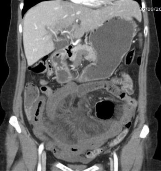
This coronal contrast-enhanced CT scan of the abdomen and pelvis illustrates a diagnostic view of mesenteric venous ischemia. The primary finding is an extensive segment of small bowel in the mid-to-lower abdomen demonstrating diffuse, circumferential wall thickening with a hypodense appearance, characteristic of intramural edema. The surrounding mesenteric fat appears hazy, consistent with mesenteric stranding often seen in venous infarction. Portions of the liver, stomach, and spleen are visible in the upper abdomen. The lack of free intraperitoneal air or extraluminal contrast suggests there is no immediate evidence of bowel perforation. This imaging is clinically significant for diagnosing acute mesenteric ischemia secondary to venous thrombosis, where venous outflow obstruction leads to significant bowel wall congestion and secondary ischemia. The visual features provided are typical for educational review of gastrointestinal emergencies and radiological manifestations of vascular compromise in the digestive tract.
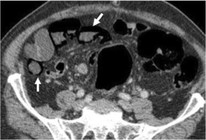
This diagnostic image is an axial contrast-enhanced CT scan of the lower abdomen demonstrating signs of advanced mesenteric ischemia. The primary finding is pneumatosis intestinalis, which is the presence of gas within the bowel wall. This gas is visible as linear and curvilinear hypoattenuating (black) areas following the contour of the intestinal wall, highlighted by white arrows. Multiple bowel loops appear dilated and contain both gas and fluid. The abdominal cavity shows evidence of significant pathology, including fat stranding and potential free fluid, consistent with mesenteric infarction. This visual is used to teach the identification of life-threatening complications in acute mesenteric ischemia, where intramural gas signifies transmural bowel necrosis. The image is relevant for gastroenterology, emergency medicine, and radiology, targeting intermediate to advanced medical learners.
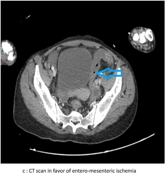
Diagnostic Imaging: This axial view Computed Tomography (CT) scan of the lower abdomen/pelvis demonstrates signs indicative of entero-mesenteric ischemia. The image reveals multiple loops of small and large bowel with heterogeneous wall enhancement and thickening. A prominent feature is pneumatosis intestinalis, indicated by a blue arrow, showing intramural gas within the bowel wall. There is evidence of mesenteric fat stranding and free fluid within the peritoneal cavity, suggesting an acute inflammatory process or infarction. Adjacent structures, including the iliac bones, pelvic musculature, and cross-sections of the proximal thighs, are visible. The scan serves as a clinical illustration of bowel ischemia, highlighting critical radiological markers such as bowel wall gas and altered morphology essential for emergency surgical evaluation. The context suggests these thromboembolic complications may be associated with a post-viral inflammatory state.
Mesenteric Ischemia
1. Classification and Pathophysiology
Acute Mesenteric Ischemia (AMI)
| Cause | Approximate Frequency |
|---|---|
| SMA embolus (SMAE) | 26-45% |
| Mesenteric arterial occlusive disease / thrombosis (MAOD/SMAT) | 25-68% |
| Non-occlusive mesenteric ischemia (NOMI) | 10-20% |
| Mesenteric venous thrombosis (MVT) | 5-10% |
| Focal segmental ischemia | ~5% |
Critical timeline: AMI can cause intestinal mucosal sloughing within 3 hours of onset and full-thickness infarction by 6 hours - Schwartz's Principles of Surgery, 11th ed.
Chronic Mesenteric Ischemia (CMI)
- Results from atherosclerotic stenosis of the main splanchnic arteries (celiac, SMA, IMA)
- Requires at least 2 of 3 major vessels to be occluded/severely stenosed for symptoms (collaterals compensate for single-vessel disease)
- Rarely leads to infarction because collaterals develop slowly
- Chronic MVT can involve the portal/splenic veins, causing portal hypertension
2. Clinical Features
Acute Mesenteric Ischemia
- Severe abdominal pain out of proportion to physical findings - the hallmark (though seen in only ~29% by modern data, as NOMI/MVT present more indolently)
- Pain is typically colicky, worst in the mid-abdomen
- Nausea, vomiting, diarrhea
- Forceful bowel evacuation early in SMAE
- Rectal bleeding (~13-16%)
- Delirium (especially in elderly)
- Fever, tachycardia, hypotension
- Abdominal distension (first sign of infarction, ~25%)
- Decreased/absent bowel sounds
- Peritonitis: rigidity, rebound tenderness, guarding
Chronic Mesenteric Ischemia
- Postprandial abdominal pain (intestinal angina) - occurs 15-30 min after eating, due to "vascular steal"
- Food fear - patients avoid eating to prevent pain
- Weight loss - often misdiagnosed as malignancy
- Often symptomatic for months before correct diagnosis
3. Diagnosis
Laboratory
- Leukocytosis (WBC >15,000) - common but nonspecific; normal WBC does not exclude early AMI
- Elevated serum lactate - nonspecific; elevated in infarction but not early ischemia
- Elevated alkaline phosphatase - released from ischemic intestinal tissue
- D-dimer - elevated in thrombotic causes (sensitive but nonspecific)
- Metabolic acidosis, elevated amylase, LDH - late findings
- Ischemia-modified albumin and procalcitonin may be helpful markers per systematic review
Imaging
- Early: nonspecific - bowel dilation, thickened folds, air-fluid levels
- Late: "Thumbprinting" (submucosal edema), pneumatosis intestinalis (intramural gas from gas-forming bacteria - sign of necrosis), portal venous gas, pneumoperitoneum
- Sensitivity ~89%, specificity ~99.5%
- Identifies: bowel wall thickening, pneumatosis intestinalis, portal venous gas, mesenteric fat stranding, segmental ischemia in vascular distribution
- Identifies the underlying cause (embolus vs. thrombosis) and location for procedural planning
- Best for occlusive disease; less reliable for NOMI
- Supplanted by CTA but still used when CTA is equivocal
- Required for NOMI diagnosis (papaverine infusion)
- Allows simultaneous therapeutic intervention
4. Management
General Principles (AMI)
- Resuscitation: IV fluids, broad-spectrum antibiotics (for bacterial translocation), correct hemodynamic instability
- Anticoagulation: Heparin for thrombotic/embolic causes (reduces clot propagation)
- Avoid vasoconstrictors if possible; treat the underlying cause of hypoperfusion
- Nasogastric decompression
Surgical vs. Endovascular Revascularization
| Cause | Primary Treatment |
|---|---|
| SMAE (embolus) | Endovascular (catheter-directed thrombolysis/thrombectomy) or surgical embolectomy; exploratory laparotomy if signs of peritonitis |
| SMAT/MAOD (thrombosis) | Endovascular angioplasty + stenting (preferred first line) or surgical bypass (aortomesenteric bypass); bowel resection if necrosis |
| NOMI | Treat underlying cause (correct hypotension/hypovolemia); intra-arterial papaverine infusion via selective angiography; laparotomy only if peritonitis |
| MVT | Anticoagulation (heparin then warfarin/LMWH/DOACs); thrombolysis in severe cases; surgical thrombectomy rarely needed |
- Peritonitis present
- Pneumatosis intestinalis or portal venous gas on CT (signs of bowel necrosis)
- Failure of endovascular therapy
- To assess bowel viability after revascularization ("second look" laparotomy at 24-48 hours is standard practice)
Chronic Mesenteric Ischemia
- Endovascular (angioplasty + stenting): First-line preferred approach; lower peri-operative morbidity and mortality; higher re-stenosis rate long-term
- Open surgical bypass (aortomesenteric bypass): More durable; reserved for patients with complex anatomy or endovascular failure
- Medical management alone is not definitive
5. Prognosis
- AMI mortality: up to 70-80% - one of the highest mortality rates of any abdominal emergency
- If revascularization is achieved before bowel infarction, mortality drops substantially
- MVT has the most favorable prognosis among AMI subtypes due to its more indolent course
- Incidence is rising due to aging populations, more patients surviving critical illness in ICUs, and increased recognition
- Schwartz's Principles of Surgery, 11th ed. - classification, pathophysiology, clinical features
- Sleisenger & Fordtran's Gastrointestinal and Liver Disease - frequencies, clinical features, diagnosis, treatment algorithm
- Frameworks for Internal Medicine - diagnostic approach, imaging roles
- Recent meta-analyses: PMID 39069018 (endovascular vs open, 2024), PMID 40275298 (AMI management, 2025), PMID 40257244 (CMI treatment, 2026)