
Ct urography my style 10 marks
CT urography technique protocol phases radiology exam 10 marks
https://appliedradiology.com/articles/ct-urography-review-of…
CT Urography (CTU)
Definition
Indications
| Category | Examples |
|---|---|
| Primary indication | Hematuria (macro/micro) - recommended by AUA & ACR as first-line imaging |
| Urolithiasis | Suspected/known renal/ureteric calculi |
| Neoplasm | Suspected renal cell carcinoma, urothelial tumors (TCC) |
| Infection | Recurrent UTI, complicated pyelonephritis, xanthogranulomatous pyelonephritis |
| Obstruction | Hydronephrosis, UPJ/UVJ obstruction |
| Trauma | Renal/ureteric injury |
| Congenital | Duplex systems, horseshoe kidney, ectopic ureter |
| Surveillance | Post-cystectomy urothelium monitoring |
Technique / Protocol
Patient Preparation
- Oral hydration: 2-3 glasses of water before the scan (aids ureteric distension)
- IV hydration optional
- IV furosemide (10-20 mg) at time of contrast injection - promotes diuresis and ureteric opacification
- Empty bladder prior to scan
Equipment
- Multidetector CT (MDCT) with thin slices (2.5-5 mm)
- Reconstructions: axial, coronal, sagittal; MIP reformats for the collecting system
Phases of Acquisition (Triphasic Protocol)
Phase 1: Non-contrast Phase
- Coverage: Top of kidneys to bladder base
- Purpose:
- Detect calculi (high attenuation without contrast)
- Identify fat-containing lesions (angiomyolipoma)
- Detect parenchymal calcifications
- Provide baseline attenuation for enhancement assessment
- Slice thickness: 2-2.5 mm
Phase 2: Nephrographic Phase
- Timing: 90-100 seconds after IV contrast injection (100-150 mL of non-ionic iodinated contrast at 2-4 mL/sec)
- Coverage: Abdomen and pelvis (kidneys to bladder)
- Purpose:
- Homogeneous cortex + medulla enhancement - best phase for detecting small renal masses
- Enhancement confirmed by comparison with non-contrast images (>20 HU increase = enhancement)
- Evaluation of renal vasculature
- Key feature: Most sensitive phase for renal cell carcinoma detection
Phase 3: Excretory (Pyelographic) Phase
- Timing: 12-15 minutes after contrast injection
- Coverage: Full abdomen and pelvis
- Purpose:
- Opacification and distension of renal calyces, pelvis, ureters, and bladder
- Best phase for urothelial tumors (TCC/UTUC)
- Evaluation of collecting system anatomy and filling defects
- Limitation: Ureteric peristalsis may cause non-opacification of segments
Split-Bolus Technique (Dose Reduction Alternative)
- Contrast given in two boluses before a single post-contrast acquisition
- 1st bolus (smaller, e.g., 50 mL) → 7-9 minute delay → 2nd bolus (larger, e.g., 75-100 mL) → 120 seconds → single scan
- Cumulative effect: early bolus provides excretory information; later bolus gives nephrographic information
- Advantage: Eliminates one acquisition - reduces radiation dose significantly
- Disadvantage: Less reliable ureteric opacification compared to triphasic
Techniques to Improve Ureteric Distension
- IV furosemide - most effective; promotes diuresis
- IV/oral hydration - increases urine flow
- Prone positioning - uses gravity to fill dependent ureters with contrast
- Compression belt - mechanical ureteric compression at the pelvic brim
- Log-rolling - patient rolled prior to excretory acquisition to redistribute contrast
Radiation Dose
| Protocol | Effective Dose |
|---|---|
| Four-phase CTU | ~25-35 mSv |
| Excretory urography (IVU) | ~3.6 mSv |
| Split-bolus CTU | ~15-20 mSv (reduced) |
Findings / Diagnostic Value
| Pathology | Best Phase |
|---|---|
| Renal calculi | Non-contrast |
| Renal mass (RCC) | Nephrographic |
| Urothelial carcinoma (TCC) | Excretory/pyelographic |
| Hydronephrosis | Excretory |
| Transitional cell filling defects | Excretory |
| Ureteric pseudodiverticulosis | Excretory (associated with risk of urothelial carcinoma) |
CT Urography vs. IVU
| Feature | CTU | IVU |
|---|---|---|
| Sensitivity for calculi | >96% | ~60% |
| Renal mass detection | Excellent | Poor |
| Urothelial TCC | Good | Limited |
| Radiation | Higher | Lower |
| Extraurinary findings | Yes | No |
| Gold standard status | Yes (upper tract) | Largely replaced |
Summary
- Calculi (non-contrast phase)
- Renal masses (nephrographic phase)
- Urothelial tumors and collecting system anatomy (excretory phase)
Isn’t there a corticomedullary phase
Complete Phases of CT Urography (Four-Phase Protocol)
Phase 1: Non-contrast Phase
- Timing: Before contrast
- Purpose: Calculi, fat lesions, baseline attenuation
Phase 2: Corticomedullary Phase (CMP)
- Timing: 25-40 seconds after contrast injection (arterial/early venous)
- Purpose:
- Renal cortex enhances brightly; medulla has not yet enhanced (clear corticomedullary differentiation)
- Renal arteries and veins are well opacified - ideal for vascular anatomy, renal artery stenosis, arteriovenous malformations
- Detection of hypervascular tumors (some RCCs, angiomyolipomas)
- Pre-surgical planning (donor kidneys, partial nephrectomy)
- Limitation: Small renal masses can be missed because the medulla is relatively unenhanced, creating a heterogeneous background that obscures isodense lesions
Phase 3: Nephrographic Phase
- Timing: 90-100 seconds
- Cortex + medulla enhance uniformly - best for renal mass detection
Phase 4: Excretory Phase
- Timing: 12-15 minutes
- Collecting system, ureters, bladder opacified - best for urothelial lesions
Practical Note
- Vascular assessment is needed (renal donor evaluation, suspected vascular lesion)
- Characterization of a known hypervascular mass
- Pre-operative planning
- Triphasic: Non-contrast + Nephrographic + Excretory (most common)
- Four-phase: Non-contrast + Corticomedullary + Nephrographic + Excretory (higher dose, ~25-35 mSv, used when vascular detail is required)
Retroperitoneal fibrosis radiology pg ans
retroperitoneal fibrosis radiology CT MRI findings postgraduate exam answer
https://radiopaedia.org/articles/retroperitoneal-fibrosis-1?…
Retroperitoneal Fibrosis - Radiology
Definition
Imaging Modalities
1. Plain X-Ray / IVU (Intravenous Urography)
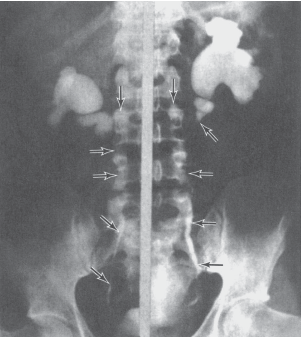
- Bilateral hydronephrosis and hydroureter (bilateral in 68%, unilateral in 20%)
- Medial deviation of the mid-ureters - the most characteristic sign
- Normally ureters course laterally; in RPF they are pulled medially toward the midline
- "Maiden waist sign" - smooth, gradual tapering of the ureter at the level of obstruction (L4-L5)
- "Pipestem ureter" - long segment of ureter is narrowed and aperistaltic due to fibrotic encasement
- Site of obstruction: usually at the level of L4-L5, where the plaque is thickest
- Proximal dilation with distal narrowing - gives the classic IVU appearance
2. Ultrasound
- Bilateral hydronephrosis - initial finding, prompts further workup
- May show a hypoechoic periaortic soft tissue mass
- Useful for monitoring treatment response - serial assessment of hydronephrosis
- Cannot characterize the fibrotic plaque well; does not differentiate benign from malignant
- Doppler: Can detect ureteric obstruction and assess renal resistive indices
3. CT Scan (Modality of Choice for Diagnosis)
| Feature | Description |
|---|---|
| Location | Periaortic/periiliac soft tissue mass centered at L4-L5 |
| Density | Soft tissue attenuation (30-60 HU), homogeneous on NECT |
| Enhancement | Variable; active disease enhances post-contrast; fibrotic/inactive disease shows less enhancement |
| Shape | Plaque-like, mantle-like, encasing the aorta, IVC and ureters |
| Ureteric involvement | Bilateral medial deviation + obstruction at the level of the plaque |
| Hydronephrosis | Bilateral > unilateral |
| Aorta | Plaque hugs anterior surface of aorta - aorta not elevated from spine (key benign feature) |
| Lymph nodes | Present in ~25% of cases |
| Extent | May extend from renal hilum to iliac vessels; rarely into mediastinum |
- Active/inflammatory RPF: enhances on arterial and venous phase
- Inactive/fibrotic RPF: little to no enhancement
- Decreasing enhancement on serial scans = treatment response
4. MRI (Superior for Follow-Up and Tissue Characterization)
| Sequence | Signal | Significance |
|---|---|---|
| T1-weighted | Low signal intensity | Fibrotic tissue (characteristic) |
| T2-weighted | Variable | Key for activity assessment |
| T2 HIGH signal | Active, inflammatory disease (edema, cellular infiltrate) | |
| T2 LOW signal | Inactive, mature fibrosis | |
| Gadolinium (Gd) post-contrast | Enhancement in active disease | |
| Decreasing Gd enhancement | Indicates response to therapy |
- Neither CT nor MRI reliably differentiates benign from malignant RPF
- Features suggesting malignant RPF:
- Anterior elevation of the aorta from the spine (suggests lymphoma pushing from behind)
- Lateral displacement of ureters (rather than medial)
- Localized bone destruction
- Infiltration of surrounding structures
- Irregular, lobulated margins
- Features suggesting benign RPF:
- Aorta hugging the spine (not elevated)
- Medial deviation of ureters
- Smooth, confluent plaque
5. Retrograde / Antegrade Pyelography
- Performed when CT/MRI is contraindicated (renal impairment, contrast allergy) or when endoscopic intervention is planned
- Shows:
- Long segment of ureteric narrowing at L4-L5
- Smooth tapering (not abrupt cutoff - helps distinguish from malignant stricture)
- Medial deviation of mid-ureter
- Proximal hydronephrosis
6. Nuclear Medicine / PET-CT
- FDG-PET/CT: Most sensitive imaging study for disease activity
- Active RPF shows increased FDG uptake in the fibrotic plaque
- Can differentiate active from inactive disease better than CT/MRI alone
- Distinguishes RPF from malignant mimics (metastatic lymphoma, etc.)
- May obviate the need for biopsy in typical cases
- Useful for monitoring treatment response
- MAG3/DTPA renography (differential scintigraphy): Assesses individual renal function - important for surgical planning (ureterolysis)
Differential Diagnosis on Imaging
| Condition | Distinguishing Feature |
|---|---|
| Lymphoma | Aorta elevated anteriorly, lateral ureteric displacement, discrete nodes |
| Metastatic disease | Prior malignancy history, bone destruction, irregular mass |
| Aortic aneurysm | Dilated aortic lumen, wall calcification, pulsatile mass |
| Amyloidosis | Diffuse retroperitoneal infiltration, other organ involvement |
| Desmoplastic reaction (post-RT/surgery) | Clinical history, localized to field |
Summary of Modality Use
| Modality | Role |
|---|---|
| IVU | Historic; shows medial ureteric deviation and hydronephrosis |
| Ultrasound | Initial screening; serial monitoring of hydronephrosis |
| CT | Gold standard for diagnosis; characterizes plaque, guides biopsy |
| MRI | Superior for disease activity and treatment monitoring (T2 signal) |
| Retrograde pyelogram | When CT/MRI contraindicated; pre-intervention planning |
| PET-CT | Most sensitive for activity; differentiates benign/malignant; monitors therapy |
Classify congenital anomalies of kidney describe imaging features of fusion anomalies of kidney radiology ans15 marks high yield Rguhs radiology pg exam style
congenital anomalies kidney classification fusion anomalies horseshoe crossed ectopia imaging radiology RGUHS PG exam
https://www.dirjournal.org/pdf/beb8919b-f013-4ea1-b1c8-40332…
Congenital Anomalies of the Kidney - Classification and Imaging Features of Fusion Anomalies
PART A: CLASSIFICATION OF CONGENITAL ANOMALIES OF THE KIDNEY
I. Anomalies of Number
| Anomaly | Details |
|---|---|
| Bilateral renal agenesis (Potter sequence) | Absent both kidneys; fatal; oligohydramnios, Potter facies |
| Unilateral renal agenesis | 1 in 1000; associated with VACTERL, mullerian anomalies |
| Supernumerary kidney | Rare; extra kidney with own ureter and blood supply |
II. Anomalies of Volume / Size
- Renal hypoplasia - small kidney, reduced nephrons, normal architecture
- Renal dysplasia - abnormal differentiation; cystic dysplastic kidney
III. Anomalies of Position (Ectopia)
| Type | Description |
|---|---|
| Simple renal ectopia | Kidney on same side but malpositioned (pelvic, lumbar, abdominal) |
| Pelvic kidney | Most common ectopia; in true pelvis |
| Thoracic (intrathoracic) kidney | Above diaphragm; 1:13,000; usually left side |
| Crossed renal ectopia | Kidney crosses midline to opposite side (with or without fusion) |
IV. Anomalies of Rotation (Malrotation)
- Normal: renal pelvis rotates 90° ventromedially during ascent
- Malrotation types (Weyrauch classification): Ventral, Ventromedial, Lateral, Dorsal positions
- Pelvis may face ventrally (non-rotation - most common), laterally, or dorsally
V. Anomalies of Fusion (Main focus - see Part B)
| Type | Example |
|---|---|
| Partial fusion | Horseshoe kidney, Crossed fused ectopia |
| Complete fusion | Cake/Lump/Disc/Pancake kidney |
VI. Anomalies of the Collecting System
- Duplex collecting system (most common renal anomaly - 2% of population)
- Calyceal diverticulum
- Congenital megacalyces
- Pelvi-ureteric junction (PUJ) obstruction
- Duplex ureter / ectopic ureter / ureterocele
VII. Cystic Anomalies (separate classification)
- Autosomal dominant/recessive polycystic kidney disease
- Multicystic dysplastic kidney (MCDK)
- Medullary sponge kidney
- Simple renal cysts
PART B: FUSION ANOMALIES OF THE KIDNEY - IMAGING FEATURES
Embryological Basis
- More common in males (2:1)
- Partial fusion - Horseshoe kidney (90% of all fusion anomalies), Crossed fused ectopia
- Complete fusion - Cake/Disc/Pancake kidney (rare, 2% of fused kidneys)
1. HORSESHOE KIDNEY
Incidence
- Most common renal fusion anomaly - 1 in 400-500 individuals (Campbell-Walsh)
- Male:Female = 2:1
- Frequently associated with other congenital anomalies - Turner syndrome, trisomy 18, VACTERL
Anatomy
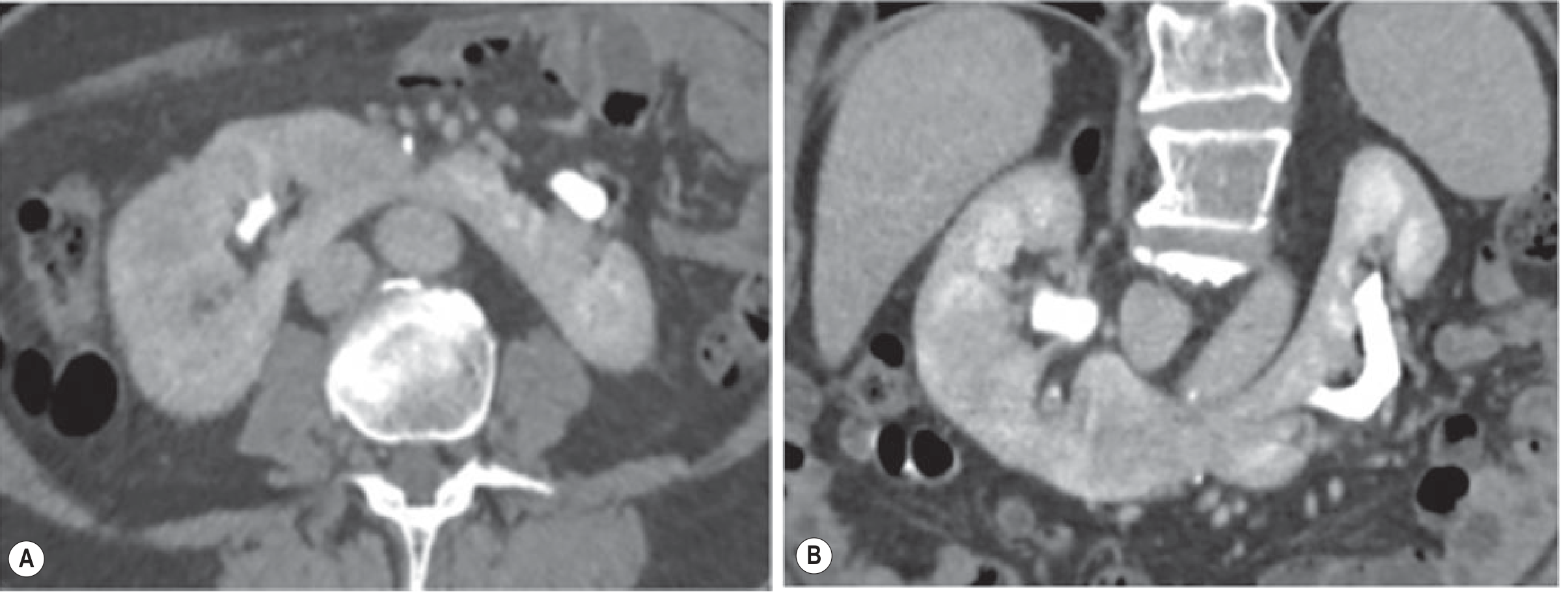
- Two distinct renal masses, one on each side of midline, connected at their lower poles (95%) by an isthmus
- Upper pole fusion: rare (5%)
- Isthmus lies at L3-L4 level, anterior to the aorta and IVC, just below the origin of the inferior mesenteric artery (IMA) - which prevents full cranial ascent
- Isthmus composition: functional renal parenchyma (85-95%) or fibrous tissue (5-15%)
- Renal pelvis: anteriorly placed (faces ventrally due to malrotation)
- Calyces: point posteriorly (fail to rotate); lowermost calyces may overlie the vertebral column
Imaging Features
A. Plain X-Ray / IVU
- Vertical or obliquely lateral axis of each renal moiety (vs. normal oblique medially inclined axis)
- Lower pole medial orientation of collecting systems - medial tilt of lower poles toward midline
- Lowered position of kidneys (below L3)
- IVU shows "inverted lily" or "flower vase" appearance - collecting systems open anteriorly/medially
- Ureters course anteriorly over the isthmus - characteristic ureteric course with a high lateral insertion into renal pelvis
- Isthmus may be visible as a soft tissue density overlying the lumbar spine
- Complications: hydronephrosis (UPJ obstruction in up to 1/3 of patients), calculi, recurrent UTI
B. Ultrasound
- Two renal masses with medially oriented lower poles
- Isthmus visible anterior to aorta and IVC - hypo/isoechoic band of tissue crossing the midline
- Renal pelvis faces anteriorly (not medially as in normal kidney)
- Doppler: variable vascular supply - multiple aberrant vessels from aorta, IMA, iliac arteries
C. CT Scan (Investigation of Choice)
| Feature | Finding |
|---|---|
| Isthmus | Parenchymal or fibrous band anterior to aorta, below IMA |
| Axis | Vertical or oblique lateral - lower poles more medial and anterior than normal |
| Position | Low lying (L3-L4), may be pelvic |
| Renal pelvis | Anterior-facing (malrotated) |
| Ureters | Cross anterior to isthmus, high lateral insertion |
| Blood supply | Multiple aberrant vessels from aorta, IMA, iliac vessels - critical for surgery |
| Isthmus density | Enhances with contrast if parenchymal; non-enhancing if fibrous |
| Complications | UPJ obstruction, hydronephrosis, calculi, Wilms tumor |
D. MRI
- MRI urography (MRU) gives equivalent anatomical information to CT
- Isthmus appears as a band of normal renal signal intensity tissue
- T2-weighted coronal images: best for showing collecting system orientation and ureteral course
- Preferred in children and pregnant women (no radiation)
- Gadolinium-enhanced: shows parenchymal enhancement of the isthmus, confirming functional tissue
E. Nuclear Medicine (MAG3/DTPA renogram)
- Assesses differential renal function of each moiety
- Evaluates drainage - identifies UPJ obstruction
- Post-operative follow-up after ureterolysis or pyeloplasty
Complications of Horseshoe Kidney (Examinable)
| Complication | Frequency | Mechanism |
|---|---|---|
| UPJ obstruction / Hydronephrosis | Up to 33% | Aberrant vessels, abnormal ureteral course over isthmus |
| Urolithiasis | ~20% | Urinary stasis, incomplete drainage |
| Recurrent UTI | Common | Stasis |
| Wilms tumor | 2x higher risk than general population | |
| Transitional cell carcinoma | Increased risk | |
| Vascular injury during aortic surgery | Important surgical hazard | Aberrant vessels around isthmus |
2. CROSSED FUSED RENAL ECTOPIA (CFRE)
Incidence
- Second most common fusion anomaly - 1 in 1300-7500
- Male predominance
Anatomy
- One kidney crosses the midline to the opposite side, and its parenchyma fuses with the other kidney
- The ureter of the crossed kidney crosses back to drain into the bladder on its own (original) side - key distinguishing feature
- Left kidney is most commonly the ectopic one (crosses to the right)
- Most common fusion pattern: upper pole of the inferior/crossed ectopic kidney fuses with the lower pole of the normally positioned kidney - produces an L-shape
McDonald and McClellan Classification of Crossed Ectopia:
| Type | Description | Frequency |
|---|---|---|
| Crossed renal ectopia with fusion | Both kidneys fused | 85% |
| Crossed renal ectopia without fusion | Crossed but separate | 10% |
| Solitary crossed renal ectopia | Only one kidney, on wrong side | Rare |
| Bilaterally crossed renal ectopia | Both kidneys cross | Rare |
Six Variants of Crossed Fused Ectopia (in decreasing order of frequency):
- Type 1 - Inferior crossed fused ectopia (most common) - crossed kidney lies inferior, fused at its upper pole to lower pole of native kidney; L-shaped configuration
- Type 2 - Sigmoid / S-shaped kidney - both kidneys on the same side, fused back-to-back with opposite pole orientation
- Type 3 - Unilateral lump kidney - both fused with multiple contact areas; globular mass
- Type 4 - Unilateral disc/shield kidney - flat, disc-shaped fusion
- Type 5 - L-shaped kidney
- Type 6 - Superior crossed fused ectopia (rarest) - crossed kidney lies superior
Imaging Features of CFRE
A. Ultrasound
- Absence of kidney in the contralateral renal fossa (empty fossa on one side)
- Large unilateral mass of renal tissue in the other fossa
- Two separate renal sinuses may be identified within the single mass
- Doppler: separate blood supplies to each moiety
- May mimic a renal tumor or retroperitoneal mass
B. IVU / Excretory Urography
- Shows two collecting systems on one side of the abdomen
- One ureter crosses the midline back to drain on its original side
- Characteristic non-opacification of contralateral renal fossa
C. CT Scan (Best modality)
- Parenchymal mass on one side containing two fused renal units
- Two distinct collecting systems visible within the fused mass
- Each renal moiety enhances normally
- One ureter crosses the midline to drain into the ipsilateral bladder
- CT angiography delineates the dual separate blood supplies
- Coronal and axial views: identify the fusion plane and configuration type
D. Nuclear Medicine (DMSA / MAG3)
- DMSA scan: shows fused uptake of both functioning renal units on one side with absent contralateral uptake - pathognomonic appearance
- MAG3 renogram: assesses drainage and identifies PUJ obstruction (common due to malrotation)
- Widely used in children as first-line functional imaging
E. MRI / MRU
- T2 coronal: clearly shows fused renal mass, collecting system anatomy
- MRU: traces both ureters - shows the crossed ureter returning to its side
- No radiation - preferred in children
3. COMPLETE FUSION ANOMALIES (Cake / Disc / Pancake Kidney)
Cake / Lump Kidney
- Complete fusion of both kidneys into a single midline mass (usually pelvic)
- 2% of all renal fusion anomalies
- No recognizable reniform shape; multiple collecting systems
- Blood supply: highly aberrant - multiple vessels from distal aorta and iliac vessels
- Imaging: pelvic soft tissue mass with renal parenchymal enhancement; bilateral ureteral drainage from single mass
- MRI best for characterization; CT angio for pre-surgical vascular mapping
Disc / Shield / Pancake Kidney
- Flat, disc-shaped bilateral fusion; both kidneys fused medially
- Located in the pelvis
- Multiple collecting systems and ureters drain normally
SUMMARY TABLE: Fusion Anomalies at a Glance
| Feature | Horseshoe Kidney | Crossed Fused Ectopia | Cake Kidney |
|---|---|---|---|
| Frequency | 1:400-500 | 1:1300-7500 | Very rare |
| % of fusion anomalies | ~90% | ~9% | ~2% |
| Position | Bilateral, low lumbar | Unilateral mass | Midline pelvic |
| Connecting bridge | Isthmus at L3-L4 | Direct parenchymal fusion | Complete midline fusion |
| Pelvis orientation | Anterior-facing | Abnormal | Multiple |
| IVU finding | Inverted lily/flower vase | Two systems on one side | Multiple systems, pelvic |
| Best modality | CT | CT / DMSA scan | CT |
| Key complication | UPJ obstruction (33%) | PUJO, VUR | Renal failure, infection |
| Associated syndrome | Turner syndrome | VACTERL | - |
HIGH-YIELD EXAM POINTS (RGUHS)
- Horseshoe kidney = most common fusion anomaly (1:400); lower pole fusion 95%; isthmus lies anterior to aorta BELOW the IMA (IMA prevents ascent)
- IVU sign: vertical axis of renal pelves + medial tilt of lower poles = "inverted lily" appearance
- UPJ obstruction in 33% of horseshoe kidneys - due to aberrant vessels or abnormal ureteral course over isthmus
- Wilms tumor risk is doubled in horseshoe kidney
- Crossed fused ectopia: left kidney crosses to right (most common); ureter crosses BACK to drain on correct side
- DMSA scan = investigation of choice in children for CFRE - shows unilateral uptake of fused mass
- Cake kidney = complete fusion; pelvic; 2% of fusion anomalies
- MRI/MRU = preferred in children and pregnant women (no radiation)
- CT angiography = mandatory before any surgery on horseshoe kidney (aberrant vasculature)
- McDonald and McClellan classification of crossed ectopia - Type 1 (inferior, L-shaped) is most common (85%)
Calcification in KUB skiagram and various modalities of used in evaluatio
calcification KUB skiagram plain radiograph types renal ureteric bladder radiology classification
https://emedicine.medscape.com/article/2165400-overview
calcifications KUB plain abdomen radiograph differential diagnosis renal nephrocalcinosis vascular prostate phlebolith urinary stones radiology
https://www.med.unc.edu/medclerk/wp-content/uploads/sites/87…
Calcifications on KUB Skiagram and Various Modalities of Evaluation
KUB Skiagram - Introduction
- Sensitivity for urinary calculi: ~45-80% (variable, depending on stone composition)
- Specificity: ~77%
- Limitation: Cannot detect radiolucent stones, cannot assess obstruction, many non-urinary calcifications mimic urinary stones
CLASSIFICATION OF CALCIFICATIONS SEEN ON KUB
A. Normal Structures That Calcify (Physiological/Incidental)
| Structure | Appearance on KUB |
|---|---|
| Costal cartilage | Bilateral, superior; may overlap renal shadows |
| Mesenteric lymph nodes | Oval, smooth, discrete opacities; near spine; can mimic ureteric stones |
| Phleboliths (pelvic vein calcifications) | Small, round, smooth opacities in pelvis; characteristic central lucency (lucent center distinguishes from stones) |
| Prostate gland calcifications | Midline or bilateral, small, dense; below pubic symphysis |
| Arterial calcifications (atherosclerosis) | Curvilinear, parallel-line "tramtrack" pattern along vessel walls |
B. Pathological Calcifications on KUB
I. RENAL CALCIFICATIONS
1. Nephrolithiasis (Renal Calculi / Urinary Stones)
| Stone Type | Composition | Radiopacity | Frequency |
|---|---|---|---|
| Calcium oxalate | CaOx monohydrate/dihydrate | Most dense, homogeneous | ~40% |
| Calcium phosphate | Apatite | Dense, may be laminated | ~20% |
| Struvite (triple phosphate) | Mg-NH4-PO4 | Moderately opaque; staghorn pattern | ~15% |
| Cystine | Cystine | Faintly opaque ("ground glass") | ~2% |
| Uric acid | Urate | Radiolucent (invisible on KUB) | ~10% |
| Indinavir (drug calculus) | Drug | Radiolucent | Rare |
- Staghorn calculus - fills entire pelvicalyceal system; branching configuration; struvite (infection stones)
- Jackstone calculus - spiculated, calcium oxalate monohydrate
- Laminated - alternating rings; calcium phosphate / mixed
- Smooth rounded - uric acid (but radiolucent)
- Renal pelvis - overlying T12-L2 region
- PUJ - at L2-L3 level, just medial to lateral border of psoas
- Pelvic brim - at sacroiliac joint level
- VUJ (Vesico-ureteric junction) - at level of ischial spine; most common site of impaction
- Ureters run along the line of transverse processes of lumbar vertebrae → key anatomical landmark to identify ureteric stones
2. Nephrocalcinosis (Renal Parenchymal Calcification)
- Calcification in the peripheral cortex and septa of Bertin
- KUB appearance: Thin peripheral lines ("tramlines"), diffusely dense renal shadow, or punctate calcifications
- Causes:
- Chronic cortical necrosis (bilateral, dense; "tram-track sign")
- Chronic glomerulonephritis
- Rejected transplant kidney
- Oxalosis (primary hyperoxaluria)
- Calcification in the medullary pyramids
- KUB appearance: Dense bilateral aggregates in the papillary/medullary regions; triangular clusters
- Causes (mnemonic: "HARD RTA"):
- Hyperparathyroidism (most common cause)
- Renal tubular acidosis (Type 1, distal)
- Medullary sponge kidney
- Hypervitaminosis D
- Sarcoidosis
- Milk-alkali syndrome
3. Dystrophic Calcification in Renal Lesions
- Renal cell carcinoma - irregular, "eggshell" or central calcification; ~10% of RCCs
- Renal cyst - thin peripheral "eggshell" calcification of cyst wall (Bosnian category)
- Wilms tumor - stippled or faint calcification in children
- Angiomyolipoma - calcification rare
- Renal TB - "putty kidney" (dystrophic calcification of entire kidney); lobar or diffuse dense calcification; associated ureteric calcification ("pipestem ureter")
- Renal abscess / Xanthogranulomatous pyelonephritis - irregular calcification within mass
II. URETERIC CALCIFICATIONS
- Calcified stones along the ureteric course
- Anatomical landmarks for tracing ureters on KUB:
- Along the tips of transverse processes L1-L5
- Across the sacroiliac joints at the pelvic brim
- Down to the ischial spine level (VUJ)
| Feature | Ureteric Stone | Phlebolith |
|---|---|---|
| Location | Along transverse processes / ureteric course | Lateral in pelvis, away from ureteric line |
| Central lucency | Absent | Present (pathognomonic) |
| Shape | Irregular, angular | Round, smooth |
| "Comet tail" sign (CT) | Absent | Present (soft tissue phlebolith streak) |
| Soft tissue rim sign (CT) | Present (edematous ureteric wall) | Absent |
III. BLADDER CALCIFICATIONS
| Type | Appearance | Cause |
|---|---|---|
| Vesical calculi | Dense, rounded/faceted opacities in midline pelvis overlying bladder | Stasis, infection, foreign body |
| Bladder wall calcification (eggshell) | Thin, curvilinear calcification outlining bladder wall | Schistosomiasis (S. haematobium) - pathognomonic; TB |
| Bladder tumor calcification | Irregular, focal; absence of previously seen wall calcification = malignancy | TCC |
| Encrusted cystitis | Dense, irregular calcification of bladder wall | Alkaline encrusting cystitis |
IV. EXTRAURINARY CALCIFICATIONS VISIBLE ON KUB (Differential Diagnosis)
| Location | Calcification | Cause |
|---|---|---|
| Vascular | Curvilinear, tramtrack | Aortic/iliac atherosclerosis; aortic aneurysm (crescent or eggshell) |
| Gallbladder | Right hypochondrium, faceted | Gallstones (only 10-15% visible) |
| Pancreas | Irregular, coarse; along pancreatic duct | Chronic pancreatitis |
| Adrenal | Triangular, above kidney | TB, Addison's disease, hemorrhage, neuroblastoma (in children) |
| Appendix | RIF, ovoid with lamination | Appendicolith (faecolith) |
| Lymph nodes | Oval, discrete; mesenteric/retroperitoneal | TB, histoplasmosis, silicosis |
| Uterine fibroid | Midline/pelvic; "whorled" or "popcorn" | Leiomyoma |
| Ovarian dermoid | Pelvic; may show teeth or bone | Teratoma |
| Prostatic calcification | Small dense clusters below bladder | Benign; chronic prostatitis |
IMAGING MODALITIES FOR EVALUATING KUB CALCIFICATIONS
1. Plain Radiograph (KUB Skiagram)
- First-line, widely available, low cost, low radiation
- Detects ~80% of urinary calculi (all radiopaque stones)
- Misses: uric acid stones, small stones, stones overlying bone/bowel gas
- Uses:
- Initial assessment of urinary calculi
- Follow-up of known stone size/burden
- Post-ESWL or surgical follow-up (stone clearance)
- Limitation: Cannot assess function or obstruction; cannot differentiate ureteric stone from phlebolith with certainty
2. Intravenous Urography / IVP (IVU)
- Historically the "gold standard" for urinary tract evaluation; now largely replaced by CT
- NECT KUB (control film) → IV contrast → serial films at 5, 15, 30 min
- Findings in urolithiasis:
- "Standing column" sign - column of contrast in ureter proximal to obstruction (dilated, non-peristaltic ureter)
- Delayed nephrogram and dense persistent nephrogram on affected side
- Crescent sign - contrast in compressed calyces around obstructing stone
- Identifies site and degree of obstruction
- Differentiates ureteric stone from phlebolith (stone seen within opacified ureter)
- Advantage over plain KUB: Confirms whether opacity is within the urinary tract
- Contraindications: Contrast allergy, renal failure, metformin use (relative), pregnancy
3. Ultrasound (USG)
| Feature | Details |
|---|---|
| Renal calculi | Echogenic foci with posterior acoustic shadowing |
| Twinkle artifact | Color Doppler - rapid color aliasing behind calculus; increases sensitivity |
| Nephrocalcinosis | Echogenic pyramids (medullary) or echogenic cortex (cortical) |
| Ureteric stones | Difficult to see (bowel gas); VUJ stones detectable via full bladder window |
| Hydronephrosis | Anechoic dilated pelvicalyceal system - identifies obstruction |
| Bladder calculi | Mobile echogenic foci with shadowing, move with posture change |
| Bladder wall calcification | Echogenic thickened wall |
Limitations: Poor for ureteric calculi (40-60% sensitivity); cannot detect small stones; operator-dependent
4. Non-Contrast CT (NCCT / CT KUB) - Gold Standard
| Parameter | Value |
|---|---|
| Sensitivity | 96-99% |
| Specificity | ~98-100% |
| Stone size detection | Down to 1 mm |
| Radiation | Higher than KUB (~3-10 mSv) |
- Detects ALL stones regardless of composition (including uric acid, indinavir)
- Soft tissue rim sign - halo of edematous ureteric wall around stone = confirms ureteric stone (vs. phlebolith)
- Comet tail sign - distinguishes phleboliths
- Identifies complications: hydronephrosis, hydroureter, perinephric fat stranding, urinoma, forniceal rupture
- Detects non-urological causes of pain (appendicitis, aortic aneurysm)
- HU (Hounsfield unit) attenuation predicts stone composition:
- Uric acid: ~200-400 HU
- Calcium oxalate: ~1000-1700 HU
- Struvite: ~600-900 HU
- Cystine: ~600-1100 HU
- Central lucency of phlebolith seen better on KUB than CT
- Stone size measurement slightly larger on KUB than CT (CT underestimates by ~12%)
5. CT Urography (CTU)
- Triphasic: NECT + Nephrographic (90s) + Excretory (12-15 min)
- Combines stone detection, mass characterization, and urothelial evaluation
- For complex cases: hematuria + suspected stones + suspected tumor
6. MRI / MR Urography (MRU)
- No ionizing radiation - preferred in pregnancy and children
- Does NOT visualize calcifications directly (calcium = signal void on MRI)
- MRU (heavily T2-weighted or Gd-enhanced): evaluates obstruction, collecting system anatomy
- Can identify the obstruction site but cannot characterize stone composition
- Role: Pregnant patients with suspected ureteric colic; children with congenital anomalies
7. Radionuclide Imaging (Nuclear Medicine)
| Agent | Function |
|---|---|
| DMSA (Tc-99m) | Cortical scarring, differential renal function, renal mass characterization |
| MAG3 / DTPA (diuretic renogram) | Obstructive uropathy - functional vs anatomical; drainage kinetics |
| Radionuclide cystography | VUR detection in children |
| PET-CT | Not routinely used for stone disease; useful for malignant/inflammatory retroperitoneal causes |
8. Retrograde Pyelography
- Invasive; endoscopic opacification of ureter/pelvis from below
- Used when: CT/IVU contraindicated, pre-stenting planning, precise anatomical mapping
- Shows site of ureteric obstruction precisely
9. Antegrade Pyelography / Nephrostomy
- Percutaneous access via dilated pelvicalyceal system
- Used for decompression + diagnostic imaging when retrograde not feasible
SUMMARY: MODALITY COMPARISON FOR KUB CALCIFICATIONS
| Modality | Sensitivity | Radiation | Detects Radiolucent Stones | Obstruction Assessment | Best Use |
|---|---|---|---|---|---|
| KUB plain | 45-80% | Low | No | No | Initial/follow-up |
| IVU | ~80% | Moderate | No | Yes (functional) | Second line (largely replaced) |
| USG | ~45% (ureteric) | None | Yes (indirect) | Yes (hydronephrosis) | Screening, pregnancy, children |
| NCCT (CT KUB) | 96-99% | Higher | Yes | Yes | Gold standard |
| CTU | Highest | Highest | Yes | Yes | Hematuria + stone + tumor |
| MRU | Moderate | None | Poor | Yes | Pregnancy, children |
| MAG3 renogram | - | Low | - | Yes (functional) | Obstruction characterization |
HIGH-YIELD EXAM POINTS
- ~85% of urinary calculi are radiopaque on KUB; uric acid and indinavir stones are radiolucent
- Staghorn calculus = struvite (infection stone); fills pelvicalyceal system; most opaque
- Phlebolith vs ureteric stone: central lucency = phlebolith; soft tissue rim sign on CT = ureteric stone
- Medullary nephrocalcinosis: causes = hyperparathyroidism (most common), RTA type 1, medullary sponge kidney
- Cortical nephrocalcinosis: tramlines on KUB; causes = cortical necrosis, oxalosis
- "Putty kidney" = TB; complete dystrophic calcification of kidney
- Schistosomiasis = eggshell bladder wall calcification on KUB; S. haematobium
- NCCT = gold standard for urinary calculi (sensitivity 96-99%); detects ALL stone types
- MRU = choice in pregnancy (no radiation); does NOT directly show stones
- KUB + USG = low-dose protocol for initial evaluation in select patients; NCCT for definitive diagnosis