
Infective endocarditis with mr tr
infective endocarditis vegetation mitral valve echocardiogram
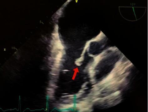
This diagnostic image is a transesophageal echocardiogram (TEE) demonstrating infective endocarditis of the mitral valve. The ultrasound shows a clear, irregular hyperechoic mass, indicated by a red arrow, which represents a vegetation attached to the valve apparatus. Specifically, the vegetation is localized to the lateral posterior (P2) segment of the mitral valve leaflet. Adjacent cardiac structures, including the mitral valve leaflets and chordae tendineae, are visible within the left heart chambers. The image illustrates the classic sonographic presentation of vegetations, which are hallmark findings in bacterial endocarditis (clinically associated here with Staphylococcus hominis). The scan provides critical diagnostic evidence for assessing valve morphology and the risk of embolic events. Educational focus includes the identification of valvular vegetations and understanding their typical location on the atrial side of atrioventricular valves in the context of systemic infection.
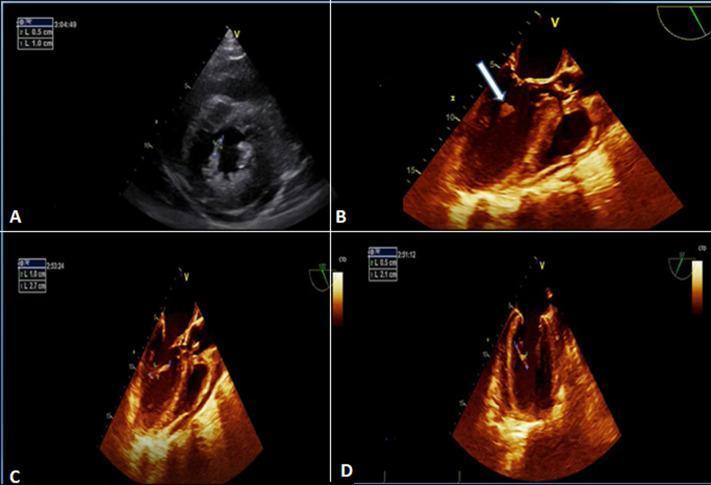
This composite of echocardiographic images demonstrates classic findings of infective endocarditis involving the mitral valve apparatus. Image A is a parasternal short-axis view from a transthoracic echocardiogram (TTE) showing an echogenic, textured mass consistent with vegetation located on the posteromedial mitral valve pillar. Images B, C, and D are transoesophageal echocardiograms (TEE) using a sepia-toned enhancement for improved tissue characterization. These views provide higher resolution of the vegetation, which appears as an irregular, hyper-echogenic, and frond-like mobile structure. In image B, an arrow specifically identifies the vegetation's attachment to the mitral valve apparatus. The diagnostic imaging highlights key features of valvular infection, including vegetation morphology, size assessment (noted in caliber measurements on the screen), and its anatomical relationship to the sub-valvular structures. This material is highly relevant for cardiology and infectious disease training regarding the diagnostic criteria for endocarditis.
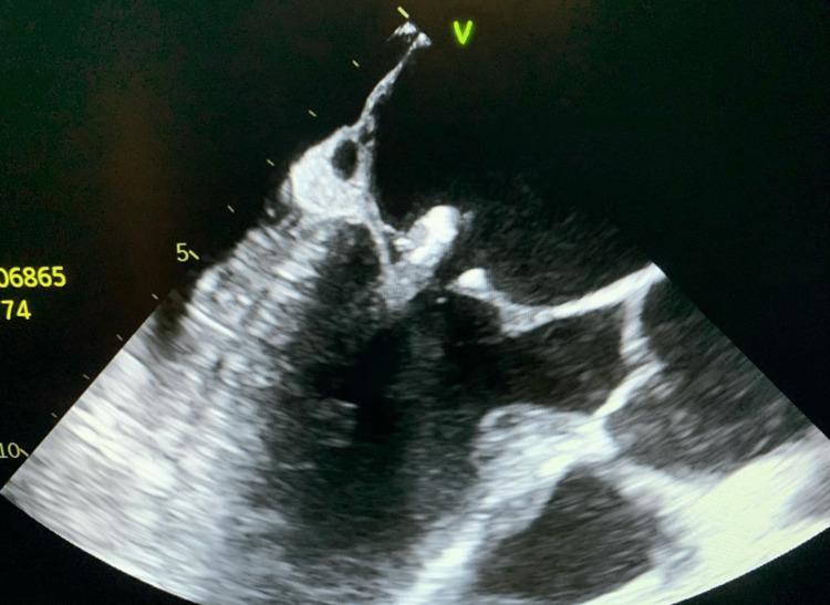
This diagnostic image is a transesophageal echocardiogram (TEE) demonstrating infective endocarditis of the mitral valve. The ultrasound shows a large, pedunculated, and irregularly shaped echogenic mass (vegetation) attached to the posterior mitral valve leaflet. The vegetation appears mobile, extending significantly into the heart chamber. The underlying mitral valve leaflets exhibit areas of increased echogenicity and thickening, consistent with valvular calcification and chronic degenerative changes, which may serve as a nidus for reinfection. This finding is clinically significant for the diagnosis of bacterial endocarditis, often necessitated when transthoracic echocardiography (TTE) is inconclusive. The image highlights the critical role of TEE in visualizing valvular vegetations, assessing their size (approximately 1.3 cm in this case), and identifying comorbid conditions like valve calcification in patients with positive blood cultures or suspected cardiac source of emboli.
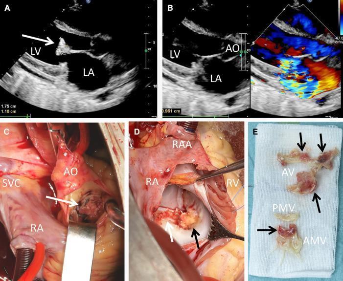
This composite figure presents clinical imaging and surgical findings of infective endocarditis. (A) Transthoracic echocardiogram (TTE) in a long-axis view shows a large, pedunculated echogenic vegetation (white arrow) measuring 1.75 cm x 1.10 cm attached to the anterior mitral valve leaflet. (B) TTE with color Doppler demonstrates a mosaic-pattern jet across the mitral valve, indicating severe mitral regurgitation. (C) Intraoperative view through the aorta (AO) reveals a yellowish vegetation (white arrow) infiltrating the aortic valve cusp. (D) Surgical exposure of the mitral valve via the right atrium (RA) shows extensive friable, yellowish vegetations (arrows) involving the anterior mitral cusp and annulus. (E) Gross specimen photograph displays the excised material on surgical gauze, with arrows pointing to the vegetations associated with the aortic valve (AV) and anterior mitral valve (AMV) tissue. The images collectively demonstrate the progression from diagnostic ultrasonography to surgical management of multi-valvular endocarditis, highlighting the morphology and destructive nature of infective vegetations.
tricuspid valve endocarditis vegetation IV drug user
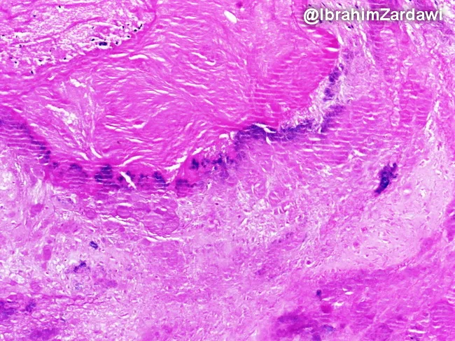
This slide is a histopathology image prepared from a cardiac valve vegetation in an intravenous drug user with infective endocarditis. The imaging modality is light microscopy of a formalin-fixed tissue section, stained with Hematoxylin and Eosin (H&E). The specimen type is a valvular vegetation consisting predominantly of a layered thrombus adherent to the valve surface, with bacterial colonies embedded within the fibrinous matrix. Under bright-field illumination, the thrombotic core appears pink to eosinophilic, while bacterial colonies stain dark purple to basophilic, forming discrete foci along the advancing edge of the vegetation. The surrounding tissue shows minimal to moderate inflammatory response, with neutrophils scattered at the periphery and scant residual myocardium at risk of ischemia. Overall architecture demonstrates an avascular, friable mass of fibrin, platelets, and microorganisms extending from the valvular surface into the endocardium. This histopathologic pattern supports a diagnosis of infective endocarditis, with organisms typical of IV drug users often Staphylococcus aureus or streptococcal species. The diagnostic significance lies in confirming infection of the endocardial surface and in guiding antimicrobial therapy, risk stratification, and surgical planning. Clinically, correlate with fever, positive blood cultures, echocardiography findings, and valve dysfunction. High-quality images enable teaching, reference, and research on endocarditis pathophysiology principles.
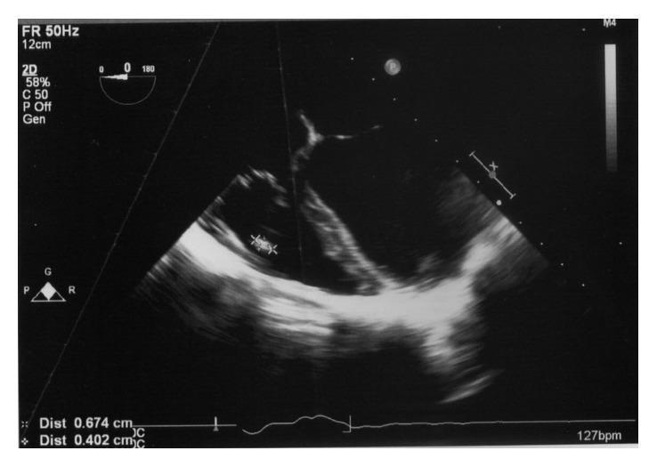
A grayscale diagnostic image from a transoesophageal echocardiogram (TOE) demonstrating a tricuspid valve vegetation in a patient with infective endocarditis. The image shows a mid-oesophageal four-chamber view focusing on the right side of the heart. A distinct, irregularly shaped, pedunculated mass measuring approximately 7 mm x 4 mm is visible, attached to the posterior leaflet of the tricuspid valve. This mass exhibits heterogeneous echogenicity, appearing more echodense than the surrounding blood pool but less uniform than the valve leaflet itself. Clinical significance includes the diagnosis of right-sided infective endocarditis, often associated in this context with pre-existing conditions like a ventricular septal defect (VSD). Key educational concepts illustrated include echocardiographic identification of valvular vegetations, assessment of mass size and mobility on heart valves, and the application of Duke's criteria for diagnosing endocarditis. The image provides critical visual evidence for managing structural heart disease and systemic infection.
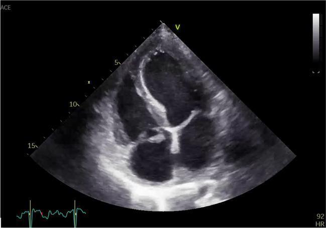
Transthoracic echocardiogram (TTE) in an apical four-chamber view demonstrating infective endocarditis of the tricuspid valve. The diagnostic image shows a prominent, irregularly shaped, and highly echogenic mass (vegetation) attached to the tricuspid valve leaflets. The vegetation exhibits a heterogeneous texture and appears to be broad-based with a possible stalk-like extension protruding into the right atrium. Notable features include thickening of the valve leaflets and evidence of valvular distortion or prolapse, characteristic of active endocarditis. The surrounding right heart chambers are visible, providing anatomical context for the right-sided valvular pathology. This image serves as a clinical example of the ultrasonic appearance of vegetations, crucial for the diagnosis of right-sided infective endocarditis, often associated with intravenous drug use or indwelling cardiac devices.
Infective Endocarditis with MR and TR
Definition & Overview
Valve Involvement and Relation to MR & TR
Frequency of valve involvement (ICE-PCS study, 2781 patients):
| Valve | Frequency |
|---|---|
| Mitral (most common left-sided) | 41% |
| Aortic | 38% |
| Tricuspid | 12% |
| Pulmonary | 1% |
- Braunwald's Heart Disease, p. 826
Why Regurgitant Valves Are Preferentially Infected
MR and Infective Endocarditis
MR as a Predisposing Factor
- Pre-existing MR is at least moderate severity, OR
- There is a flail mitral leaflet
IE Causing Acute MR
- Leaflet perforation by the vegetation
- Rupture of chordae tendineae from tissue destruction
- Annular abscess extending to undermine valve support
- Vegetation attached to leaflet, usually on atrial surface
- Flail leaflet or chordal rupture
- Color Doppler showing eccentric regurgitant jet
- Signs of acute LV volume overload
Tricuspid Regurgitation and Infective Endocarditis
TR as a Result of IE
- Direct leaflet destruction by the vegetation
- Perforation of tricuspid leaflets
- Chordal rupture
- S. aureus is the predominant organism (tricuspid valve involvement in 78% of IVDU cases, mitral in only 24%) - Goldman-Cecil, p. 1422
- Right-sided IE presents with septic pulmonary emboli - producing multiple cavitating pulmonary lesions, hemoptysis, and pleuritic chest pain
Primary vs Secondary TR in IE
- Primary TR (from IE): Direct valve infection and destruction, more common with IVDU, Staphylococcal endocarditis
- Secondary (functional) TR: After left-sided IE causes LV failure → pulmonary hypertension → RV dilation → annular dilation with functional TR - Miller's Anesthesia, Morgan & Mikhail
Clinical Features
Symptoms (Braunwald's):
| Symptom | % Affected |
|---|---|
| Fever | 80-95% |
| Chills | 40-70% |
| Weakness/malaise | 20-50% |
| Dyspnea | 20-40% |
| Stroke | 10-20% |
| Chest pain | 5-15% |
| Hemoptysis (right-sided) | 5-10% |
Physical Signs:
| Sign | % Affected |
|---|---|
| Fever | 80-90% |
| Heart murmur | 75-85% |
| New or changing murmur | 10-50% |
| Splenomegaly | 10-40% |
| Petechiae / conjunctival hemorrhage | 10-40% |
| Splinter hemorrhages | 5-15% |
| Janeway lesions (painless, palms/soles) | 5-10% |
| Osler nodes (painful, fingertips) | 3-10% |
| Roth spots | 2-10% |
Diagnosis: Modified Duke Criteria
Major Criteria
- Positive blood cultures - typical organisms (S. viridans, S. aureus, HACEK, Enterococcus) in ≥2 separate cultures; or persistently positive blood cultures ≥12h apart
- Echocardiographic evidence - oscillating intracardiac mass (vegetation), abscess, new partial dehiscence of prosthetic valve, or new valvular regurgitation (MR or TR)
Minor Criteria
- Predisposing heart condition or IVDU
- Fever ≥38°C
- Vascular phenomena (arterial emboli, septic pulmonary infarcts, Janeway lesions)
- Immunologic phenomena (Osler nodes, Roth spots, positive RF)
- Microbiologic evidence (positive blood culture not meeting major criteria)
Echocardiography
- TTE first for all suspected IE
- TEE when TTE is non-diagnostic, prosthetic valve is involved, or high clinical suspicion persists
- TEE is mandatory for intraoperative guidance during valve surgery for IE
- Vegetation (oscillating mass, independent motion from leaflet)
- New valvular regurgitation (MR, TR by color Doppler)
- Perivalvular abscess (especially aortic root)
- Fistulas, perforation, leaflet aneurysm
Microbiology
| Setting | Organism | Valve |
|---|---|---|
| Community-acquired (native valve) | Streptococcus viridans (50-60%) | Mitral/Aortic |
| Healthcare-associated / IVDU | S. aureus (most common overall in high-income countries) | Any, especially tricuspid (IVDU) |
| Elderly / GU procedures | Enterococcus faecalis | Mitral/Aortic |
| Subacute with large vegetations | HACEK organisms | Left-sided |
| Culture-negative | ~10% of all IE | - |
- Robbins & Kumar, p. 368-369; Braunwald's Heart Disease, p. 825-826
Management
Antibiotic Therapy
- Streptococcal NVE: Penicillin G or amoxicillin ± gentamicin for 4 weeks (2 weeks if susceptible)
- Staphylococcal NVE: Nafcillin/oxacillin (MSSA) or vancomycin (MRSA) × 6 weeks; right-sided IVDU IE may be treated with shorter 2-week course if uncomplicated
- Enterococcal: Ampicillin + gentamicin × 4-6 weeks
- HACEK: Ceftriaxone × 4 weeks
Indications for Surgery (AHA/ESC Guidelines)
- Acute severe MR or TR causing heart failure / cardiogenic shock / pulmonary edema refractory to medical therapy
- Valve dehiscence or obstruction causing hemodynamic instability
- Perivalvular abscess
- Heart failure secondary to new severe MR or TR
- Large vegetation (>10 mm) especially with embolism
- Recurrent emboli despite appropriate antibiotics
- Persistent bacteremia after 5-7 days of appropriate therapy
- Mobile vegetations >10 mm
Special Considerations for Right-sided IE (TR)
- Often managed medically (antibiotics alone) in IVDU with right-sided S. aureus IE without complications
- Surgery for TR in IE may involve tricuspid valve repair (valvuloplasty) rather than replacement to avoid prosthetic valve infection risk in ongoing IVDU
- Tricuspid valve excision (valvectomy) without replacement has been done in selected IVDU cases, taking advantage of the fact that TR is hemodynamically better tolerated than left-sided disease
Neurological Events and Surgery Timing
- Major ischemic stroke: delay valve surgery at least 4 weeks if clinically feasible (Class IIa)
- Intracranial hemorrhage: delay surgery at least 4 weeks
Complications
| Complication | Details |
|---|---|
| Acute MR | Leaflet perforation, chordal rupture → acute pulmonary edema, emergency surgery |
| Severe TR | Right heart failure, hepatic congestion, septic pulmonary emboli |
| Perivalvular abscess | Most common with aortic valve IE; causes heart block |
| Embolic stroke | 10-20% overall; higher with large/mobile vegetations |
| Septic pulmonary emboli | Right-sided IE; multiple cavitating lung lesions |
| Splenic/renal infarction | Left-sided emboli |
| Mycotic aneurysm | 2-4%; intracranial most dangerous |
| Renal failure | Immune complex deposition (focal GN), septic emboli, nephrotoxic drugs |
Prevention
- Prosthetic cardiac valves
- Previous IE
- Certain congenital heart defects (unrepaired cyanotic CHD, repaired CHD with residual defects)
- Cardiac transplant with valvulopathy
- Ventricular assist devices
Summary: IE, MR, and TR - Key Points
- MR predisposes to IE (especially MVP with MR/flail leaflet) - and IE causes acute MR through leaflet perforation or chordal rupture
- TR in IE is the hallmark of right-sided endocarditis, overwhelmingly in IVDU with S. aureus; presents with septic pulmonary emboli rather than systemic emboli
- New or changing MR/TR murmur is a major Duke criterion - always prompt echocardiographic evaluation
- Acute severe MR from IE = surgical emergency; severe TR from IE can sometimes be medically managed if right heart function is preserved
- A multispecialty heart valve team (ID, cardiology, cardiac surgery) is mandatory for all IE management