
Treatment for sprain
"sprains and strains"[MeSH Terms] AND "therapy"[Subheading]
PMID: 41023742
Treatment of a sprain (mainly ankle, the most common)
Immediate care: PRICE
- Ice: Apply for up to 20 minutes at a time, and don't leave it on longer, to avoid nerve injury (Swanson's Family Medicine Review).
- Compression: Use an elastic bandage or air-filled splint and wear it through the day to control swelling (Swanson's Family Medicine Review).
- Elevation: Keep the limb raised above heart level when possible.
- Pain control: Acetaminophen or an oral NSAID is usually enough. This is standard practice, not taken from the sources I pulled.
Treatment by grade (lateral ankle)
| Grade | Findings | Treatment |
|---|---|---|
| 1 (microscopic fiber tearing) | Minimal tenderness, little or no effusion, can bear weight within 24 hours | PRICE, weight bearing as tolerated, early range-of-motion and proprioceptive exercises |
| 2 (partial tear) | Moderate tenderness and hematoma, unable to bear weight for more than 24 hours | PRICE, air-cast immobilization, range-of-motion and proprioceptive exercises as tolerated |
| 3 (complete tear) | Large effusion, positive anterior drawer and talar tilt tests | PRICE, immobilization, delayed rehabilitation (prolonged), with or without delayed surgery |
Rehabilitation
- Early, functional rehabilitation is better than prolonged immobilization for most sprains.
- Bailey and Love's describes limited initial weight bearing, pain control and proprioceptive exercises. Peroneal strengthening and stretching follow once the pain allows.
- Physical therapy should focus on balance and proprioception (Miller's Review of Orthopaedics).
- A 2025 meta-analysis of 10 RCTs (565 patients) found physical therapy gave modestly better pain relief than conventional care for grade I and II sprains. The pain-score difference was small (WMD -0.46 on VAS), and the authors call for larger trials.
When to see a doctor or get imaging
- Ottawa ankle rules (from my own knowledge): get an X-ray if there is bone tenderness at the malleoli or the base of the 5th metatarsal or navicular, or if the patient can't take 4 steps.
- High ankle (syndesmosis) sprains, grade 3 sprains and associated fractures need orthopaedic input. Syndesmotic injury can significantly prolong recovery (Rosen's).
- Reassess if pain or instability persists beyond 6 to 8 weeks. Chronic instability may need surgical stabilization.
Other sites
Make a presentation on ankle sprain management
lateral ankle ligaments anatomy anterior talofibular calcaneofibular
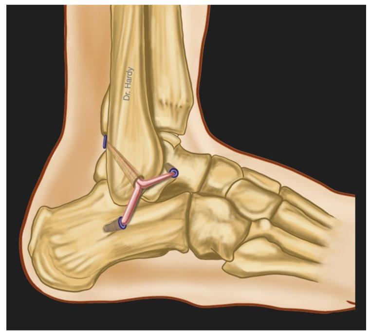
This medical anatomical diagram illustrates an anatomic surgical reconstruction of the lateral ankle ligament complex from a lateral view. The diagram depicts the skeletal structures of the distal fibula, talus, calcaneus, and tarsal bones. The primary focus is the surgical reconstruction of the three main lateral ligaments: the Anterior Talofibular Ligament (ATFL), the Calcaneofibular Ligament (CFL), and the Posterior Talofibular Ligament (PTFL). The reconstruction utilizes a split tendon autograft (such as a gracilis tendon) that is anchored to the distal fibula. The graft bifurcates and extends into three distinct limbs, which are secured into bone tunnels on the talus and calcaneus using blue-colored interference screws or anchors. This illustration demonstrates the surgical technique used to manage chronic lateral ankle instability by replicating the native anatomy of the lateral ligament complex to restore joint stability and biomechanical function.
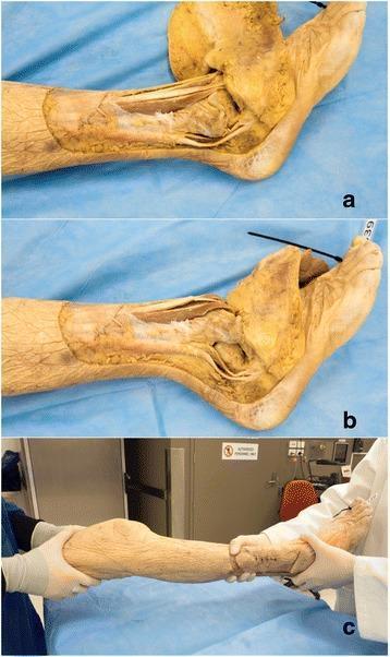
This composite of three clinical photographs displays a human cadaveric prosection used as an ankle dislocation and relocation model. Panels 'a' and 'b' show lateral views of a dissected lower leg and foot. In panel 'a', an extensive surgical incision around the medial and lateral malleoli reveals the underlying anatomy; the lateral ankle ligaments (anterior talofibular, calcaneofibular, and posterior talofibular) and joint capsule have been divided to destabilize the joint. Panel 'b' demonstrates the resulting ankle dislocation, showing a clear loss of congruity between the talus and the tibial mortise, with the foot displaced anteriorly and superiorly. Panel 'c' illustrates the clinical application, showing medical personnel in gloves performing a manual relocation procedure on the intact skin of the specimen. The series serves as an educational tool for orthopedic trainees to visualize the internal ligamentous disruption required for dislocation and the physical maneuvers necessary for reduction of the talocrural joint.
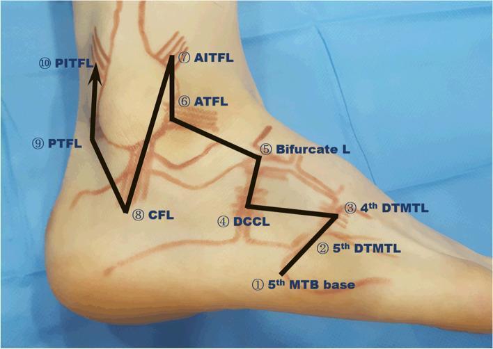
This clinical photograph illustrates the surface anatomy and systematic ultrasound examination protocol for the lateral aspect of the human ankle and foot. Bony landmarks and ligamentous structures are marked with skin-ink outlines, while a black zigzag arrow indicates a 10-step sequential scanning pathway. The numbered sequence identifies: (1) 5th metatarsal bone (MTB) base, (2) 5th and (3) 4th dorsal tarsometatarsal ligaments (DTMTL), (4) dorsal calcaneocuboid ligament (DCCL), (5) bifurcate ligament, (6) anterior talofibular ligament (ATFL), (7) anterior inferior tibiofibular ligament (AITFL), (8) calcaneofibular ligament (CFL), (9) posterior talofibular ligament (PTFL), and (10) posterior inferior tibiofibular ligament (PITFL). This educational resource demonstrates the clinical surface projection of the lateral ligament complex and syndesmotic structures, providing a standardized diagnostic algorithm for evaluating lateral ankle sprains and midtarsal joint injuries. The image serves as a guide for probe positioning during musculoskeletal ultrasonography to ensure comprehensive assessment of potential ligamentous pathology and occult avulsion fractures.
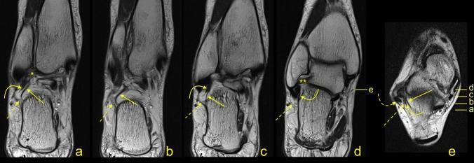
This diagnostic image set consists of MRI scans of a human ankle, including four oblique coronal-oblique sections (a–d) and one axial cross-section (e), illustrating the complex ligamentous anatomy of the lateral ankle compartment. The images focus on the Calcaneofibular Ligament (CFL), indicated by a curved yellow arrow, and its interconnections with surrounding structures. Key anatomical landmarks identified include the Fibulotalocalcaneal Ligament (FTCL, straight arrow), the Peroneal Tendon Sheath (PTS, dashed arrow), and the Superior Peroneal Retinaculum (SPR, dashed curved arrow). Additionally, the spatial relationship of the CFL with the Posterior Talofibular Ligament (PTFL, single asterisk *) and the Anterior Talofibular Ligament (ATFL, double asterisks **) is demonstrated. The axial section (e) serves as a reference for the levels of the oblique sections, progressing from distal (a) to proximal (d). This series highlights the structural continuity and fascial connections between these ligaments, which are critical for understanding ankle stability and pathology during diagnostic evaluation.
ls -la /tmp/workspace/ ; mkdir -p /tmp/workspace/ankle-sprain-ppt/assets && cd /tmp/workspace/ankle-sprain-ppt/assets && python /tmp/skills/shared/scripts/fetch_images.py "https://cdn.orris.care/cdss_images/a72598e0ba0c161ea2ad73b5d8ee6abade22d9f89d9f0522bd6d9457b5848f83.png" "https://cdn.orris.care/cdss_images/fc277906f802ef28f7a0b019e30f0f923014e463220543385c297b8fa016ab93.png" > imgs.json; python -c " import json,base64 d=json.load(open('imgs.json')) for i,x in enumerate(d): print(x['error'], x['base64'][:30] if x['base64'] else None) if x['base64']: open(f'img{i}.png','wb').write(base64.b64decode(x['base64'].split(',',1)[1])) "; python -c " from PIL import Image for f in ['img0.png','img1.png']: print(f, Image.open('/tmp/workspace/ankle-sprain-ppt/assets/'+f).size)"
mkdir -p /tmp/workspace/ankle-sprain-ppt/src /tmp/workspace/ankle-sprain-ppt/output && cat > /tmp/workspace/ankle-sprain-ppt/src/build.py <<'EOF' from pptx import Presentation from pptx.util import Inches, Pt from pptx.dml.color import RGBColor from pptx.enum.text import PP_ALIGN, MSO_ANCHOR from pptx.enum.shapes import MSO_SHAPE NAVY=RGBColor(0x0B,0x2A,0x3F); TEAL=RGBColor(0x12,0x7C,0x8A); ICE=RGBColor(0xE6,0xF2,0xF4) CORAL=RGBColor(0xE8,0x5D,0x4A); WHITE=RGBColor(255,255,255); INK=RGBColor(0x1F,0x2D,0x3A); GREY=RGBColor(0x5F,0x6F,0x7A) FONT="Calibri" A="/tmp/workspace/ankle-sprain-ppt/assets/" prs=Presentation(); prs.slide_width=Inches(13.333); prs.slide_height=Inches(7.5) blank=prs.slide_layouts[6] def rect(s,x,y,w,h,fill,shape=MSO_SHAPE.RECTANGLE): r=s.shapes.add_shape(shape,Inches(x),Inches(y),Inches(w),Inches(h)) r.fill.solid(); r.fill.fore_color.rgb=fill; r.line.fill.background(); r.shadow.inherit=False return r def text(s,x,y,w,h,t,size=18,color=INK,bold=False,align=PP_ALIGN.LEFT,anchor=MSO_ANCHOR.TOP): tb=s.shapes.add_textbox(Inches(x),Inches(y),Inches(w),Inches(h)); tf=tb.text_frame; tf.word_wrap=True tf.vertical_anchor=anchor tf.margin_left=tf.margin_right=Inches(0.05); tf.margin_top=tf.margin_bottom=Inches(0.03) lines=t if isinstance(t,list) else [t] for i,l in enumerate(lines): p=tf.paragraphs[0] if i==0 else tf.add_paragraph() p.alignment=align; r=p.add_run(); r.text=l r.font.size=Pt(size); r.font.bold=bold; r.font.color.rgb=color; r.font.name=FONT return tb def bullets(s,x,y,w,h,items,size=18,color=INK,gap=8): tb=s.shapes.add_textbox(Inches(x),Inches(y),Inches(w),Inches(h)); tf=tb.text_frame; tf.word_wrap=True tf.margin_left=tf.margin_right=Inches(0.05) for i,it in enumerate(items): p=tf.paragraphs[0] if i==0 else tf.add_paragraph() p.space_after=Pt(gap) if isinstance(it,tuple): r=p.add_run(); r.text=it[0]+" "; r.font.bold=True; r.font.size=Pt(size); r.font.color.rgb=TEAL; r.font.name=FONT r=p.add_run(); r.text=it[1]; r.font.size=Pt(size); r.font.color.rgb=color; r.font.name=FONT else: r=p.add_run(); r.text=it; r.font.size=Pt(size); r.font.color.rgb=color; r.font.name=FONT return tb def header(s,title,sub=None): rect(s,0,0,13.333,1.15,NAVY); rect(s,0,1.15,13.333,0.06,CORAL) text(s,0.6,0.15,12,0.85,title,32,WHITE,True,anchor=MSO_ANCHOR.MIDDLE) if sub: text(s,0.6,1.3,12,0.45,sub,16,GREY) def footer(s,src,n): text(s,0.6,7.05,11.5,0.35,src,11,GREY) text(s,12.2,7.05,0.6,0.35,str(n),11,GREY,align=PP_ALIGN.RIGHT) def notes(s,t): s.notes_slide.notes_text_frame.text=t def card(s,x,y,w,h,title,body,accent=TEAL,size=15): rect(s,x,y,w,h,ICE); rect(s,x,y,0.09,h,accent) text(s,x+0.25,y+0.1,w-0.4,0.45,title,18,NAVY,True) bullets(s,x+0.25,y+0.6,w-0.4,h-0.7,body,size,gap=4) # 1 Title s=prs.slides.add_slide(blank); rect(s,0,0,13.333,7.5,NAVY); rect(s,0.8,3.55,1.6,0.08,CORAL) text(s,0.8,1.7,11,1.8,["Ankle Sprain","Management"],54,WHITE,True) text(s,0.8,3.85,11,0.6,"Assessment, acute care, rehabilitation and when to escalate",22,RGBColor(0xCA,0xE6,0xEA)) text(s,0.8,6.5,11,0.4,"For clinical education. Based on Rosen's Emergency Medicine, Textbook of Family Medicine 9e, Bailey & Love 28e, Miller's Review of Orthopaedics 9e",12,RGBColor(0x9F,0xB8,0xC2)) notes(s,"Overview of ankle sprain management from assessment through rehabilitation, drawn from standard textbooks plus a 2025 meta-analysis.") # 2 Anatomy s=prs.slides.add_slide(blank); header(s,"Anatomy and epidemiology") s.shapes.add_picture(A+"img0.png",Inches(0.6),Inches(1.55),height=Inches(5.3)) text(s,0.6,6.85,5.6,0.25,"Lateral ankle ligaments (Textbook of Family Medicine 9e, Fig. 30-47)",10,GREY) bullets(s,6.8,1.6,6.0,5.3,[ ("85%","of ankle sprains involve the lateral ligaments; medial and syndesmotic sprains make up the other 15%."), ("ATFL:","restricts anterior talar motion. Injured first and most often (about two thirds are isolated ATFL tears)."), ("CFL:","restricts inversion. Injured with ATFL in about 20%."), ("PTFL:","strongest of the three, rarely involved."), ("Deltoid:","medial, under 5% of sprains, often with a lateral malleolar fracture."), ("Syndesmosis:","10 to 20% of sprains in competitive athletes ('high' ankle sprain)."), ],17,gap=10) footer(s,"Sources: Textbook of Family Medicine 9e p. 838-839; Rosen's Emergency Medicine, Ch. 49; Bailey & Love 28e p. 526",2) notes(s,"Most sprains are inversion injuries with the ankle plantarflexed, injuring ATFL first. Dorsiflexed inversion tends to injure the CFL.") # 3 Mechanism & grading s=prs.slides.add_slide(blank); header(s,"Mechanism and grading","Inversion with plantar flexion is the classic mechanism") cols=[("Grade 1","Microscopic fiber tearing",["Minimal ATFL tenderness","Little or no effusion","Weight bearing at once or within 24 h"],TEAL), ("Grade 2","Partial tear",["Moderate tenderness, hematoma","Small to moderate effusion","Cannot bear weight for over 24 h"],RGBColor(0xE0,0x9A,0x2B)), ("Grade 3","Complete tear",["Marked tenderness, hematoma","Large effusion","Positive anterior drawer and talar tilt"],CORAL)] for i,(g,p,items,c) in enumerate(cols): x=0.6+i*4.1 rect(s,x,2.0,3.85,1.2,c); text(s,x+0.2,2.05,3.4,0.6,g,28,WHITE,True); text(s,x+0.2,2.65,3.4,0.45,p,17,WHITE) rect(s,x,3.2,3.85,2.9,ICE); bullets(s,x+0.2,3.4,3.5,2.6,items,17,gap=12) text(s,0.6,6.3,12,0.6,"Grading is clinical and guides treatment. Function (weight bearing, stability) matters more than the label.",16,NAVY,True) footer(s,"Source: Rosen's Emergency Medicine, Table 49.1",3) notes(s,"Grade classification from Rosen Table 49.1.") # 4 Assessment s=prs.slides.add_slide(blank); header(s,"Clinical assessment") card(s,0.6,1.6,6.0,2.5,"History",["Inversion injury, often a felt 'roll' or pop","Swelling, bruising tracking down the foot","Ability to bear weight immediately"],TEAL,16) card(s,6.8,1.6,6.0,2.5,"Inspection and palpation",["Observe gait, look for a limp","Palpate lateral ligaments, both malleoli, 5th metatarsal base, navicular, proximal fibula","Compare with the other ankle"],TEAL,16) card(s,0.6,4.3,6.0,2.6,"Stability tests",["Anterior drawer: ATFL (excess anterior talar translation)","Talar tilt: ATFL in plantar flexion, CFL in slight dorsiflexion","Best done after swelling settles (days 4 to 7)"],CORAL,16) card(s,6.8,4.3,6.0,2.6,"Look for the mimics",["Syndesmosis: squeeze and external rotation tests","Fractures: malleoli, lateral process of talus, 5th metatarsal","Peroneal tendon injury, osteochondral talar lesion"],CORAL,16) footer(s,"Sources: Bailey & Love 28e p. 526; Textbook of Family Medicine 9e. Timing of stability tests is standard practice",4) notes(s,"The delayed-examination timing is general clinical practice rather than from the cited textbook passages.") # 5 Imaging s=prs.slides.add_slide(blank); header(s,"Imaging: Ottawa ankle rules","Order an ankle X-ray only if one of these is present") rect(s,0.6,1.9,7.4,4.9,ICE) text(s,0.9,2.0,6.8,0.5,"Ankle series if:",20,NAVY,True) bullets(s,0.9,2.6,6.9,2.0,["Bone tenderness at the posterior edge or tip of the lateral malleolus (distal 6 cm)","Bone tenderness at the posterior edge or tip of the medial malleolus (distal 6 cm)","Unable to bear weight at injury or in the clinic"],17,gap=10) text(s,0.9,4.7,6.8,0.5,"Foot series if:",20,NAVY,True) bullets(s,0.9,5.2,6.9,1.4,["Tenderness at the base of the 5th metatarsal or at the navicular","Unable to bear weight"],17,gap=10) rect(s,8.4,1.9,4.35,4.9,NAVY) text(s,8.65,2.0,3.9,0.5,"Beyond plain films",20,WHITE,True) bullets(s,8.65,2.6,3.9,4.1,["External rotation view if a high ankle sprain is suspected","MRI is not routine. Use it for chronic pain or instability: osteochondral lesions, tendon injury, impingement, syndesmosis injury","Rules have very high sensitivity, so a negative result reliably excludes a fracture"],15,WHITE,gap=10) footer(s,"Sources: Bailey & Love 28e Table 36.1; Rheumatology 2022 Box 84.1. Sensitivity comment from general knowledge",5) notes(s,"Ottawa rules per Bailey & Love Table 36.1.") # 6 Acute PRICE s=prs.slides.add_slide(blank); header(s,"Acute management: PRICE","First 24 to 72 hours") items=[("P","Protection","Brace, air-cast or elastic wrap; crutches if walking hurts"), ("R","Rest","Relative rest. Limit weight bearing early, then progress"), ("I","Ice","Up to 20 minutes at a time. Longer risks nerve injury"), ("C","Compression","Elastic bandage or air splint, worn through the day"), ("E","Elevation","Raise the foot above heart level to reduce swelling")] for i,(l,t,d) in enumerate(items): y=1.55+i*1.0 c=rect(s,0.8,y,0.8,0.8,CORAL,MSO_SHAPE.OVAL); text(s,0.8,y,0.8,0.8,l,30,WHITE,True,PP_ALIGN.CENTER,MSO_ANCHOR.MIDDLE) text(s,1.9,y,2.5,0.8,t,22,NAVY,True,anchor=MSO_ANCHOR.MIDDLE) text(s,4.3,y,4.6,0.8,d,15,INK,anchor=MSO_ANCHOR.MIDDLE) rect(s,9.3,1.55,3.5,4.8,ICE); rect(s,9.3,1.55,3.5,0.08,TEAL) text(s,9.5,1.75,3.1,0.5,"Pain relief",20,NAVY,True) bullets(s,9.5,2.3,3.1,4.0,["Acetaminophen or an oral NSAID is usually enough","Check contraindications (renal, GI, anticoagulants)","Avoid heat and alcohol early, as they increase swelling"],15,gap=10) text(s,0.8,6.55,12,0.4,"Early protected movement beats prolonged immobilization for most sprains.",16,NAVY,True) footer(s,"Sources: Rosen's Table 49.1; Swanson's Family Medicine Review; Miller's Review of Orthopaedics. Analgesia advice is standard practice",6) notes(s,"PRICE appears in Rosen, Miller, Bailey and Love and Swanson. Analgesic detail is general practice.") # 7 Management by grade s=prs.slides.add_slide(blank); header(s,"Treatment by grade") rows=[("Grade","Immobilization / support","Weight bearing","Rehabilitation"), ("1","PRICE, simple support","As tolerated","Early range of motion, proprioceptive and functional exercises"), ("2","PRICE, air-cast","Protected, then progress","Range of motion and proprioceptive exercises as tolerated"), ("3","PRICE, immobilization","Limited initially","Delayed, longer program. Delayed surgery in some")] tbl=s.shapes.add_table(4,4,Inches(0.6),Inches(1.7),Inches(12.1),Inches(3.6)).table for j,w in enumerate([1.3,3.4,2.8,4.6]): tbl.columns[j].width=Inches(w) for i,r in enumerate(rows): for j,v in enumerate(r): c=tbl.cell(i,j); c.text=v; p=c.text_frame.paragraphs[0]; p.runs[0].font.size=Pt(17 if i else 18); p.runs[0].font.name=FONT p.runs[0].font.bold=(i==0 or j==0); c.vertical_anchor=MSO_ANCHOR.MIDDLE c.fill.solid() if i==0: c.fill.fore_color.rgb=NAVY; p.runs[0].font.color.rgb=WHITE else: c.fill.fore_color.rgb=ICE if i%2 else WHITE; p.runs[0].font.color.rgb=INK rect(s,0.6,5.6,12.1,1.2,NAVY) text(s,0.9,5.65,11.5,1.1,"Most sprains do well without surgery. Many patients return to sport within a few weeks, depending on severity.",19,WHITE,anchor=MSO_ANCHOR.MIDDLE) footer(s,"Sources: Rosen's Emergency Medicine Table 49.1; Bailey & Love 28e p. 527. Weight-bearing wording condensed",7) notes(s,"Table adapted from Rosen Table 49.1.") # 8 Rehab s=prs.slides.add_slide(blank); header(s,"Rehabilitation phases","General framework; progress by function, not the calendar") ph=[("1","Protect","Days 0 to 3","Control pain and swelling. Protected weight bearing. Gentle range of motion."), ("2","Restore motion","Days 3 to 14","Full range of motion, normal gait, start isometric work."), ("3","Strengthen","Weeks 2 to 6","Peroneal strengthening, calf work, stretching, balance and proprioception."), ("4","Return to activity","Weeks 4 to 8+","Sport-specific drills, hop tests, brace or tape for return to sport.")] for i,(n,t,d,b) in enumerate(ph): x=0.6+i*3.1 rect(s,x,1.9,2.9,4.6,ICE); rect(s,x,1.9,2.9,1.3,TEAL if i<3 else CORAL) text(s,x+0.15,1.95,0.7,1.2,n,44,WHITE,True,anchor=MSO_ANCHOR.MIDDLE) text(s,x+0.8,2.0,2.05,0.6,t,19,WHITE,True,anchor=MSO_ANCHOR.MIDDLE); text(s,x+0.8,2.6,2.05,0.5,d,14,WHITE) text(s,x+0.2,3.4,2.5,3.0,b,16,INK) text(s,0.6,6.6,12.2,0.4,"Supervised physical therapy gave modestly better pain relief in grade 1 and 2 sprains (2025 meta-analysis, 10 RCTs, n=565).",14,NAVY,True) footer(s,"Sources: Bailey & Love 28e; Miller's Review; Yang 2025 (PMID 41023742). Time frames are typical, not textbook-specified",8) notes(s,"Phase timings are a typical clinical framework. The meta-analysis showed WMD -0.46 on VAS; effect is small, authors call for larger trials.") # 9 Special sprains s=prs.slides.add_slide(blank); header(s,"Medial and high ankle sprains") card(s,0.6,1.6,6.0,4.9,"Syndesmotic (high) sprain",["Dorsiflexion plus external rotation","10 to 20% of sprains in competitive athletes","Pain above the ankle, positive squeeze and external rotation tests","Recovery is significantly longer","Unstable or widened mortise: orthopaedic referral, possible fixation"],CORAL,17) card(s,6.8,1.6,6.0,4.9,"Medial (deltoid) sprain",["Eversion force, under 5% of sprains","Usually with a lateral malleolar fracture","Medial plus lateral ligament injury or ecchymosis suggests bimalleolar instability","Needs imaging and often orthopaedic input","Isolated mild injury: PRICE and functional rehab"],TEAL,17) footer(s,"Sources: Rosen's Emergency Medicine; Textbook of Family Medicine 9e (Syndesmosis and Medial Ankle Sprains)",9) notes(s,"Management of grade 1 to 2 medial sprains follows the same functional principles.") # 10 Escalation s=prs.slides.add_slide(blank); header(s,"Refer, image or operate") card(s,0.6,1.6,6.0,2.5,"Escalate early",["Fails Ottawa rules, or suspected fracture","Syndesmosis or bimalleolar injury","Neurovascular compromise","Grade 3 with gross instability"],CORAL,16) card(s,6.8,1.6,6.0,2.5,"Reassess if",["Pain or swelling persists beyond 6 to 8 weeks","Recurrent giving way","Locking, catching or deep pain (osteochondral lesion)"],TEAL,16) rect(s,0.6,4.35,12.2,2.45,NAVY) text(s,0.9,4.45,11.6,0.5,"Surgery",22,WHITE,True) bullets(s,0.9,5.0,11.6,1.8,["Indicated for persistent pain and recurrent sprains (chronic ankle instability)","Anatomic repair: imbrication of the attenuated ATFL, with or without inferior extensor retinaculum reinforcement","Non-anatomic reconstruction techniques exist for chronic instability"],16,WHITE,gap=6) footer(s,"Sources: Bailey & Love 28e p. 527; Miller's Review; Rheumatology 2022",10) notes(s,"Referral thresholds combine textbook content with general clinical practice.") # 11 Takeaways s=prs.slides.add_slide(blank); rect(s,0,0,13.333,7.5,NAVY); rect(s,0.8,1.5,1.2,0.08,CORAL) text(s,0.8,0.6,11,0.8,"Key points",38,WHITE,True) bullets(s,0.8,1.9,11.7,4.5,[ "Most sprains are lateral inversion injuries involving the ATFL. Grade by function and stability.", "Use the Ottawa ankle rules to decide on X-ray. Do not image everyone.", "Start PRICE and analgesia at once, then move to early protected motion.", "Rehabilitation (range of motion, peroneal strength, balance) prevents re-injury.", "Think syndesmosis, medial ligament and fracture when the picture does not fit.", "Surgery is reserved for persistent pain or recurrent instability."],20,WHITE,gap=14) text(s,0.8,6.6,11.7,0.6,"Key references: Rosen's Emergency Medicine Ch. 49; Textbook of Family Medicine 9e Ch. 30; Bailey & Love 28e Ch. 36; Miller's Review of Orthopaedics 9e; Yang C et al. J Orthop Surg Res 2025 (PMID 41023742)",11,RGBColor(0x9F,0xB8,0xC2)) notes(s,"Summary slide.") prs.save("/tmp/workspace/ankle-sprain-ppt/output/Ankle_Sprain_Management.pptx") EOF cd /tmp/workspace/ankle-sprain-ppt && python src/build.py && ls -la output
Ankle Sprain Management
Presentation · PPTX
- Title
- Anatomy and epidemiology, with the lateral ligament diagram from Textbook of Family Medicine 9e
- Mechanism and grades 1 to 3
- Clinical assessment, including the anterior drawer and talar tilt tests and conditions that mimic a sprain
- Imaging and the Ottawa ankle rules
- Acute management with PRICE and pain relief
- Treatment by grade, in a table
- Four rehabilitation phases
- Syndesmotic (high) and medial (deltoid) sprains
- When to refer, when to reassess, and surgery for chronic instability
- Key points and references
I suggest relispray
Relispray composition uses topical spray
- The US DailyMed listing gives the active ingredient as menthol 4% w/w, a topical analgesic (counterirritant) labelled for minor pain from sprains, strains, arthritis and backache.
- The Indian range is marketed as an Ayurvedic, drug-free spray, and there is also a Nitron variant with an "icepac" cooling action. I couldn't verify the ingredient lists of the Indian variants, so check the label on the pack you're using.
- Menthol gives a cooling sensation that relieves pain. It doesn't treat the torn ligament, reduce swelling or speed healing.
- From my own knowledge, not the textbooks I searched: topical NSAIDs such as diclofenac gel have better trial evidence than menthol or other counterirritants for acute sprain pain. If the user wants a topical, a topical NSAID is the better-supported option.
- It can be used on top of oral analgesia, ice and rehab. Don't let it delay an Ottawa-rule X-ray or referral.
- For external use only. Avoid the eyes, mucous membranes, wounds and irritated skin.
- Do not use it with a heating pad, and do not bandage over it. That conflicts with the compression step in PRICE, so let the spray dry fully and don't wrap tightly over freshly sprayed skin.
- It is flammable and under pressure. Keep it away from flame and heat.
- Stop if the skin becomes irritated.