
Lower Limb high yield summary
lower limb nerve supply lumbosacral plexus diagram
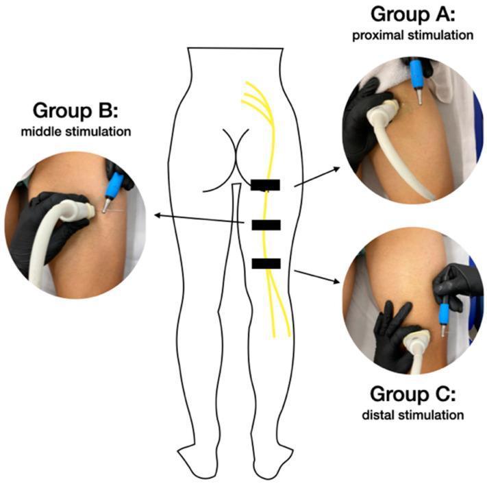
This composite educational image illustrates ultrasound-guided percutaneous neuromodulation of the sciatic nerve. Centrally, a schematic diagram of the posterior human lower body shows the lumbosacral plexus and the vertical descent of the sciatic nerve down the right thigh. Three black rectangular markers indicate the study's target stimulation zones: proximal (Group A), middle (Group B), and distal (Group C). Flanking the diagram are clinical photographs demonstrating the procedural setup for each group. Each photograph shows a clinician's gloved hands performing the technique on a patient in the prone position. A high-frequency linear ultrasound transducer is held in a transverse orientation to locate the sciatic nerve, while an acupuncture needle with a blue-handled applicator is inserted using a long-axis approach to reach the perineurium. The primary educational focus is the anatomical positioning and procedural alignment required for targeted electrical stimulation of the sciatic nerve at varying levels of the posterior thigh for clinical intervention.
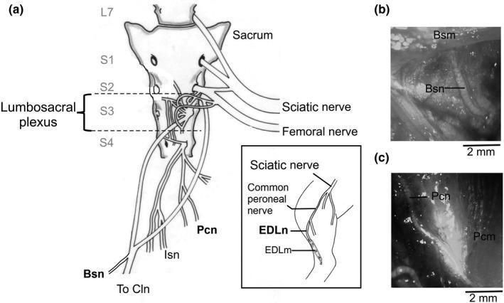
This composite educational graphic details the neuroanatomy of the lumbosacral plexus and associated pelvic floor nerves, using a female rabbit model as a translational surrogate. Panel (a) presents a schematic anatomical diagram of the sacrum and lumbosacral plexus, highlighting the plexus's origin between vertebral levels S2 and S4. Key motor nerves depicted include the bulbospongiosus nerve (Bsn) and the pubococcygeus nerve (Pcn), which are critical for pelvic floor function. An inset illustrates the extensor digitorum longus nerve (EDLn) branching from the sciatic nerve system, serving as a motor nerve control. Panels (b) and (c) provide macro-photographs of surgical dissections, showing the physical morphology of the Bsn and Pcn as elongated, light-colored fibers situated near their respective muscle insertions (Bsm and Pcm). Scale bars of 2 mm are provided for anatomical reference. The content is designed to illustrate the structural pathways and clinical relevance of pelvic innervation for studies regarding pelvic floor dysfunction and neuromuscular outcomes.
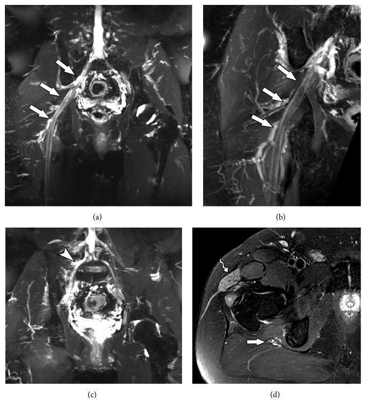
This set of four Magnetic Resonance Neurography (MRN) images illustrates post-traumatic lumbosacral plexus injury and associated muscle denervation. (a) Coronal STIR 3D SPACE and (b) Coronal T2 with fat suppression demonstrate diffuse thickening and increased signal intensity of the right sciatic nerve (white arrows) extending from the lumbosacral plexus to the thigh. (c) Coronal STIR 3D SPACE shows focal thickening of a lumbar plexus trunk (white arrowhead), consistent with neural stretching or traction injury. (d) Axial T2-weighted image reveals diffuse muscle edema, characterized by hyperintense signal within the tensor fascia lata (wavy arrow) and gluteus minimus (star) muscles. This pattern of muscular edema is a secondary sign of acute denervation or neuropathy involving the superior gluteal nerve branches. These diagnostic images are clinically relevant for evaluating peripheral nerve trauma and distinguishing between structural nerve damage and secondary muscular changes in patients presenting with lower limb weakness or drop foot.
knee joint ligaments meniscus anatomy
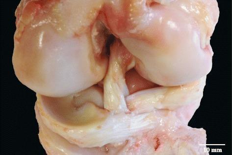
A clinical photograph of a dissected porcine (pig) stifle joint, serving as a comparative anatomical model for the human knee. The image illustrates the complex spatial relationship between the cruciate ligaments and meniscal attachments. Specifically, it demonstrates the two distinct fiber bundles of the anterior cruciate ligament (ACL) which are physically separated by the interposition of the anterior horn of the lateral meniscus. The ACL bundles exhibit a characteristic white-to-yellowish fibrous texture, while the meniscal tissue appears more translucent and slightly yellow. The underlying tibial plateau and femoral condyles are visible, providing anatomical context for the ligamentous insertions. This visual resource is used to teach comparative anatomy and to understand the variations in ligamentous and fibrocartilaginous arrangements within mammalian joints, highlighting the structural differences in meniscal anchoring between species.
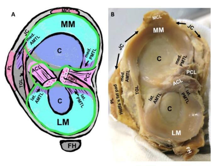
Educational visual showing the gross anatomy and schematic representation of the human tibial plateau and its intraarticular structures. The image is divided into a labeled schematic (A) and a corresponding clinical photograph of a cadaveric specimen (B), demonstrating the spatial relationships of the knee joint. Key anatomical features identified include the medial meniscus (MM) and lateral meniscus (LM) resting on the tibial articular cartilage (C). The cruciate ligaments—anterior (ACL) and posterior (PCL)—are shown centrally. Detailed focus is given to the meniscotibial ligaments, specifically the medial and lateral anterior meniscotibial ligaments (AMTL) and posterior meniscotibial ligaments (PMTL), which provide elastic fixation for the meniscal horns. Additional structures visible include the transverse genicular ligament (TGL) connecting the menisci, the medial collateral ligament (MCL), the joint capsule (JC), and the fibular head (FH). The schematic further illustrates the synovial membrane (green line) and its relation to the joint cavity. This material is designed to teach orthopedic anatomy, emphasizing the complex ligamentous network supporting knee joint stability and the entheses involved in degenerative conditions like osteoarthritis.
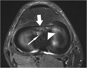
This diagnostic image is an axial proton-density-weighted, fat-suppressed fast spin echo (FSE) MRI of a human knee joint. The imaging modality highlights musculoskeletal structures by suppressing high-signal fat, allowing for detailed visualization of ligaments and meniscal attachments. The image demonstrates three key anatomical features: the anterior intermeniscal (transverse) ligament, indicated by a thick white arrow, which appears as a hypointense band connecting the anterior horns of the menisci; the anterior root of the medial meniscus, marked by a thin white arrow; and the anterior root of the lateral meniscus, indicated by a white arrowhead. These structures exhibit characteristic low signal intensity against the intermediate signal of surrounding tissues. The scan illustrates normal meniscal anatomy and root insertions, which are critical for maintaining meniscal stability and load distribution within the knee. This high-resolution axial section is essential for identifying meniscal root tears or variations in the intermeniscal ligament that could be misinterpreted as pathology in other planes.
Lower Limb - High Yield Anatomy Summary
1. REGIONS & AREAS OF TRANSITION
- Gluteal region - posterolateral, between iliac crest and gluteal fold
- Thigh - inguinal ligament to knee (anterior); gluteal fold to knee (posterior)
- Leg - knee to ankle
- Foot - distal to ankle joint
- Femoral triangle (proximal thigh) - entry point for femoral nerve, artery, vein from abdomen
- Popliteal fossa (posterior knee) - diamond-shaped; major vessels/nerves pass between thigh and leg
- Tarsal tunnel (posteromedial ankle) - formed by bone + flexor retinaculum; transmits flexor tendons, tibial nerve, and posterior tibial vessels
2. BONES
Femur
- Longest bone in the body
- Neck-shaft angle: ~126° (coxa valga if >130°, coxa vara if <120°)
- Greater trochanter - attachment of gluteus medius, minimus, and short lateral rotators
- Lesser trochanter - attachment of iliopsoas
- Ischial tuberosity landmarks:
- Upper medial: origin of semitendinosus + long head of biceps femoris
- Upper lateral: origin of semimembranosus
- Lower lateral: part of adductor magnus
Patella
- Largest sesamoid bone; embedded in quadriceps tendon
- Directs quadriceps pull anteriorly over the knee without tendon wear
3. MUSCLE COMPARTMENTS
| Region | Compartment | Key Muscles | Nerve | Action |
|---|---|---|---|---|
| Thigh | Anterior | Quadriceps femoris (rectus femoris, vastus medialis/lateralis/intermedius), sartorius, iliopsoas | Femoral (L2-L4) | Knee extension, hip flexion |
| Thigh | Medial | Adductors (longus, brevis, magnus), gracilis, pectineus | Obturator (L2-L4); pectineus by femoral; ischial part of adductor magnus by tibial division of sciatic | Adduction |
| Thigh | Posterior | Hamstrings (biceps femoris long head, semitendinosus, semimembranosus) | Tibial part of sciatic (L4-S3) | Hip extension, knee flexion |
| Gluteal | - | Gluteus maximus, medius, minimus; piriformis; obturator internus/externus; gemelli; quadratus femoris | Inferior gluteal (gluteus maximus); Superior gluteal (gluteus medius, minimus, TFL) | Extension/abduction/rotation |
| Leg | Anterior | Tibialis anterior, extensor hallucis longus, extensor digitorum longus, fibularis tertius | Deep fibular (peroneal) nerve | Dorsiflexion, toe extension |
| Leg | Lateral | Fibularis longus, fibularis brevis | Superficial fibular nerve | Eversion, weak plantarflexion |
| Leg | Posterior (superficial) | Gastrocnemius, soleus, plantaris | Tibial nerve (S1-S2) | Plantarflexion |
| Leg | Posterior (deep) | Tibialis posterior, flexor digitorum longus, flexor hallucis longus, popliteus | Tibial nerve | Inversion, toe flexion |
- Anterior thigh = SARVIS (Sartorius, Rectus femoris, Vastus lateralis/intermedius/medialis, Iliopsoas)
- Hamstrings = "Pes Anserinus" at knee: Sartorius, Gracilis, Semitendinosus (all insert via pes anserinus on medial tibia)
4. NERVE SUPPLY - LUMBOSACRAL PLEXUS
Major Nerves Summary
| Nerve | Root | Motor | Sensory |
|---|---|---|---|
| Femoral | L2-L4 | Anterior compartment of thigh + iliacus, pectineus | Anterior thigh, anteromedial knee, medial leg, medial foot |
| Obturator | L2-L4 | Medial compartment (except pectineus, ischial part adductor magnus) + obturator externus | Medial upper thigh |
| Sciatic | L4-S3 | Posterior thigh, ALL leg and foot muscles | Lateral leg, lateral and plantar foot |
| Superior gluteal | L4-S1 | Gluteus medius, minimus, TFL | None |
| Inferior gluteal | L5-S2 | Gluteus maximus | None |
| Lat. cutaneous n. of thigh | L2-L3 | None | Lateral thigh |
| Posterior cutaneous n. of thigh | S1-S3 | None | Posterior thigh, gluteal fold |
Sciatic Nerve - Key Points
- Largest nerve in the body (L4-S3)
- Exits pelvis via greater sciatic foramen, inferior to piriformis
- Divides into:
- Tibial nerve (anterior divisions L4-S3) - posterior compartments
- Common fibular (peroneal) nerve (posterior divisions L4-S2) - anterior and lateral compartments of leg
Common Fibular Nerve - Clinically Important
- Wraps around the neck of the fibula - can be injured by:
- Fractures of the fibular head/neck
- Plaster casts placed too high
- Direct trauma
- Injury causes foot drop (loss of dorsiflexion + eversion) + sensory loss over lateral leg and dorsum of foot
Femoral Nerve Injury
- Loses knee extension (quadriceps) - unable to climb stairs or kick
- Loss of knee jerk reflex (L3-L4)
- Sensory loss over anterior thigh, medial leg/foot
5. DERMATOMES (Autonomous Zones)
| Spinal Level | Test Area |
|---|---|
| L1 | Skin over inguinal ligament |
| L2 | Lateral side of thigh |
| L3 | Lower medial thigh |
| L4 | Medial side of great toe (digit I) |
| L5 | Medial side of digit II (web space) |
| S1 | Little toe (digit V) |
| S2 | Back of thigh |
| S3 | Skin over gluteal fold |
6. MYOTOMES & REFLEXES
| Movement | Level |
|---|---|
| Hip flexion | L1-L2 |
| Knee extension | L3-L4 |
| Knee flexion | L5-S2 |
| Plantarflexion | S1-S2 |
| Digit adduction | S2-S3 |
| Knee jerk (patellar reflex) | L3-L4 |
| Ankle jerk (Achilles reflex) | S1-S2 |
7. HIP JOINT
- Type: Multiaxial ball-and-socket synovial joint
- Stability > Mobility (compared to shoulder)
- Articular surfaces: spherical head of femur + lunate surface of acetabulum
- Acetabular labrum - fibrocartilage rim that deepens the socket
- Transverse acetabular ligament - bridges the acetabular notch inferiorly
- Ligament of the head of femur (ligamentum teres) - carries small branch of obturator artery to femoral head; clinically important in children (avascular necrosis)
Hip Joint Ligaments
| Ligament | Attachments | Limits |
|---|---|---|
| Iliofemoral (Y-ligament of Bigelow) | AIIS → intertrochanteric line | Extension and lateral rotation |
| Pubofemoral | Pubic part of acetabular rim → lower femoral neck | Abduction and extension |
| Ischiofemoral | Ischial acetabular rim → greater trochanter | Extension, medial rotation |
Blood Supply to Femoral Head
- Medial circumflex femoral artery (branch of profunda femoris) - most important
- Obturator artery branch (via ligamentum teres) - minor; important only in children
- Lateral circumflex femoral artery
- Clinical: Femoral neck fractures risk AVN of femoral head by disrupting medial circumflex femoral artery
8. KNEE JOINT
- Largest synovial joint in the body
- Modified hinge joint (flexion/extension + slight rotation)
Key Structures
| Structure | Details |
|---|---|
| Medial meniscus | C-shaped; attached to tibial collateral ligament - less mobile, more commonly torn |
| Lateral meniscus | More circular; not attached to fibular collateral ligament - more mobile, less torn |
| ACL | Tibia → femur; prevents anterior tibial displacement; taut in extension; most commonly injured ligament in knee |
| PCL | Stronger than ACL; prevents posterior tibial displacement; taut in flexion |
| Tibial (medial) collateral ligament | Prevents valgus stress; fused to medial meniscus |
| Fibular (lateral) collateral ligament | Prevents varus stress; cord-like, NOT attached to lateral meniscus |
"Unhappy Triad" (O'Donoghue)
- ACL + MCL + Medial meniscus torn together (valgus force + external rotation)
- Classic in skiing injuries and football tackles
Knee Locking Mechanism
- At full extension, lateral femoral condyle "screws home" - the knee locks
- Popliteus muscle unlocks the knee (laterally rotates femur on fixed tibia)
9. ANKLE JOINT
- Synovial hinge joint - primarily dorsiflexion and plantarflexion
- Bones: talus + tibia (medial malleolus + roof) + fibula (lateral malleolus)
- Most stable position: dorsiflexion (wider anterior part of talus is in the socket)
- Most susceptible to injury: plantarflexion (narrower part of talus - the mortise is loose)
Ankle Ligaments
- Medial (deltoid) ligament - very strong; resists eversion; rarely isolated tear
- Lateral ligaments (3 bands) - commonly torn in inversion sprains:
- Anterior talofibular ligament (most commonly torn - Grade 1-3 sprain)
- Calcaneofibular ligament
- Posterior talofibular ligament
10. BLOOD SUPPLY
Arterial Supply
| Artery | Territory |
|---|---|
| Femoral artery (continuation of external iliac, passes under inguinal ligament) | Main supply; enters femoral triangle lateral to femoral vein |
| Profunda femoris (deep femoral artery, largest branch of femoral) | Most of thigh; medial + lateral circumflex femoral arteries |
| Popliteal artery (femoral → adductor hiatus) | Knee + leg; divides into anterior and posterior tibial arteries |
| Anterior tibial | Anterior compartment; becomes dorsalis pedis artery at foot |
| Posterior tibial | Posterior and lateral compartments; gives off fibular (peroneal) artery; continues to sole |
| Superior/inferior gluteal arteries | Gluteal region (from internal iliac) |
Venous Drainage
| Vein | Course |
|---|---|
| Great saphenous vein | Medial dorsal venous arch → medial leg → medial thigh → joins femoral vein just below inguinal ligament |
| Small saphenous vein | Lateral dorsal venous arch → posterior leg → popliteal vein |
| Femoral vein | Deep; follows femoral artery; becomes external iliac under inguinal ligament |
11. FEMORAL TRIANGLE
- Boundaries:
- Superior (base): inguinal ligament
- Medial: adductor longus
- Lateral: sartorius
- Floor: iliopsoas (laterally), pectineus (medially)
- Contents (lateral to medial): NAVEL = Nerve (femoral), Artery (femoral), Vein (femoral), Empty space, Lymphatics
- The femoral sheath encloses femoral artery, vein, and lymphatics (NOT the nerve)
- Femoral canal is medial compartment of femoral sheath - site of femoral hernias (more common in women due to wider pelvis)
12. ADDUCTOR (SUBSARTORIAL) CANAL
- Transmits femoral artery and vein + saphenous nerve (sensory branch of femoral nerve) from femoral triangle to popliteal fossa
- Passes under sartorius, between vastus medialis and adductor longus/magnus
- Femoral artery exits through adductor hiatus in adductor magnus → becomes popliteal artery
13. POPLITEAL FOSSA
- Boundaries: Semimembranosus + semitendinosus (superomedial); biceps femoris (superolateral); gastrocnemius medial and lateral heads (inferiorly)
- Contents (superficial to deep): Tibial nerve → popliteal vein → popliteal artery (deepest)
- Common fibular nerve runs along medial border of biceps femoris tendon
14. CLINICAL HIGH YIELD POINTS
| Condition | Nerve/Structure | Key Feature |
|---|---|---|
| Foot drop | Common fibular nerve (neck of fibula) | Loss of dorsiflexion + eversion; high-stepping gait; cast or fracture |
| Meralgia paresthetica | Lateral cutaneous nerve of thigh (L2-L3) | Burning/numbness lateral thigh; compression at ASIS; no motor loss |
| Trendelenburg sign | Superior gluteal nerve / gluteus medius | Pelvis drops to opposite side when standing on affected leg |
| Femoral neck fracture | Medial circumflex femoral artery | AVN of femoral head; intracapsular = higher risk |
| Unhappy triad | ACL + MCL + medial meniscus | Valgus + external rotation force |
| Femoral hernia | Femoral canal | Below and lateral to pubic tubercle; more common in women |
| Inguinal hernia | Inguinal canal | Above and medial to pubic tubercle; more common in men |
| DVT | Deep leg veins (especially popliteal) | Homans' sign; risk of PE |
| Compartment syndrome | Any leg compartment | Anterior most common; increased pressure; 5 Ps |
| ACL injury | ACL | Anterior drawer test / Lachman test positive; immediate hemarthrosis |
| Sciatic nerve injury (piriformis) | Sciatic nerve | Pain down posterior thigh + lateral leg + foot |
15. QUICK NERVE INJURY SUMMARY
COMMON FIBULAR (peroneal) nerve injury (neck of fibula):
- Foot drop (loss of dorsiflexion + eversion)
- Sensory loss: lateral leg + dorsum of foot
- High-stepping gait

FEMORAL nerve injury:
- Loss of knee extension (quadriceps)
- Loss of knee jerk
- Sensory loss: anterior thigh + medial leg

SCIATIC nerve injury (high):
- All below knee lost (+ posterior thigh hamstrings)
- Foot drop + plantar flexion loss

TIBIAL nerve injury:
- Loss of plantarflexion + inversion + toe flexion
- Sensory: sole of foot
- "Calcaneovalgus" deformity

OBTURATOR nerve injury:
- Loss of adduction (thigh sways out when walking)
- Sensory: medial thigh

SUPERIOR GLUTEAL nerve:
- Trendelenburg gait (pelvis drops contralaterally)

INFERIOR GLUTEAL nerve:
- Weak hip extension (difficulty climbing stairs/rising from chair)
16. KEY NUMBERS TO REMEMBER
| Fact | Value |
|---|---|
| Femoral neck-shaft angle (normal) | ~126° |
| Sciatic nerve roots | L4-S3 |
| Femoral nerve roots | L2-L4 |
| Knee jerk (patellar reflex) | L3-L4 |
| Ankle jerk (Achilles) | S1-S2 |
| Largest nerve in body | Sciatic |
| Largest synovial joint | Knee |
| Most commonly torn knee ligament | ACL |
| Most commonly torn ankle ligament | Anterior talofibular |
| Most stable ankle position | Dorsiflexion |
| Blood supply at risk in femoral neck # | Medial circumflex femoral a. |
| Great saphenous vein drains into | Femoral vein (below inguinal lig.) |