
Burns according to Swarts explain everything with guidelines parameters
burn wound depth classification first second third degree
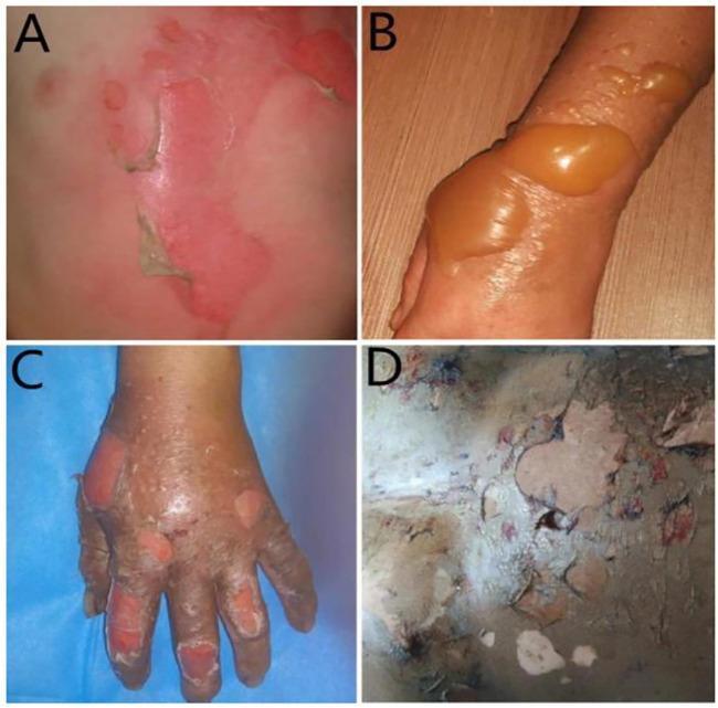
A composite of four clinical photographs demonstrating the progression and classification of burn wound severity. Image A illustrates a first-degree (superficial) burn, showing blanching erythema of the skin with areas of peeling epidermis. Image B depicts a superficial second-degree (partial-thickness) burn on an extremity, characterized by large, tense, fluid-filled bullae with translucent amber-colored serous fluid and surrounding erythematous inflammation. Image C shows a deep second-degree burn on the hand, featuring mottled red and white tissue with ruptured blisters and areas of exposed dermis. Image D represents a third-degree (full-thickness) burn, displaying a dry, leathery, and inelastic eschar with a variegated color palette of white, grey, and brown, indicating destruction of the epidermis, dermis, and subcutaneous tissue. This comparison chart serves as a diagnostic educational tool for identifying burn depth based on visual morphological features such as blistering, tissue color, and texture.

This clinical series demonstrates the progression of burn severity categorized by depth and tissue involvement. (a) First-degree burn (superficial): Presents as erythema of the epidermis without blistering, consistent with a sunburn. (b) Second-degree superficial partial-thickness burn (SPTB): Displays clear blistering (bullae) and moist, red tissue, indicating damage to the papillary dermis. (c) Second-degree deep partial-thickness burn (DPTB): Shows a mottled appearance with ruptured blisters and more significant exudate, extending into the reticular dermis. (d) Third-degree burn (full-thickness): Characterized by a central area of leathery, dark eschar formation surrounded by a zone of stasis, indicating total destruction of the epidermis and dermis into the subcutaneous fat. (e) Fourth-degree burn: Illustrates the most severe injury, extending through all skin layers to involve underlying fascia, skeletal muscle (rectus abdominis), and potentially bone, appearing charred and necrotic. This visual comparison serves as an educational tool for clinical triage and the classification of thermal injuries.
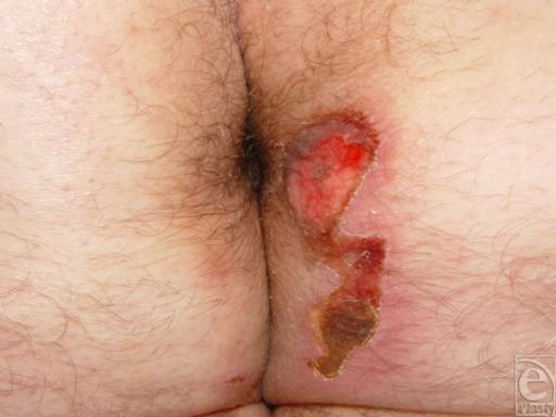
Clinical photograph showing a mixed-depth thermal contact burn on the right perianal region. The lesion is vertically oriented and exhibits features consistent with second- and third-degree burns. The superior aspect of the wound presents as a bright red, moist, well-demarcated area of deep partial-thickness injury (second-degree), likely indicating exposed dermis. Moving inferiorly, the wound transitions into a full-thickness injury (third-degree) characterized by a tan, leathery appearance and a dark brown, opaque eschar at the lower pole. The surrounding skin displays significant blanching erythema and inflammation, with clear margins between the viable tissue and the necrotic burn site. The clinical context involves a paraplegic patient with sensory deficits, highlighting the risk of thermal injury from external sources like car seat heaters. This image serves as an educational example of thermal trauma morphology, burn staging, and the importance of pressure and temperature awareness in neurologically impaired patients.
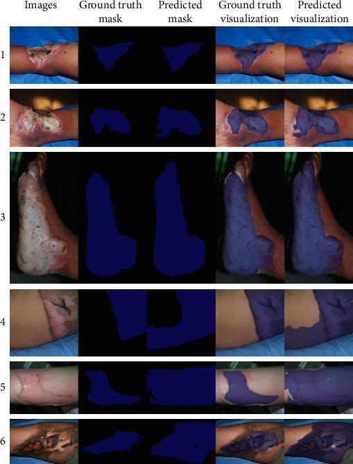
This clinical dataset collage demonstrates the application of deep learning for burn wound segmentation. The image is structured in a 6x5 grid. The first column displays clinical photographs of various burn injuries on human limbs, showing a spectrum of wound morphology: superficial redness, blistering, deep partial-thickness wounds with white/pink slough, and full-thickness burns characterized by charred eschar and exposed subcutaneous tissue. The second and third columns present 'Ground truth mask' and 'Predicted mask' as binary blue-on-black segmentations, representing expert labels versus AI-generated area detections. The fourth and fifth columns, 'Ground truth visualization' and 'Predicted visualization,' show these masks overlaid as semi-transparent blue filters on the original clinical images to illustrate the accuracy of the automated segmentation. The examples highlight the challenges in defining burn boundaries, especially in cases with transitional zones between burned and healthy skin. This material is designed for medical informatics and dermatology education, focusing on automated diagnostic imaging and the assessment of burn surface area and depth.
rule of nines burn surface area TBSA estimation Lund Browder
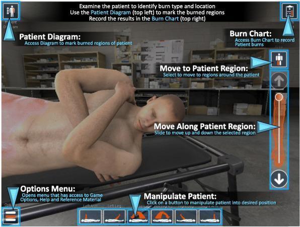
This infographic depicts the user interface of a virtual medical simulation for burn care assessment. The central focus is a 3D-rendered male virtual patient lying on a cot, exhibiting visible erythematous skin changes consistent with thermal injury. The interface features several educational and diagnostic tools: 1) a 'Patient Diagram' for marking burned regions, 2) a 'Burn Chart' for recording data, 3) navigation controls ('Move to Patient Region' and 'Move Along Patient Region') to examine the body in detail, and 4) 'Manipulate Patient' icons to change body positioning for better visualization. Textual instructions at the top guide the user through the clinical workflow: identifying burn type and location, marking regions on a diagram, and calculating Total Body Surface Area (TBSA). The tool is designed for medical training to improve clinical accuracy in burn staging and surface area estimation using standard methodologies like the Lund-Browder chart. The background mimics a clinical environment with stocked medical supplies.
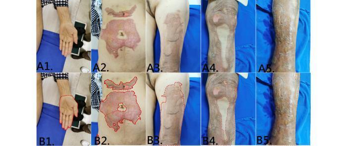
This composite of clinical photographs illustrates various burn injuries and skin lesions across multiple anatomical regions, paired with corresponding segmentation masks (B1-B5) used for Total Body Surface Area (TBSA) estimation. A1 displays a supinated left hand and palm used as an anatomical reference for sizing. A2 focuses on the abdomen, showing a large, irregularly shaped partial-thickness burn characterized by a pink-to-red erythematous base with areas of central pallor, suggesting varying burn depths. A3, A4, and A5 depict injuries on the left thigh, right leg/knee, and left lower leg, respectively. These areas exhibit extensive post-inflammatory hyperpigmentation, mottled tan-to-brown discoloration, and varying textures ranging from smooth to glossy or dry, characteristic of healing burn wounds or hypertrophic scarring. The series demonstrates the application of machine learning (Mask R-CNN) in dermatology and burn surgery to accurately delineate wound boundaries (red dashed lines in row B) for objective clinical assessment and treatment planning.
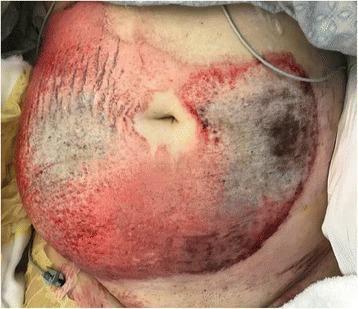
This clinical photograph displays a large, mixed-depth friction burn on the anterior abdomen of a patient following a motor vehicle accident. The injury covers a significant Total Burn Surface Area (TBSA), estimated at 13%. The wound exhibits heterogeneous characteristics: peripheral regions show intense erythema and mottled pink-to-red discoloration consistent with partial-thickness (second-degree) burns. Central and dependent areas of the wound demonstrate darker, grayish-black eschar and leathery texture, indicating full-thickness (third-degree) tissue damage covering approximately 4% of the TBSA. The burn follows a circular pattern around the umbilicus, which remains relatively spared. Visual signs of mechanical friction, such as linear abrasions and uneven texture, are evident throughout the injured area. This image serves as an educational example of high-energy friction trauma, illustrating the variable depths of thermal and mechanical skin injury and the clinical indicators used to differentiate between partial and full-thickness burns in a trauma setting.
Burns - Schwartz's Principles of Surgery (11th Edition)
BACKGROUND
- Schwartz's Principles of Surgery, 11e, p. 251
INITIAL EVALUATION
- Airway assessment
- Estimation of burn size (%TBSA)
- Diagnosis of CO and cyanide poisoning
- Follow ABA criteria for referral to a regional burn center
- Never administer prophylactic antibiotics other than tetanus vaccination
- Early excision and grafting of full-thickness and deep partial-thickness burns improve outcomes
- IV fluid resuscitation for burns >20% TBSA (children >15% TBSA) should be titrated to MAP >60 mmHg and appropriate urine output
ABA CRITERIA - REFERRAL TO A BURN CENTER
| Criterion |
|---|
| Partial-thickness burns >10% TBSA |
| Burns involving the face, hands, feet, genitalia, perineum, or major joints |
| Third-degree burns in any age group |
| Electrical burns, including lightning injury |
| Chemical burns |
| Inhalation injury |
| Burns in patients with complicated preexisting medical disorders |
| Burns with concomitant trauma where burn is the greater risk |
| Burned children in hospitals without qualified pediatric personnel |
| Burns requiring special social, emotional, or rehabilitative intervention |
BURN CLASSIFICATION (Etiology)
| Type | Details |
|---|---|
| Thermal | Flame (most common for hospital admission, highest mortality - associated with inhalation injury/CO poisoning), contact, scald |
| Electrical | 3% of US admissions; cardiac arrhythmia risk, compartment syndrome with rhabdomyolysis; baseline ECG required in all; fasciotomy for high-voltage injuries |
| Chemical | 3% of admitted burn patients; acid = coagulation necrosis; alkali = liquefactive necrosis; hydrofluoric acid = liquefactive necrosis + hypocalcemia; treat with calcium gluconate topically/IV/intra-arterial |
BURN DEPTH
| Degree | Depth | Clinical Features | Healing |
|---|---|---|---|
| 1st degree (Superficial) | Epidermis only | Painful, erythema, no blisters, blanches | Heals in 3-5 days, no scarring |
| 2nd degree - Superficial Partial Thickness | Superficial dermis (papillary dermis) | Extremely painful, weeping, blisters, moist pink base | Usually heals without surgery in 14-21 days |
| 2nd degree - Deep Partial Thickness | Deep dermis (reticular dermis) | Less painful, mottled red/white, ruptured blisters | Often requires excision & grafting |
| 3rd degree (Full Thickness) | Full dermis | Leathery, painless, non-blanching, waxy/charred | Requires excision and grafting |
| 4th degree | Soft tissue, muscle | Extends into underlying soft tissue | Requires excision; possible amputation |
| 5th degree | Through muscle to bone | Charred muscle | Major reconstruction |
| 6th degree | Bone charring | Charred bone | Extensive reconstruction |
Note: First-degree (superficial) burns should NOT be included when calculating %TBSA for resuscitation.
Jackson's Three Zones of Burn Injury
| Zone | Description | Outcome |
|---|---|---|
| Zone of Coagulation | Center; coagulated/necrotic tissue | Needs excision and grafting |
| Zone of Stasis | Peripheral; variable vasoconstriction/ischemia | Appropriate resuscitation may prevent conversion to deeper wound |
| Zone of Hyperemia | Outermost; like superficial partial-thickness | Heals with minimal/no scarring |
BURN SIZE ESTIMATION (%TBSA)
Rule of Nines (Adults)
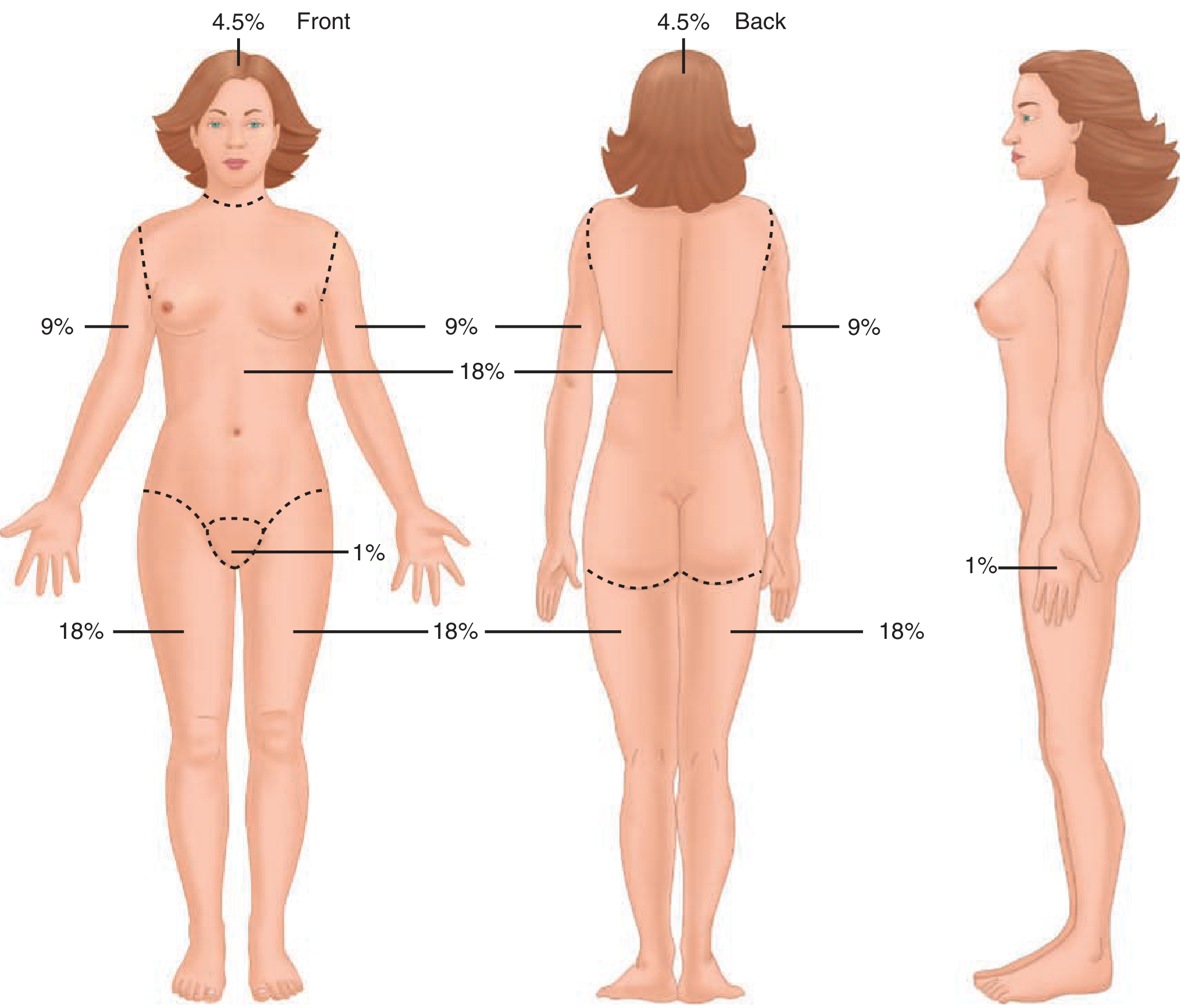
| Region | %TBSA |
|---|---|
| Anterior trunk | 18% |
| Posterior trunk | 18% |
| Each upper extremity | 9% |
| Each lower extremity | 18% |
| Head | 9% (4.5% front + 4.5% back) |
| Genitalia/perineum | 1% |
- In children under 3 years: head accounts for a larger relative surface area (adjust accordingly)
- Rule of the palm: The palmar surface of the hand including digits = 1% TBSA (useful for smaller/odd-shaped burns)
- Lund and Browder chart: More accurate for children and adults (adjusts for age-related proportional differences)
PROGNOSIS
- Age
- %TBSA
- Inhalation injury
- Coexistent trauma
- Pneumonia
- Burn injury reduces short-term QoL by ~30% and long-term QoL by ~11%
- ~28% of burn survivors never return to work
- Itching is a late, bothersome consequence affecting both adult and pediatric populations
RESUSCITATION
Parkland (Baxter) Formula - Most Commonly Used
3-4 mL × kg body weight × %TBSA burned of Lactated Ringer's solution over 24 hours
- Half given in the first 8 hours (from time of injury, not hospital arrival)
- Remaining half over the next 16 hours
- Urine output: 0.5-1.0 mL/kg/hr (adults); 1 mL/kg/hr (children)
- MAP >60 mmHg
| Agent | Role |
|---|---|
| Albumin | May reduce total fluid load; many burn centers use as adjunct during resuscitation |
| Hypertonic saline | Decreases total fluid load over first 24h; risk of hyperchloremic acidosis |
| High-dose Ascorbic acid (Vit C) | May decrease fluid volume requirements |
| Plasmapheresis | Associated with decreased fluid requirements in patients requiring higher-than-predicted volumes |
| Blood transfusion | Restrictive strategy (Hb target 7-8 g/dL) generally acceptable |
INHALATION INJURY & VENTILATOR MANAGEMENT
- Increased incidence of ARDS
- Increased ventilator days
- Higher rate of multiple organ dysfunction syndrome
- Higher mortality
- Aggressive pulmonary toilet
- Nebulized albuterol (bronchodilator)
- Nebulized N-acetylcysteine (antioxidant/free radical scavenger)
- Aerosolized heparin (prevents fibrin plugs/airway casts; associated with increased ventilator-free days)
- Avoid steroids (associated with worse outcomes; exception: septic shock requiring vasopressors)
Carbon Monoxide (CO) Poisoning
- CO has 200-250x greater affinity for hemoglobin than oxygen
- Causes carboxyhemoglobinemia, anoxia, uncoupling of oxidative phosphorylation, free radical generation, platelet activation
- Pulse oximetry is falsely elevated - must obtain arterial carboxyhemoglobin level
- Treatment: 100% normobaric oxygen (gold standard) - reduces CO half-life from ~5 hours to ~60 minutes
- Hyperbaric oxygen may be considered for severe poisoning
Cyanide Poisoning
- Consider in patients with altered mental status following burn/smoke inhalation
- Treat with hydroxocobalamin or sodium thiosulfate
TREATMENT OF THE BURN WOUND - TOPICAL ANTIMICROBIALS
| Agent | Advantages | Disadvantages |
|---|---|---|
| Silver sulfadiazine | Broad-spectrum; painless | Contraindicated near skin grafts; may retard epithelial migration in partial-thickness wounds |
| Mafenide acetate | Penetrates eschar; effective for wound infections; solution excellent for fresh skin grafts | Painful on partial-thickness burns; carbonic anhydrase inhibitor (historically: metabolic acidosis) |
| Silver nitrate (0.5% solution) | Broad-spectrum | Electrolyte extravasation (hyponatremia); rare methemoglobinemia; black staining |
| Silver-impregnated dressings | Reduce dressing changes; shorter hospital stay | Limit serial wound examination |
| Biobrane | Prolonged barrier; wound heals underneath | Only for fresh, uncontaminated superficial partial-thickness burns |
| Bacitracin/Neomycin/Polymyxin B | Useful for small burns, facial burns, near-healed wounds | Potential nephrotoxicity in large burns |
NUTRITION
- Nutritional support is more critical in large burns than almost any other patient population
- Hypermetabolic response in burn injury may raise baseline metabolic rates by up to 200%
- Leads to catabolism of muscle proteins and decreased lean body mass
- Early enteral feeding for burns >20% TBSA is safe and may:
- Reduce loss of lean body mass
- Slow the hypermetabolic response
- Result in more efficient protein metabolism
- Shorten ICU stay
- Decrease wound infection rates
- If enteral feeds started within the first few hours of admission, gastric ileus may be avoided
- Metoclopramide can be used to promote GI motility
SURGERY
- Early excision and grafting is the cornerstone of surgical management for full-thickness and deep partial-thickness burns
- Tangential excision: Sequential removal of thin layers of necrotic eschar until viable tissue is reached
- Fascial excision: Removal of all tissue down to the fascia (used for very deep burns or infected wounds; results in worse cosmesis)
- Deep partial-thickness burns benefit from excision and skin grafting
- Superficial partial-thickness burns typically heal with nonoperative management
- Zone of stasis may convert to deeper injury if infection occurs or perfusion is suboptimal
WOUND COVERAGE
- Split-thickness skin grafts (STSG): Standard for coverage after excision
- Meshed grafts: Allow larger coverage; interstices fill by re-epithelialization; use bacitracin/neomycin/polymyxin B with greasy gauze on near-closed interstices
- Biologic membranes: Temporary wound coverage options
LATE COMPLICATIONS
| Complication | Description |
|---|---|
| Hypertrophic scarring | Common after burns; treated with pressure garments, silicone, intralesional steroids |
| Contractures | Particularly around joints; require early rehabilitation and splinting, later surgical release |
| Heterotopic ossification | Ectopic bone formation; managed with excision once mature |
| Itching | Late but highly prevalent and bothersome; affects both adults and children |
PSYCHOLOGICAL RECOVERY
- Post-traumatic stress disorder (PTSD)
- Depression and anxiety
- Body image disturbance
- Pain-related psychological distress
BURN DISASTERS / MASS CASUALTY EVENTS
- Triage based on survivability using Revised Baux Score
- Maintain airway in all patients with signs of inhalation injury
- Initiate Parkland formula fluid resuscitation
- Prioritize early excision in patients most likely to survive
- Transfer to burn centers as capacity allows
Taneo, John Vic 21/M Pending room Impression: 4% TBSA Scald Burn injury (hot oil) dorsum of both feet NOI: Work Related Injury POI: Capitol (Abli Cebu) Restaurant DOI: 8/4/26 TOI: 9:40 PM Stable Multiple first degree burn on legs +blisters on dorsum of both feet For silver sulfa wet to dry dressing on blisters Pain control and TT TIG
BURN INJURY CLINICAL NOTE
PATIENT INFORMATION
| Name | Taneo, John Vic |
| Age/Sex | 21 years old / Male |
| Status | Pending Room |
| Date of Injury | August 4, 2026 |
| Time of Injury | 9:40 PM |
HISTORY OF PRESENT ILLNESS
PHYSICAL EXAMINATION
| Parameter | Findings |
|---|---|
| Mechanism | Scald — hot oil |
| %TBSA | 4% |
| Body parts affected | Dorsum of both feet; bilateral legs |
| Burn depth - legs | First-degree (superficial) — erythema, no blistering |
| Burn depth - dorsum of feet | Second-degree superficial partial-thickness — multiple blisters present |
IMPRESSION
- First-degree (superficial) burns — bilateral legs
- Second-degree superficial partial-thickness burns — dorsum of both feet (blistering present)
PLAN
1. Wound Care
- Silver sulfadiazine 1% cream wet-to-dry dressing over blistered areas (dorsum of both feet)
- Dressing change once daily or as needed
- Do NOT rupture intact blisters - leave intact to serve as natural biologic barrier
- Gentle cleansing with normal saline prior to each dressing change
2. Pain Control
- Oral analgesics (e.g., Paracetamol 500-1000 mg q6h PRN pain, or Ibuprofen 400 mg q8h with food if no contraindication)
- Reassess pain at each dressing change; opioid analgesia may be considered for procedural pain during dressing changes
3. Tetanus Prophylaxis
- Tetanus Toxoid (TT) - administer per immunization status
- Tetanus Immune Globulin (TIG) - administer as indicated (uncertain or incomplete immunization history)
4. Monitoring
- Monitor for signs of wound infection (increased erythema, purulent discharge, fever, worsening pain)
- Daily wound inspection with dressing changes
- Monitor blisters for integrity and signs of deepening wound
5. Disposition
- For room admission (pending bed)
- Elevation of both lower extremities to reduce edema
- Ambulation restricted until pain controlled and wound assessed
NOTES & CONSIDERATIONS
Silver sulfadiazine is appropriate for partial-thickness burns with blistering. However, note per Schwartz's that silver sulfadiazine may retard epithelial migration in healing partial-thickness wounds and must be kept away from any skin graft sites if surgical intervention becomes necessary.
No prophylactic systemic antibiotics are indicated (per ABA guidelines and Schwartz's — prophylactic antibiotics promote fungal infections and resistant organisms).
At 4% TBSA in an otherwise healthy 21-year-old male, this patient does not meet ABA criteria for burn center transfer. Outpatient or general ward management is appropriate with close follow-up.
Revised Baux Score = 21 (age) + 4 (%TBSA) = 25 — Very low predicted mortality. Excellent prognosis for full recovery.
First-degree burns on the legs are NOT counted in the 4% TBSA calculation per Schwartz's guidelines (superficial/first-degree burns excluded from TBSA estimation for resuscitation purposes).
At 4% TBSA, IV fluid resuscitation is not indicated — patient can hydrate orally (Schwartz's: IV resuscitation rarely indicated for burns <15% TBSA in adults who can hydrate orally).
How to approach a trauma burn patient
burn patient primary survey airway management algorithm
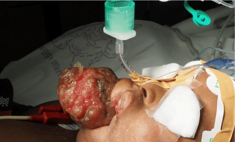
This clinical photograph shows a pediatric patient undergoing airway management for a surgical procedure. The primary pathology is a large, exophytic, and multinodular mass originating from the left facial/maxillary region. The lesion exhibits varied morphology with areas of erythema, surface ulceration, and fleshy protuberances that distort the facial anatomy. Regarding clinical intervention, the patient has been successfully intubated via the nasotracheal route. A narrow-diameter endotracheal tube is visible entering the right naris, secured with medical adhesive tape and padding. The tube is connected to a green anesthetic circuit adapter, indicating ongoing mechanical ventilation or anesthesia delivery. Monitoring equipment is evident, including electrocardiogram (ECG) leads adhered to the forehead and chest area. The image demonstrates the complexities of securing a definitive airway in a patient with significant facial pathology and highlights the importance of specialized techniques, such as videolaryngoscopy or fiberoptic-assisted intubation, in pediatric cases with anatomical distortion.
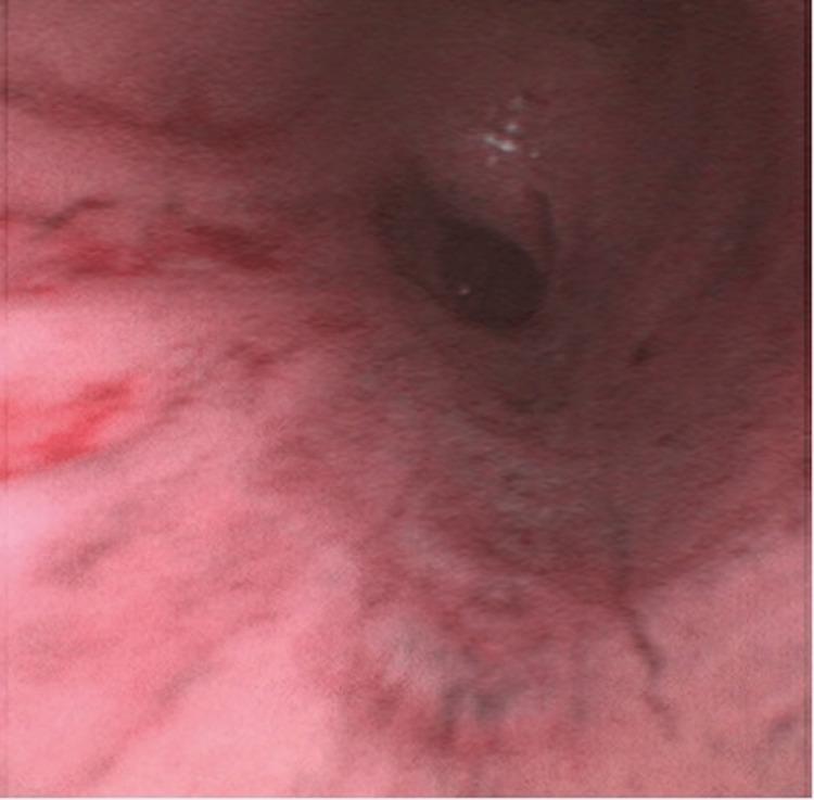
This bronchoscopic image shows the distal airway of a patient with severe inhalation injury. The primary finding is a denuded and erythematous epithelium, where the normal smooth mucosal lining has sloughed off due to thermal or chemical damage. The underlying tissue appears raw, swollen, and predominantly red, indicating significant inflammation and vascular congestion. A focal dark lesion or area of soot deposition is visible within the lumen, further characterizing the severity of the injury. The clinical presentation is consistent with a high-grade inhalation injury, commonly seen in burn patients. This image demonstrates the rapid progression from initial mucosal blistering to circumferential epithelial denudation, a critical factor in airway management and the pathophysiology of post-burn respiratory complications.
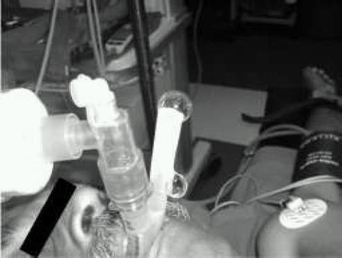
This clinical photograph demonstrates a ProSeal Laryngeal Mask Airway (PLMA) in situ on a patient, illustrating the 'gel displacement test' for airway seal evaluation. The airway management apparatus consists of a primary ventilation tube and a secondary, parallel gastric drain tube exiting the patient's mouth. In the image, a column of clear, water-soluble lubricant gel is visible within the proximal end of the gastric drain tube. The test is used to detect leaks; during positive pressure ventilation, the displacement or ejection of this gel column signifies an incompetent seal at the hypopharynx or malposition of the device's distal cuff. This procedural image highlights a critical bedside assessment technique in anesthesiology and emergency medicine to ensure correct supraglottic airway placement and the separation of respiratory and gastrointestinal tracts. The patient is also shown with standard monitoring equipment, including an ECG electrode and a blood pressure cuff.
escharotomy incision lines circumferential burn extremity chest
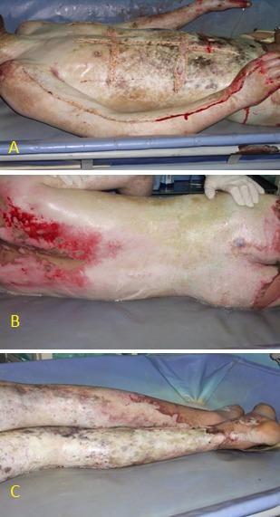
A three-panel clinical photograph (A, B, and C) illustrating a pediatric patient with near-total body surface area (approximately 95% TBSA) thermal injuries. Panel A displays the anterior torso and upper extremity, showing mixed-depth burns characterized by a leathery, charred, and blanched appearance with deep brown and white patches. Several linear escharotomy incisions are visible across the chest, abdomen, and along the medial aspect of the arm to relieve pressure from circumferential burns and prevent compartment syndrome. Panel B shows the posterior aspect of the torso and buttocks, demonstrating extensive full-thickness and deep partial-thickness burns with areas of erythema and skin sloughing. Panel C focuses on the lower extremities, depicting widespread full-thickness burns on both legs with a pale, avascular, and mottled texture, extending to the feet and toes. This series serves as a critical educational example of severe burn management, highlighting the characteristic appearance of eschar in full-thickness injuries and the necessary emergency surgical interventions required in critical care trauma settings.
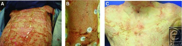
A series of three clinical photographs (A, B, and C) illustrating the progression of burn wound healing and surgical intervention on the anterior trunk. Image A shows acute deep-partial to full-thickness burns with significant inflammation, mottled red and pink tissue, and horizontal escharotomy incisions across the thorax and abdomen to improve compliance. Small white wound dressings or surgical clips are visible along the incision lines. Image B depicts an intermediate stage of healing where a large-area meshed skin graft has been applied to the trunk, showing a characteristic geometric pattern over the underlying wound bed; circular ECG monitoring electrodes are adhered to the right side. Image C shows the later stages of healing and re-epithelialization on the upper chest. The skin appears lighter and more uniform but exhibits residual erythematous scarring, texture irregularities, and hypertrophic changes. A tracheostomy site is visible at the midline of the neck, and the overall integrity of the skin barrier shows significant improvement from the initial presentation.
Approach to the Trauma Burn Patient
OVERVIEW - THE THREE PHASES
- Prehospital Care
- ED Resuscitation and Stabilization
- Admission or Transfer to a Burn Center
PHASE 1 - PREHOSPITAL CARE
| Step | Action |
|---|---|
| 1. Stop the burning process | Remove from burning environment; remove burning clothing, rings, watches, jewelry, belts (retain heat; cause tourniquet effect) |
| 2. Airway | Assess and secure immediately; prophylactic intubation for perioral burns in closed-space fires |
| 3. Fluid resuscitation | IV isotonic crystalloid (Lactated Ringer's) |
| 4. Relieve pain | Analgesia per protocol |
| 5. Protect the wound | Cover with clean sheets |
| 6. Transport | To the nearest appropriate facility or burn center |
Cooling: Early wound cooling can reduce burn depth and pain - but uncontrolled cooling causes hypothermia. Only cool small burns; do NOT cool burns >20% TBSA. Wrap in clean dry blankets for transport.
PHASE 2 - ED MANAGEMENT
PRIMARY SURVEY - "ABCDE"
A - Airway (highest priority)
- Facial burns
- Singed nasal hair
- Soot in mouth or nose
- Hoarseness or stridor
- Carbonaceous (sooty) sputum
- Expiratory wheezing
- Subjective dyspnea
- Oropharyngeal mucosal burns
Rule: If ANY sign of airway compromise is present - swelling of the neck, burns inside the mouth, wheezing - perform early endotracheal intubation immediately. Do not wait for deterioration.
- Orotracheal intubation is preferred
- Nasotracheal only if facial trauma + experienced provider present
- Perioral burns + singed nasal hairs alone do NOT confirm inhalation injury but mandate oropharyngeal examination
- Administer 100% humidified oxygen by face mask to all burn patients
B - Breathing
| Action | Purpose |
|---|---|
| Continuous pulse oximetry + supplemental O₂ | Monitor oxygenation |
| Arterial blood gas (with carboxyhemoglobin level) | Pulse oximetry is falsely elevated in CO poisoning |
| Bronchoscopy if inhalation injury suspected | Diagnostic + therapeutic |
| Chest X-ray | Baseline (may be normal initially) |
| Mechanical ventilation as needed | Lung-protective settings: tidal volume <7 mL/kg, plateau pressure <31 cm H₂O |
- Supraglottic: Heat injury to upper airway - rapid edema
- Subglottic: Smoke/chemical injury to lower airway - may not manifest for up to 24 hours
- Systemic: CO and cyanide poisoning

C - Circulation
| Action | Details |
|---|---|
| 2 large-bore peripheral IVs | In unburned skin if possible; IV through burned skin is acceptable |
| For burns >40% TBSA | Two large-bore IVs mandatory |
| Cardiac monitoring | Especially for electrical burns |
| Heart rate 100-120 bpm | Normal in burn patients due to catecholamine response - do not over-interpret |
| Foley catheter | Monitor urine output; also prevents urinary retention in perineal burns |
Note: IV fluid resuscitation is generally required for burns >20% TBSA in adults, >10% TBSA in children. Burns <15% TBSA in adults can usually hydrate orally.
D - Disability (Neurological)
- Assess GCS, mental status
- Altered mental status should raise suspicion for CO poisoning, cyanide poisoning, or associated head trauma
- Assess need for cervical spine immobilization (especially in explosion, fall, or motor vehicle collision patients)
E - Exposure / Environment
- Full exposure for complete burn assessment
- Prevent hypothermia - warm blankets, warmed IV fluids, warm environment
- Burned patients are especially vulnerable to hypothermia due to impaired thermoregulation
- Continuous core temperature monitoring for large burns
SECONDARY SURVEY
- Burning agent(s) - thermal / chemical / electrical
- Duration of exposure
- Open vs. enclosed space (critical for inhalation injury suspicion)
- Loss of consciousness
- Risk of blast injury, explosion, fall (associated trauma)
- Contact with electricity
- PMH, medications, allergies
- Tetanus immunization status
- Eyes: examine for corneal burns
- Estimate and record burn size (%TBSA) and burn depth
- Check for circumferential burns on extremities, chest, abdomen, neck
BURN SIZE ESTIMATION
| Region | %TBSA |
|---|---|
| Head | 9% |
| Each arm | 9% |
| Anterior trunk | 18% |
| Posterior trunk | 18% |
| Each leg | 18% |
| Genitalia | 1% |
- Rule of Palm: 1 palm surface = 1% TBSA (for small/irregular burns)
- Lund & Browder chart: More accurate, especially in children (adjusts for age)
- Do NOT include first-degree burns in TBSA calculation
FLUID RESUSCITATION
| Formula | Dose | Fluid |
|---|---|---|
| Parkland (Baxter) | 4 mL × kg × %TBSA | Lactated Ringer's |
| Modified Parkland | 3-4 mL × kg × %TBSA | LR |
| Modified Brooke | 2 mL × kg × %TBSA | LR |
| ABA Pediatric (≤30 kg) | 3 mL × kg × %TBSA + D5LR maintenance | LR |
- Half in the first 8 hours from time of injury (not from hospital arrival)
- Remaining half over the next 16 hours
- Urine output: 0.5-1.0 mL/kg/hr (adults); 1 mL/kg/hr (children)
- MAP: >60 mmHg
"Fluid Creep" Warning: Over-resuscitation can cause burn wound conversion, extremity compartment syndrome, abdominal compartment syndrome, and pulmonary edema. Adjust to patient response - do not rigidly follow formula alone.
| Formula | First 24h |
|---|---|
| Parkland | LR 4 mL/kg/%TBSA; half in first 8h |
| Modified Parkland | LR 3-4 mL/kg/%TBSA; half in first 8h |
| Evans | Crystalloid 1 mL/kg/%TBSA + colloid 1 mL/kg/%TBSA + 2000 mL D5W |
| Brooke | LR 1.5 mL/kg/%TBSA + colloid 0.5 mL/kg/%TBSA + 2000 mL D5W |
ADJUNCT INTERVENTIONS
| Intervention | Indication |
|---|---|
| Nasogastric tube | Partial-thickness burns >20% TBSA (frequent ileus) |
| Foley catheter | All major burns - hourly urine output monitoring |
| Tetanus Toxoid (TT) | All burn patients |
| Tetanus Immune Globulin (TIG) | Uncertain or incomplete immunization |
| Pain control | Multimodal - opioids ± NSAIDs ± adjuncts; plan for weaning |
| CBC, BMP, creatinine, glucose | All burns |
| ABG + carboxyhemoglobin | Moderate/severe burns or inhalation injury |
| Serum CK + urine myoglobin | Electrical burns, suspected rhabdomyolysis |
| ECG | All electrical burns; baseline cardiac assessment |
| Chest X-ray | Suspected inhalation; baseline |
| Pregnancy test | All females of reproductive age |
| No prophylactic antibiotics | Promotes fungal infections and resistant organisms |
ESCHAROTOMY
| Location | Indications |
|---|---|
| Limbs | The 6 P's: Pain, Pallor, Paresthesia, Paralysis, Poikilothermia, Pulselessness; absent oximetry signal; compartment pressure >30 mmHg |
| Chest | Increased airway pressures, decreased tidal volumes, decreased preload, tachycardia |
| Abdomen | Poor respiratory compliance, hemodynamic instability, tube feed intolerance, decreased urine output, bladder pressure >25 mmHg |
| Face/Periorbital | Increased intraocular pressure >30 mmHg |
| Neck | Airway compromise |
Timing: Escharotomy is rarely needed in the first few hours. Increased tissue edema from resuscitation usually brings on the need for it later. Monitor closely.
- Incisions through eschar down to subcutaneous level (eschar is necrotic - minimal pain/bleeding)
- Extend slightly into normal tissue proximally and distally
- Extremities: incisions along medial and lateral aspects; extend into fingers for hand burns
- Use electrocautery to minimize blood loss
- If escharotomy alone is insufficient: proceed to fasciotomy or decompressive laparotomy as needed
LOCAL WOUND CARE IN ED
| Burn type | Local Care |
|---|---|
| First-degree | No dressing needed; topical NSAID or aloe vera for pain |
| Superficial partial-thickness (blisters) | Gentle cleansing with soap and water; intact blisters - leave intact (natural biologic barrier); silver sulfadiazine or other topical antimicrobial + dressing |
| Deep partial/full-thickness | Topical antimicrobial dressing; plan for early surgical excision |
| Chemical burns | Copious water irrigation for minimum 30 minutes; specific antidotes (e.g., calcium gluconate for HF acid) |
| Electrical burns | Wound care + monitor for rhabdomyolysis, arrhythmia, compartment syndrome |
PHASE 3 - DISPOSITION
ABA Criteria for Burn Center Transfer
| Criterion |
|---|
| Partial-thickness burns >10% TBSA |
| Burns involving face, hands, feet, genitalia, perineum, major joints |
| Third-degree burns in any age group |
| Electrical burns (including lightning) |
| Chemical burns |
| Inhalation injury |
| Burns with preexisting medical comorbidities |
| Burns + concomitant trauma where burn is the primary risk |
| Children in hospitals without qualified pediatric care |
| Burns <10 years or >50 years of age |
| Burns requiring special social/emotional/rehabilitative intervention |
SPECIAL POPULATIONS
| Population | Key Modification |
|---|---|
| Pregnant | Fetal monitoring; resuscitation requirements may exceed formula estimates; early OB + burn specialist consult; spontaneous abortion is common in large TBSA burns |
| Pediatric | Head = larger relative TBSA; use Lund & Browder chart; IO access if IV unobtainable; ABA pediatric fluid formula |
| Elderly | Age is independent mortality predictor; aggressive resuscitation monitoring |
| Electrical burns | Cardiac monitoring; vigilance for compartment syndrome/rhabdomyolysis; ophthalmology and neurology consult for high-voltage injuries |
QUICK REFERENCE - SUMMARY ALGORITHM
BURN PATIENT ARRIVES
        |
  STOP burning process
  Remove clothing/jewelry
        |
   PRIMARY SURVEY
   A → Airway: any sign? → Intubate early
   B → 100% O₂, ABG, carboxyhemoglobin, bronchoscopy PRN
   C → 2 large-bore IVs, start LR, cardiac monitor, Foley
   D → GCS, suspect CO/CN/head injury
   E → Expose fully, prevent hypothermia
        |
   DIRECTED HISTORY
   Agent / Duration / Space / Trauma / TT status
        |
   SECONDARY SURVEY
   %TBSA (Rule of Nines) + Burn Depth
   Check circumferential burns → Escharotomy?
   Corneal exam / Perineal / Joint involvement
        |
   RESUSCITATION
   Parkland: 3-4 mL × kg × %TBSA LR over 24h
   Half in first 8h from injury
   Target UO: 0.5-1.0 mL/kg/hr, MAP >60
        |
   ADJUNCTS
   NGT (>20% TBSA) / TT+TIG / Pain control
   Labs: CBC, BMP, ABG, CK, urine myoglobin, ECG
   NO prophylactic antibiotics
        |
   WOUND CARE
   Clean → Topical antimicrobial → Dressing
        |
   DISPOSITION
   Admit vs. Transfer to burn center (ABA criteria)
Criteria
BURN CRITERIA — COMPLETE REFERENCE
1. ABA CRITERIA FOR BURN CENTER REFERRAL/TRANSFER
| # | Criterion |
|---|---|
| 1 | Partial-thickness burns >10% TBSA |
| 2 | Burns involving face, hands, feet, genitalia, perineum, or major joints |
| 3 | Third-degree (full-thickness) burns in any age group |
| 4 | Electrical burns, including lightning injury |
| 5 | Chemical burns |
| 6 | Inhalation injury |
| 7 | Burns in patients with complicated preexisting medical disorders |
| 8 | Burns + concomitant trauma where burn is the greater immediate risk |
| 9 | Burned children in hospitals without qualified pediatric personnel |
| 10 | Burns in patients requiring special social, emotional, or rehabilitative intervention |
| 11 | Burns in children <10 years and adults >50 years of age (Tintinalli's) |
2. CRITERIA FOR IV FLUID RESUSCITATION
| Population | Threshold |
|---|---|
| Adults | Burns >20% TBSA |
| Children | Burns >15% TBSA (Schwartz's) / >10% TBSA (Rosen's) |
| Adults <15% TBSA | IV resuscitation rarely needed — oral hydration usually sufficient |
3. PARKLAND FORMULA PARAMETERS
| Parameter | Value |
|---|---|
| Formula | 3-4 mL × kg × %TBSA of Lactated Ringer's over 24 hours |
| First 8 hours | Half of total volume (from time of injury, not arrival) |
| Next 16 hours | Remaining half |
| Target urine output (adults) | 0.5-1.0 mL/kg/hr |
| Target urine output (children) | 1 mL/kg/hr |
| Target MAP | >60 mmHg |
4. CRITERIA FOR EARLY INTUBATION
| Sign | Meaning |
|---|---|
| Hoarseness / stridor | Impending airway obstruction |
| Expiratory wheezing | Lower airway injury |
| Subjective dyspnea | High-risk indicator |
| Burns inside the mouth / oropharyngeal mucosal burns | Direct thermal injury |
| Swelling of the neck | Impending obstruction |
| Carbonaceous (sooty) sputum | Inhalation confirmed |
| Perioral burns + closed-space fire | Prophylactic intubation |
| Facial burns + singed nasal hairs | Warrant further evaluation (not alone sufficient) |
5. CRITERIA FOR ESCHAROTOMY
| Location | Indications |
|---|---|
| Extremities | Pain, Pallor, Paresthesia, Paralysis, Poikilothermia, Pulselessness (6 P's); absent oximetry signal; compartment pressure >30 mmHg |
| Chest | Increased airway pressures, decreased tidal volumes, decreased preload, tachycardia |
| Abdomen | Hemodynamic instability, poor respiratory compliance, tube feed intolerance, decreased urine output, bladder pressure >25 mmHg |
| Periorbital | Intraocular pressure >30 mmHg |
| Neck | Airway compromise |
6. CRITERIA FOR NASOGASTRIC TUBE INSERTION
| Indication |
|---|
| Partial-thickness burns >20% TBSA (frequent ileus) |
| Decreased level of consciousness |
| Inability to tolerate oral intake |
7. CRITERIA FOR INHALATION INJURY DIAGNOSIS
| Clinical Clue | Significance |
|---|---|
| History of fire in enclosed space | Highest risk factor |
| Facial burns | Suggestive |
| Singed nasal hair | Suggestive |
| Soot in mouth/nose | Strongly suggestive |
| Hoarseness | Upper airway involvement |
| Carbonaceous sputum | Confirmed inhalation |
| Expiratory wheezing | Lower airway involvement |
| Carboxyhemoglobin level (arterial) | Documents CO exposure; pulse oximetry is falsely elevated |
| PaO₂:FiO₂ ratio <350 | Predicts inhalation injury + increased fluid needs |
| Bronchoscopy | Gold standard for diagnosis and grading |
8. CRITERIA FOR TETANUS PROPHYLAXIS IN BURNS
| Immunization History | Action |
|---|---|
| Up to date (booster within 5 years) | None needed |
| Incomplete or uncertain | TT (Tetanus Toxoid) |
| Unknown / no prior immunization | TT + TIG (Tetanus Immune Globulin) |
| All burn patients (general rule) | Tetanus booster should be administered in the ED |
9. REVISED BAUX SCORE — MORTALITY PREDICTION
| Component | Value |
|---|---|
| Formula | Age + %TBSA (original Baux) |
| Revised formula | Age + %TBSA + 17 (if inhalation injury present) |
| Interpretation | Higher score = higher predicted mortality |
| Current standard | Revised Baux Score (accounts for inhalation injury) |
Example — John Vic Taneo: 21 + 4 = Baux 25 → Excellent prognosis
10. CRITERIA TO NOT INCLUDE IN %TBSA CALCULATION
| Excluded | Reason |
|---|---|
| First-degree (superficial) burns | Do not drive fluid resuscitation |
| Erythema only (no dermal involvement) | Superficial; no systemic effect |
11. CRITERIA FOR PROPHYLACTIC ANTIBIOTIC — CONTRAINDICATION
NEVER give prophylactic systemic antibiotics in burn patients.
- Promotes fungal infections
- Promotes resistant organisms
- Abandoned since the mid-1980s
- Exception: Tetanus vaccination only
- Mupirocin: reserved for culture-positive MRSA burns only
12. CRITERIA FOR COOLING A BURN WOUND
| Condition | Action |
|---|---|
| Small burns (<20% TBSA), early presentation | Cooling acceptable — reduces depth and pain |
| Burns >20% TBSA | Avoid cooling — risk of hypothermia |
| All patients in transport | Wrap in clean dry blankets |