
Pharyngeal abscess in detail according to standard ENT textbook in simplified manner
Reading File
Now let me search the medical library for pharyngeal abscess content.
Finding Sources
Finding Sources
Finding Sources
Your credits are exhausted. You can purchase a top-up or subscribe to a plan to continue using Orris. If you're already subscribed, your daily credits will reset tomorrow.
Pharyngeal abscess in detail according to standard ENT textbook in simplified manner
Pharyngeal abscesses are collections of pus in the deep spaces around the pharynx. The main types are peritonsillar, retropharyngeal, and parapharyngeal abscesses. They present with fever, sore throat, dysphagia, and can be life-threatening if not treated promptly with drainage and antibiotics.
Searching sources...
Finding Sources
Finding Sources
Finding Sources
Excellent - I have great sources. Let me now read the key ENT textbook sections simultaneously.
Reading File
Reading File
Searching Images
pharyngeal abscess peritonsillar retropharyngeal parapharyngeal diagram
Now let me read more from the Cummings ENT textbook on the full retropharyngeal section, and also read deeper Cummings peritonsillar content and the Bailey & Love content:
Reading File
Reading File
Finding Sources
Let me now read the most detailed peritonsillar abscess section from KJ Lee's Essential Otolaryngology and also the chronic retropharyngeal and images:
Reading File
Reading File
Searching Images
peritonsillar abscess quinsy tonsil swelling clinical photo
Let me get a bit more from KJ Lee's for parapharyngeal space infections and also fetch the key CT images found in the textbooks:
Reading File
Loading Image
Loading Image
I now have comprehensive data from multiple standard ENT textbooks. Let me compile the full detailed, simplified response.
Pharyngeal Abscess - A Complete ENT Guide
(Sources: Scott-Brown's Otorhinolaryngology, KJ Lee's Essential Otolaryngology, Cummings Otolaryngology, Bailey & Love's Surgery)
Overview
Pharyngeal abscesses are collections of pus in the deep fascial spaces around the pharynx. They are serious, potentially life-threatening conditions requiring prompt recognition and treatment. There are three main types:
| Type | Location | Typical Age |
|---|---|---|
| Peritonsillar (Quinsy) | Lateral to tonsil | Older children, young adults |
| Retropharyngeal | Behind pharyngeal wall (between middle and deep cervical fascia layers) | Infants & children < 5 years |
| Parapharyngeal | Lateral pharyngeal space | Young children |
1. Peritonsillar Abscess (Quinsy)
What is it?
A collection of pus in the potential space between the tonsil and the pharyngeal constrictor muscle (specifically around the Weber glands). It is the most common deep neck space infection.
How does it form?
- Starts as bacterial tonsillitis/pharyngitis
- Infection spreads beyond the tonsil capsule into surrounding space
- Caused by a mixed aerobic + anaerobic bacterial infection
- Common organisms: Group A Streptococcus, Staphylococcus aureus, anaerobes
Symptoms (Classic Triad)
- Trismus - difficulty opening the mouth (best clinical indicator)
- Severe unilateral sore throat + odynophagia (painful swallowing)
- Uvula deviated to the opposite side (pushed away by swelling)
- Fever, drooling, muffled "hot potato" voice
- Swollen lymph nodes in the neck
- Antecedent history of pharyngitis/tonsillitis
Examination Findings
- Unilateral palatal (soft palate) enlargement
- Uvula displaced to the contralateral side
- Bulging of anterior pillar
- Trismus makes examination difficult
Diagnosis
- Mainly clinical (trismus + uvular deviation + unilateral bulge = classic)
- Needle aspiration - both diagnostic and therapeutic (if uncertain whether cellulitis vs abscess)
- CT neck with contrast - if clinical picture is unclear OR if spread to deeper spaces is suspected
Treatment
- IV antibiotics - high-dose penicillin or cephalosporin (first line); add metronidazole for anaerobes
- Steroids (IV dexamethasone) - helps reduce swelling and pain
- Drainage - mainstay of treatment:
- Needle aspiration (preferred initial approach - safer, effective)
- Incision and drainage (I&D) if needle aspiration fails
- Adequate IV fluids and pain control
⚠️ Tonsillectomy during acute attack is NOT popular - risk of pus aspiration. However, "quinsy tonsillectomy" may be done in experienced hands.
Recurrence & Follow-up
- Recurrence rate = 17%
- Interval tonsillectomy recommended after:
- Second episode of quinsy on the same side
- Background history of recurrent tonsillitis
2. Retropharyngeal Abscess
What is it?
Pus collection in the retropharyngeal space - the space between the posterior pharyngeal wall and the prevertebral fascia. This space extends from the skull base down to the mediastinum, making it a dangerous conduit for spread.
How does it form?
Acute type:
- Suppuration (breakdown) of the lymph nodes of Rouviere in the retropharyngeal space
- These nodes receive lymphatic drainage from nasopharynx, sinuses, and middle ear
- The nodes naturally disappear by age 5, explaining why this is mainly a childhood disease
- Also from: foreign body perforation of the pharynx/esophagus, or spread from adjacent spaces
Chronic type (now rare):
- Extension of tuberculosis of the cervical spine through the anterior longitudinal ligament into the prevertebral space
- Occurs mainly in adults
Common Organisms
- Beta-hemolytic Streptococcus
- Anaerobic Streptococci
- Staphylococcus aureus
Symptoms
- Torticollis (head tilted to one side) - best clinical indicator
- High fever, systemically very unwell
- Dysphagia (difficulty swallowing), drooling, sialorrhea
- Neck rigidity and stiffness
- Croupy cough, altered cry (in infants)
- Dyspnoea / airway compromise (can be the most prominent symptom)
- Febrile convulsions and vomiting
- Bulge in posterior pharyngeal wall (best seen on nasopharyngoscopy)
- External neck swelling/abscess possible
🚨 These children must always be examined by the most senior clinician available. Airway compromise is a real risk.
Diagnosis
1. Lateral Neck X-ray (Plain Film)
- Shows thickening of prevertebral soft tissues
- Anterior displacement of airway
- Loss of normal cervical lordosis (due to prevertebral muscle spasm)
- Occasional gas bubbles within the abscess
- Normal limits:
- At C2: retropharyngeal space ≤ 7 mm
- At C6: ≤ 14 mm in children, ≤ 22 mm in adults
- Pathological if thickening > ½ the width of the vertebral body at the same level
- ⚠️ Neck must be extended during X-ray - flexion gives false appearance of thickening
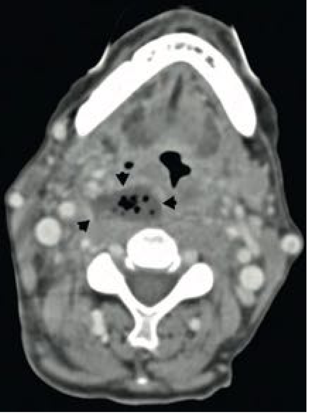
2. CT Neck with Contrast (Gold Standard)
- Distinguishes phlegmon (cellulitis - not drainable) from abscess (drainable)
- Abscess: hypodense/low attenuation with ring enhancement on contrast
- Shows gas bubbles if present
- Maps the full extent before surgery
3. MRI - superior soft tissue detail but less practical in acute setting
Fig. 104.9 - CT neck showing retropharyngeal abscess: fluid collection with gas in the right retropharyngeal space (arrowheads):
Treatment
- Immediate high-dose IV antibiotics (broad-spectrum)
- If pus collection is confirmed: urgent surgical drainage under GA
- Route: usually peroral (through the mouth) - using dressing forceps guided by a finger, head-down position to prevent aspiration
- Occasionally: external cervical approach (anterior to carotid sheath via neck incision)
- Experienced anaesthetist mandatory - induction is risky (risk of abscess rupture on intubation)
- Tracheostomy is very rarely necessary for severe airway compromise
- Chronic (TB) retropharyngeal abscess: must NOT be opened into the mouth (risk of TB dissemination) - requires external drainage + specific anti-TB treatment
3. Parapharyngeal Abscess
What is it?
Pus in the parapharyngeal (lateral pharyngeal) space - the potential space lateral to the pharynx, shaped like an inverted pyramid.
How does it form?
- Most commonly spreads from a peritonsillar or retropharyngeal abscess
- Also from dental infections, tonsillitis, or parotitis
- Contains important structures: carotid artery, internal jugular vein, cranial nerves IX-XII
How to distinguish from Peritonsillar Abscess?
- Maximal swelling is behind the posterior faucial pillar (not at the soft palate)
- Little or no soft palate oedema (unlike peritonsillar abscess)
- Neck swelling is prominent externally
- Patient is usually a young child with severe general malaise
Symptoms
- Severe trismus (jaw stiffness)
- Possible airway compromise and difficulty swallowing
- Systemically very unwell
- Obvious neck swelling externally
- Fever, malaise
Diagnosis
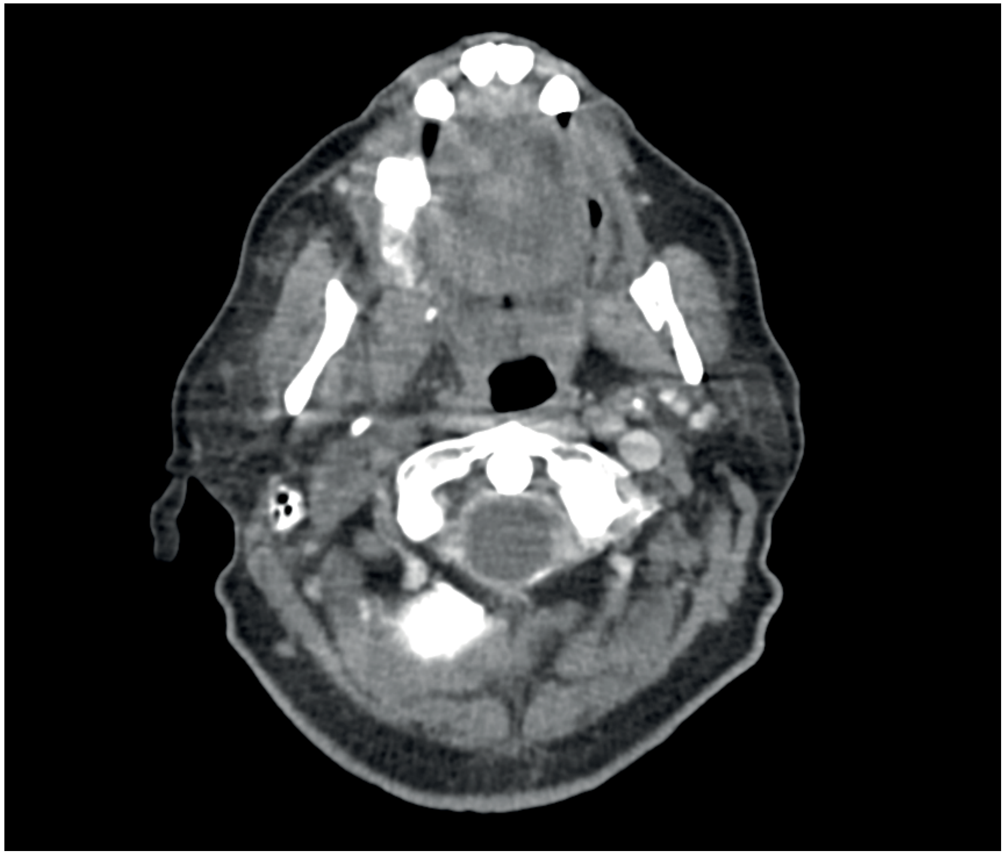
- Ultrasound or CT scan - confirms diagnosis + helps plan surgical approach
- CT shows ring-enhancing collection lateral to the pharynx
Fig. 52.29 - Axial CT neck showing right parapharyngeal abscess:
Treatment
- High-dose broad-spectrum IV antibiotics immediately
- IV fluid resuscitation
- If collection evident: Surgical drainage (mandatory)
- Transcervical drainage (external neck incision) - standard approach
- If abscess points into oropharynx: intraoral drainage with blunt instrument
- Requires senior anaesthetist due to airway risk
- In early cases without frank abscess: antibiotics alone may resolve it
4. Dangerous Complications of All Pharyngeal Abscesses
These are life-threatening and require urgent intervention:
| Complication | Mechanism |
|---|---|
| Airway obstruction | Direct compression by expanding abscess |
| Mediastinitis | Infection tracks down retropharyngeal/parapharyngeal space into chest |
| Lemierre's Syndrome | Septic thrombophlebitis of the internal jugular vein with metastatic abscesses (rare but fatal) |
| Aspiration pneumonia | Spontaneous or iatrogenic rupture of abscess |
| Cervical osteomyelitis | Spread to vertebral bodies |
| Retroperitoneal sepsis | Extension below diaphragm (very rare) |
| Carotid artery erosion | In parapharyngeal abscess (surgical emergency) |
⚠️ It is essential to be proactive in managing deep neck space infections - delays can be fatal.
5. Quick Comparison Summary
| Feature | Peritonsillar | Retropharyngeal | Parapharyngeal |
|---|---|---|---|
| Age | Older children/adults | Infants & <5 yrs | Young children |
| Best indicator | Trismus | Torticollis | Neck swelling + trismus |
| Uvula deviation | Yes, to opposite side | No | No |
| Airway risk | Moderate | High | High |
| Key imaging | Clinical ± CT | Lateral X-ray + CT | CT / Ultrasound |
| Drainage route | Oral (needle/I&D) | Oral (peroral) ± cervical | Transcervical (mostly) |
| TB association | No | Yes (chronic type) | No |
6. Key Organisms
- Group A beta-hemolytic Streptococcus - most common
- Staphylococcus aureus (including MRSA in some settings)
- Anaerobic Streptococci
- Mixed flora is common in all types
7. Antibiotic Principles
- First line: High-dose IV Penicillin (amoxicillin-clavulanate) or Cephalosporin
- Add metronidazole for anaerobic cover in parapharyngeal/retropharyngeal
- MRSA cover (vancomycin/clindamycin) if risk factors present
- Duration: typically 10-14 days, transition to oral once improving
Sources: Scott-Brown's Otorhinolaryngology Head & Neck Surgery Vol 2, KJ Lee's Essential Otolaryngology, Cummings Otolaryngology Head & Neck Surgery, Bailey & Love's Short Practice of Surgery 28th Ed.
This is a shared conversation. Sign in to Orris to start your own chat.