
1. why does right sided heart failure causes peripheral edema while left sided heart failure causes pulmonary edemaa. 2. what will happen when hydrostatic pressure increases and oncotic pressure decreases together in pathology of edema ? 3. how does mosquito and insect bite causes edema 4. since nephrotic syndrome leads to edema, can nephritc syndrome also causes edema 5. if there is slight chnage in the presssure at the veonous end will it also result in edema 6. what happens when oncotic pressure will remain and hydrostatic pressure will remain unchnged. 7. elaborate on cardogenic and non-cardiogenic edema 8. Why does decreased albumin causes generalized edema? 9. how does inflammation or an allergic reaction increases capillary permeability and cause edema? 10. why does standing for a long time cause swelling mainly in feet and ankles? 11. if lymphatic drainage system can normally remove excess interstitial fluid, why does edema become severe when lymphatic drainage is blocked? answer the questions simply just based on what's asked and not the entire detailed history of the question. Keep it simple and easy to understand.
1. Why does right-sided heart failure cause peripheral edema while left-sided causes pulmonary edema?
-
The right heart receives blood returning from the body (systemic veins) and pumps it to the lungs. When it fails, blood backs up into the systemic veins - raising venous (hydrostatic) pressure throughout the body. This pushes fluid out of capillaries into tissues, especially in the legs and ankles (gravity-dependent areas). Result: peripheral/dependent edema.
-
The left heart receives blood from the lungs and pumps it to the body. When it fails, blood backs up into the pulmonary veins - raising pressure in lung capillaries. Fluid leaks into the lung tissue and air sacs. Result: pulmonary edema (breathlessness, wet lungs).
2. What happens when both hydrostatic pressure increases AND oncotic pressure decreases together?
- Increased hydrostatic pressure actively pushes fluid out.
- Decreased oncotic pressure means there is less "pulling force" to keep fluid inside or bring it back.
3. How do mosquito/insect bites cause edema?
- Immune cells (mast cells) release histamine and other mediators.
- Histamine causes local capillaries to dilate and become "leaky" (increased permeability).
- Fluid, proteins, and immune cells pour out of these leaky capillaries into the surrounding tissue.
- This shows up as the classic red, swollen, itchy bump at the bite site.
4. Can nephritic syndrome also cause edema?
| Feature | Nephrotic | Nephritic |
|---|---|---|
| Edema | ++++ (severe) | ++ (mild-moderate) |
| Serum albumin | Low | Normal/slightly low |
5. Will a slight change in venous-end pressure also cause edema?
6. What happens when both oncotic pressure AND hydrostatic pressure remain unchanged?
- Fluid filtered at the arterial end = fluid reabsorbed at the venous end + fluid cleared by lymphatics.
- No excess fluid accumulates in the tissues.
7. Cardiogenic vs. Non-Cardiogenic Edema
Cardiogenic Edema
- Cause: The heart is failing, raising venous back-pressure.
- Mechanism: Increased hydrostatic pressure in capillaries pushes fluid out.
- Where: Pulmonary edema (left heart failure) or peripheral/dependent edema (right heart failure).
- Clues: Elevated JVP, third heart sound, cardiac history, responds to diuretics.
Non-Cardiogenic Edema
| Type | Mechanism | Example |
|---|---|---|
| Low oncotic pressure | Decreased albumin → less "pull" to retain fluid | Nephrotic syndrome, cirrhosis, malnutrition |
| Increased permeability | Leaky capillaries let protein and fluid escape | Sepsis, burns, ARDS, inflammation, allergic reactions |
| Lymphatic obstruction | Lymphatics can't drain excess fluid | Filariasis, post-mastectomy, cancer |
| Venous obstruction | Local back-pressure without heart failure | DVT, tumor compressing a vein |
8. Why does decreased albumin cause generalized edema?
- The pulling force inside vessels is weakened.
- At the venous end of capillaries, less fluid is reabsorbed back into the blood.
- Fluid accumulates in tissues everywhere in the body simultaneously.
9. How do inflammation/allergic reactions increase capillary permeability and cause edema?
- Immune cells (especially mast cells) release histamine, bradykinin, and prostaglandins.
- These mediators cause the endothelial cells lining capillaries to contract and pull apart, creating gaps.
- Now not just water, but also plasma proteins leak out into the interstitium.
- Proteins in the interstitium raise interstitial oncotic pressure, which pulls even more fluid out of capillaries.
- Result: localized swelling (edema) at the site of inflammation or allergic reaction.
10. Why does standing for a long time cause swelling in the feet and ankles?
- Blood pools in leg veins → venous (hydrostatic) pressure rises in leg capillaries.
- This pushes more fluid out of capillaries into the tissues of the feet and ankles.
- The lymphatics can handle some of it, but prolonged standing overwhelms them.
- Result: bilateral, dependent, pitting edema in the feet and ankles.
11. If lymphatics normally remove excess fluid, why does edema become severe when they are blocked?
- Fluid accumulates - the overflow drain is gone.
- More critically, proteins that normally leak into the interstitium cannot be cleared. These proteins raise interstitial oncotic pressure.
- Higher interstitial oncotic pressure actively pulls more fluid out of capillaries, creating a vicious cycle.
- The edema keeps worsening and becomes protein-rich, firm, and non-pitting over time (true lymphedema).
is there any treatment tog Milroys disease
Milroy disease treatment management
primary lymphedema FLT4 VEGFR3 treatment
Treatment of Milroy's Disease
1. Complete Decongestive Therapy (CDT) - The Main Treatment
- Manual Lymph Drainage (MLD) - a specialist massage technique that moves fluid from the swollen area toward functioning lymph basins elsewhere in the body
- Short-stretch compression bandaging - applied immediately after MLD to hold the reduction
- Therapeutic exercises - specific movements to drive lymph flow through any collateral pathways
- Skin care - keeping skin intact to prevent infections (cellulitis is a major risk)
- Wearing compression garments (stockings/sleeves) daily - these are graded by pressure (20-60 mmHg depending on severity)
- Continuing exercises
- Skin hygiene
2. Compression Garments
| Class | Pressure | Use |
|---|---|---|
| I | 20-30 mmHg | Simple/mild edema |
| II | 30-40 mmHg | Moderate edema |
| III | 40-50 mmHg | Severe edema |
| IV | 50-60 mmHg | Elephantiasis/severe lymphedema |
3. Intermittent Pneumatic Compression (IPC)
4. Antibiotics (for infection prevention)
5. Surgical Options (limited, selected cases)
- Debulking surgery (Charles procedure) - removes excess fibrotic and fatty tissue in severely affected limbs
- Lymphovenous anastomosis - microsurgical connections between lymphatics and small veins to create a bypass
- Vascularized lymph node transfer - transplanting healthy lymph nodes to the affected area
6. What About Diuretics?
7. Emerging/Experimental Treatments
In Summary
| Approach | Role |
|---|---|
| Manual lymph drainage + bandaging | Main treatment - reduces swelling |
| Compression garments | Lifelong maintenance |
| Skin care + antibiotics | Prevents cellulitis |
| Intermittent pneumatic compression | Adjunct device |
| Surgery | Last resort for severe cases |
| Curative/gene therapy | Not yet available |
lip edema
lip angioedema swelling
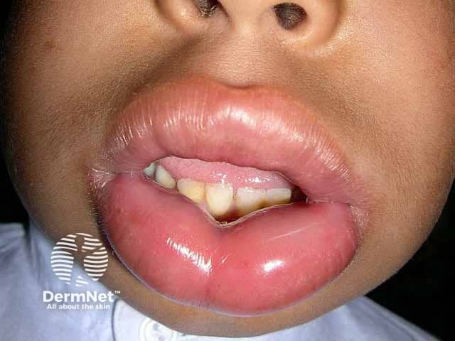
Clinical photography of the lips (frontal close-up) showing severe bilateral lip edema. The vermilion and perioral soft tissues are markedly swollen, producing tense, shiny contours that obscure the normal landmarks. Mucosal surfaces appear moist with no visible ulcers or vesicles; dentition is partially masked by swelling. There is symmetry across the upper and lower lips, with involvement of perioral skin but without obvious surrounding erythema or warmth visible in the image. No airway compromise is evident in the photograph, but lip angioedema can progress rapidly and mandating urgent clinical assessment. Pathology: edema of subcutaneous and submucosal tissues consistent with angioedema; no infectious purulence or necrosis seen. Diagnostic significance: lip angioedema is a potentially life-threatening condition due to airway risk; requires prompt evaluation, segregation from cellulitis, and identification of trigger type. Differential considerations: allergic angioedema, bradykinin-mediated hereditary angioedema, medication-induced edema (e.g., ACE inhibitors), infectious cellulitis, trauma, or angioedema with urticaria. Clinical correlation: obtain exposure history, past angioedema episodes, medication use; management includes airway monitoring, antihistamines, corticosteroids, with epinephrine if signs of systemic reaction or airway threat; specialist consultation as needed. This image serves educationally for recognition, triage, and discussion of management strategies in acute lip swelling and potential airway emergencies today.
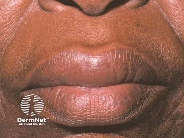
This is a high-resolution clinical photograph of the perioral region focusing on the lips, captured as a frontal close-up for documentation of acute lip edema. Imaging modality: Clinical photography; technique: standardized lighting, macro-close-up view, frontal orientation, minimal compression. Magnification approximately 1:1 to 2:1. Anatomical location: lips (labial mucosa) within the orolabial region of the face, part of the integumentary system. Visual features: bilateral lip swelling involving both the upper and lower lips with soft-tissue edema that distends the vermilion border; surface skin appears smooth without discrete vesicles, ulceration, ecchymosis, or erosions; mild hue change may reflect venous congestion. Clinical interpretation: edema is transient and compatible with angioedema, a rapid-onset swelling of subcutaneous and submucosal tissues. Differential considerations include allergic angioedema, hereditary angioedema, angiotensin-converting enzyme inhibitor–associated angioedema, trauma-related edema, and infection-related swelling. Clinical significance: lip angioedema can compromise the airway in severe cases; recognizing transient edema is essential for timely treatment and monitoring. Potential use cases: medical education, dermatology and allergy case reviews, pharmacovigilance, and AI-assisted image retrieval for perioral edema. This description emphasizes reproducible terminology to improve retrieval across platforms and support longitudinal comparisons in clinical documentation, telemedicine consultations, and multicenter edema datasets.
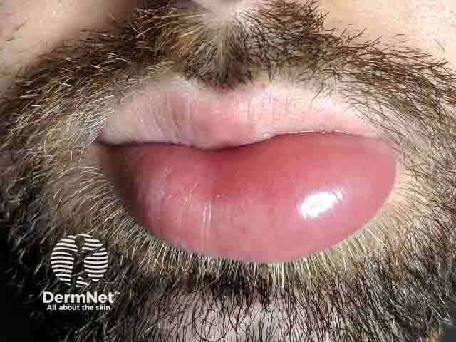
Imaging modality: Clinical photograph; technique: high-resolution frontal close-up; view: frontal view of the perioral region focusing on the lips. Anatomical location: lower lip prominence with surrounding mucosal lip tissue; context: soft tissue edema consistent with angioedema of the lips. Visual features: bright pink-red, smooth, expanded lip contour; surface unbroken with no ulceration; turgor and elasticity reduced due to edema; perioral skin shows fine hairs present, no purpura, no vesiculation; the chin and surrounding beard are visible, providing scale. Pathology: edema of the subcutaneous tissue without overt dermal necrosis; non-pitting, non-inflammatory swelling. Diagnostic significance: supportive of acute angioedema; helps differentiate from cellulitis (more erythema, warmth, induration) and allergic dermatitis (more surface involvement). Clinical correlation: correlate with onset, exposure to allergens/ACE inhibitors; assess airway risk if swelling progresses; monitor for tongue or laryngeal involvement; required management may include antihistamines, corticosteroids, or epinephrine in an anaphylactic scenario; potential uses: educational reference for dermatology, emergency medicine, allergology, and primary care; differential diagnoses include hereditary angioedema, medication-induced edema, infectious edema; imaging is not required for routine angioedema but clinical photography documents severity and treatment response. This image is suitable for case-based teaching, triage decision-making, patient education on recognizing lip swelling and seeking prompt medical care.
Lip Edema
Causes
1. Allergic Angioedema (Most Common Acute Cause)
- Triggered by food (nuts, shellfish, eggs), drugs (NSAIDs, penicillin), insect stings, latex
- Mechanism: mast cells release histamine → capillaries become leaky → fluid pours into lip tissue
- Usually comes with urticaria (hives) and itching
- Can progress to involve tongue, throat, and larynx → airway emergency
- Onset: within minutes of exposure
2. ACE Inhibitor-Induced Angioedema
- Caused by drugs like enalapril, lisinopril, ramipril (very common blood pressure medications)
- Mechanism: ACE normally breaks down bradykinin. When blocked, bradykinin accumulates → vasodilation + leaky capillaries
- No hives, no itching - just swelling (painful/pressure sensation)
- Has a predilection for lips, tongue, eyelids
- Can occur any time - even years after starting the drug
- 3-4x more common in African Americans; 50% more common in women
- Does NOT respond to antihistamines or steroids well
3. Hereditary Angioedema (HAE)
- Genetic condition - C1 esterase inhibitor deficiency (autosomal dominant)
- Without C1-INH, the kallikrein-kinin system runs unchecked → excess bradykinin produced
- Recurrent episodes of lip, face, abdominal, or throat swelling
- No hives - distinguishes it from allergic angioedema
- Triggered by stress, trauma, infection, estrogen (worse in pregnancy/OCP use)
- Onset usually in childhood/teens
4. Melkersson-Rosenthal Syndrome
- A rare chronic condition causing recurrent, persistent lip swelling (orofacial granulomatosis)
- Classic triad: recurrent orofacial edema + facial nerve palsy + fissured tongue (full triad in only 25%)
- Lips become permanently enlarged and deformed over time - "cobblestone" or chapped, fissured appearance
- Biopsy shows non-caseating granulomas (like sarcoid/Crohn's)
- Cause unknown; thought to involve lymphatic/vascular disruption by granulomas
- Distinguished from angioedema by its chronic, non-transient nature
5. Infections
- Cellulitis - bacterial skin infection spreading to lip tissue (more erythema, warmth, induration)
- Dental abscess - infection spreads from tooth root into buccal/lip tissue
- Herpes labialis (cold sores) - HSV-1 causes localized swelling with vesicles/crusting
6. Trauma / Physical
- Lip biting, insect bites, burns, lip piercings
- Localized, usually obvious history
7. Contact Dermatitis
- Reaction to lipstick, lip balm, toothpaste, food contact
- Causes itchy, swollen, sometimes crusted lips
Key Differences at a Glance
| Feature | Allergic Angioedema | HAE / ACE-inhibitor | Melkersson-Rosenthal | Cellulitis |
|---|---|---|---|---|
| Onset | Minutes | Hours | Gradual/recurrent | Hours-days |
| Hives? | Yes | No | No | No |
| Itchy? | Yes | No (pressure/pain) | No | No |
| Recurrent? | Maybe | Yes | Yes (chronic) | Sometimes |
| Responds to antihistamines? | Yes | No | Partly (steroids) | No (antibiotics) |
| Fever? | No | No | No | Yes |
Treatment
Allergic (Histaminergic) Angioedema
- Mild: Oral antihistamines (cetirizine, loratadine) - 1st line; H2-blockers can be added; short course of oral steroids (prednisone) for 2nd line
- Severe (airway threatened): Epinephrine IM (adrenaline) immediately - this is the priority
- Avoid the trigger going forward; patient should carry an EpiPen
ACE Inhibitor Angioedema
- Stop the ACE inhibitor - this is the most important step
- Antihistamines/steroids are largely ineffective (bradykinin-mediated, not histamine)
- Supportive care; monitor airway closely
- Fresh frozen plasma (FFP) has been used in severe cases (contains C1-INH)
- Agents like icatibant (bradykinin-2 receptor blocker) and ecallantide (kallikrein inhibitor) have been tried with mixed results
Hereditary Angioedema (HAE) - Acute Attack
| Drug | Mechanism | Route | Time to Relief |
|---|---|---|---|
| Berinert / Rucorest | C1-INH replacement | IV | ~48-90 min |
| Ecallantide | Plasma kallikrein inhibitor | SC | ~67 min |
| Icatibant | Bradykinin-2 receptor blocker | SC | ~2 hours |
| Fresh frozen plasma | Contains C1-INH | IV | Variable |
Melkersson-Rosenthal
- No consistent cure - treatment is difficult
- Systemic corticosteroids (most commonly used), dapsone, metronidazole, methotrexate - none reliably effective
- Intralesional steroid injections into the lips for cosmetic improvement
- Surgery (lip reduction) for severe cosmetic deformity
- Manage facial nerve palsy symptomatically
Infections
- Cellulitis: antibiotics (amoxicillin-clavulanate or clindamycin)
- Dental abscess: drainage + antibiotics
- Herpes labialis: acyclovir/valacyclovir
When to Worry (Red Flags)
- Swelling extends to the tongue, floor of mouth, throat, or larynx
- Patient has difficulty breathing, stridor, muffled voice, or drooling
- Swelling is rapidly progressive