
Types of non union
Reading File
Non-union (fracture non-union) refers to failure of a fracture to heal within the expected time frame. The classic classification divides it into two broad categories: hypertrophic (viable/vascular) and atrophic (non-viable/avascular), with several subtypes in between.
Searching sources...
Finding Sources
Finding Sources
Reading File
Reading File
Finding Sources
Finding Sources
Searching the Web
types of non-union fracture classification Weber Cech hypertrophic atrophic
Reading File
Reading File
Reading File
Loading Image
Reading File
Reading File
Here is a comprehensive overview of the types of non-union based on the Weber & Cech classification (1976), which remains the most widely used system worldwide.
Definition
Non-union is a fracture that has failed to heal and shows no potential to heal without further intervention. It can also be defined as a fracture failing to show clinical or radiological improvement over 3 months. In practice, the term is generally not applied until 6 months after injury. - Bailey & Love's Short Practice of Surgery, 28th Ed.
Classification (Weber & Cech, 1976)
Non-unions are divided into two broad groups based on biological viability (vascularity) at the fracture site:
GROUP 1: VIABLE (Hypervascular / Reactive) Non-Unions
Biology is intact - the fracture ends have adequate blood supply and bone-forming capacity. The problem is mechanical (insufficient stability).
1. Hypertrophic Non-Union ("Elephant Foot")
- Appearance: Abundant callus formation around both bone ends, producing a bulbous "elephant foot" shape on X-ray
- Vascularity: Rich - good blood supply
- Cause: Excessive micromotion / inadequate stabilization
- Bone scan: Hot (increased uptake)
- Treatment: Stabilization alone (plate, IM nail) - no bone graft needed. Biology is intact; rigid fixation allows fibrocartilage to mineralize, typically within 8 weeks
- Campbell's Operative Orthopaedics, 15th Ed.
2. Hypertrophic Non-Union ("Horse Hoof")
- Appearance: Moderate callus (less than elephant foot), persistent fracture line
- Vascularity: Adequate
- Cause: Less severe mechanical instability than elephant foot
- Treatment: Stabilization +/- bone graft
3. Oligotrophic Non-Union
- Appearance: Minimal or no callus, but bone ends are still viable
- Vascularity: Adequate (confirmed by bone scan - shows increased uptake, unlike atrophic)
- Cause: Distraction of bone ends, soft tissue interposition, or malreduction - the gap prevents contact even though biology is intact
- Distinction from atrophic: Bone scan shows increased uptake (oligotrophic) vs. "cold cleft" (atrophic)
- Treatment: Mechanical compression or bone grafting to restore contact + stabilization
- Rockwood & Green's Fractures in Adults, 10th Ed. 2025
GROUP 2: NON-VIABLE (Avascular / Areactive) Non-Unions
Biology is compromised - poor vascularity, little/no bone-forming capacity. The problem is both biological AND mechanical.
4. Torsion Wedge Non-Union
- Appearance: A butterfly/wedge fragment with impaired vascularity
- Cause: Butterfly fragment loses its blood supply, preventing incorporation
- Treatment: Stabilization + bone graft
5. Comminuted Non-Union
- Appearance: Multiple fragments, some or all avascular
- Cause: High-energy injury with extensive devascularization
- Treatment: Stabilization + aggressive bone grafting
6. Defect Non-Union
- Appearance: A segment of bone is missing (from trauma, infection, or previous surgery)
- Cause: Bone loss
- Treatment: Bone transport (Ilizarov), vascularized fibular graft, or Masquelet technique
7. Atrophic Non-Union (True Atrophic)
- Appearance: No callus at all, tapered/rounded bone ends, sclerosis and medullary canal closure
- Vascularity: Poor to absent
- Cause: Host factors (diabetes, smoking, malnutrition, radiation), severe soft tissue stripping, open fractures
- Bone scan: Cold ("cold cleft" between areas of activity)
- Treatment: Mandatory debridement/excision of avascular bone ends + aggressive biological stimulus (autograft, BMP) + rigid stabilization
- Rockwood & Green's Fractures in Adults, 10th Ed. 2025
5. Pseudoarthrosis (Synovial / False Joint)
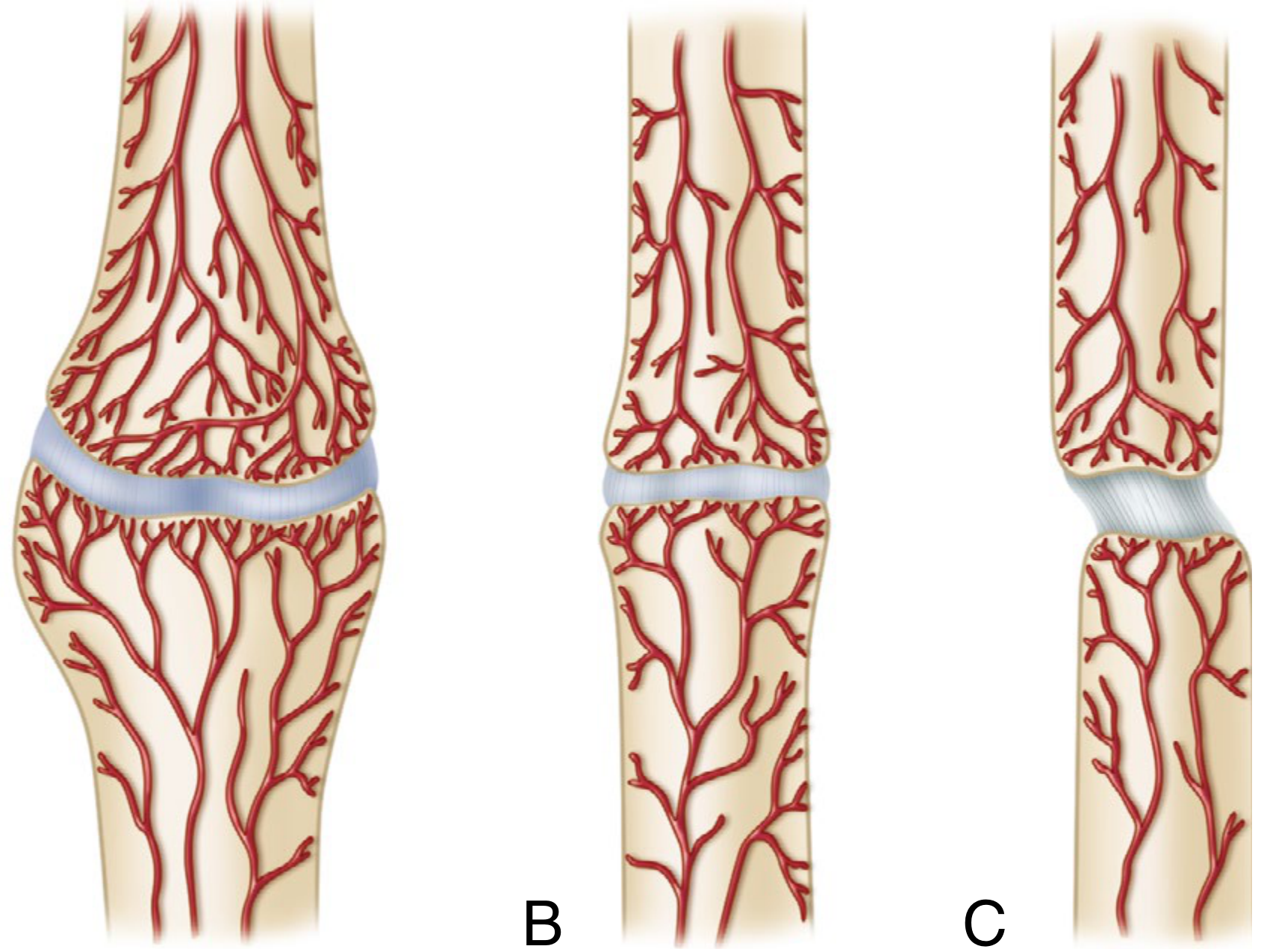
A special subtype - considered an extreme form of hypertrophic non-union in which chronic excessive motion leads to the formation of a synovial pseudocapsule around the fracture gap containing fluid, mimicking a true synovial joint. Medullary canals become sealed. On bone scan, a "cold cleft" appears between two "hot" areas.
- Rockwood & Green's Fractures in Adults, 10th Ed. 2025; Campbell's Operative Orthopaedics, 15th Ed.
Infected (Septic) Non-Union
A separate classification dimension - any of the above types can be septic or aseptic. Septic non-unions are far more difficult to treat, typically requiring staged management: infection control first (with debridement and external fixation), then bone grafting after eradication of infection.
Summary Table
| Type | Callus | Vascularity | Core Problem | Key Treatment |
|---|---|---|---|---|
| Hypertrophic (Elephant Foot) | Abundant | Good | Instability | Stabilize only |
| Hypertrophic (Horse Hoof) | Moderate | Good | Instability | Stabilize ± graft |
| Oligotrophic | Minimal | Adequate | Gap/distraction | Contact + stabilize ± graft |
| Torsion Wedge | Variable | Poor (fragment) | Avascular fragment | Stabilize + graft |
| Comminuted | Absent | Poor | Devascularization | Stabilize + graft |
| Defect | Absent | Poor | Bone loss | Bone transport / Masquelet |
| Atrophic | Absent | Poor/absent | Biology failure | Debride + graft + stabilize |
| Pseudoarthrosis | Variable | Variable | Chronic motion/false joint | Resect pseudocapsule + stabilize |
Key clinical principle: Hypertrophic non-unions are a mechanical problem - stabilization alone is curative. Atrophic non-unions are a biological problem - both stability and biology (bone graft) must be restored. Oligotrophic sits between the two and often requires both.
- Sources: Rockwood & Green's Fractures in Adults, 10th Ed. 2025; Campbell's Operative Orthopaedics, 15th Ed. 2026; Bailey & Love's, 28th Ed.; Pye's Surgical Handicraft, 22nd Ed.
This is a shared conversation. Sign in to Orris to start your own chat.