


Solve the 2 papers very accurately get me images ,flow charts and diagrams where necessary
fetal circulation changes at birth ductus arteriosus foramen ovale
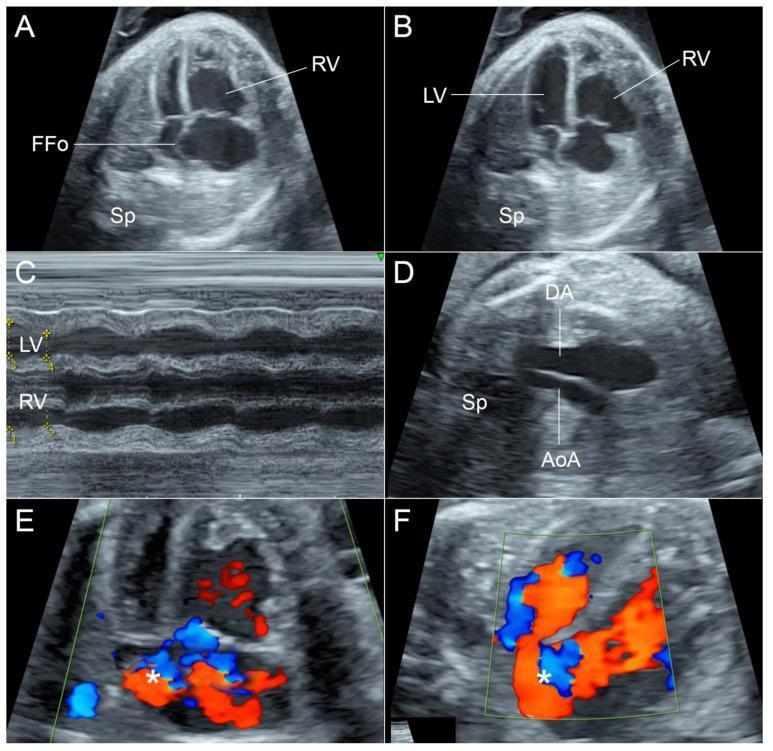
This composite figure presents a detailed fetal echocardiographic evaluation at 35 weeks of gestation. Panels A and B (four-chamber views) demonstrate significant chamber disproportion, with a dilated right ventricle (RV) compared to the left ventricle (LV). The flap of the foramen ovale (FFo) appears relatively fixed and exhibits tense bulging throughout both systole (A) and diastole (B). Panel C (M-mode) confirms impaired RV contractility, showing a reduced shortening fraction compared to the LV. Panel D (three-vessel and trachea view) reveals a hypoplastic aortic arch (AoA) in comparison to the larger ductus arteriosus (DA). Panels E and F utilize color Doppler imaging to illustrate abnormal hemodynamics at the interatrial septum, characterized by turbulent blood flow (marked with asterisks) and restricted passage across the foramen ovale. These findings are clinically significant for identifying secondary cardiac changes due to fetal anemia and altered preload conditions, emphasizing the assessment of the foramen ovale and ventricular function in high-risk pregnancies.
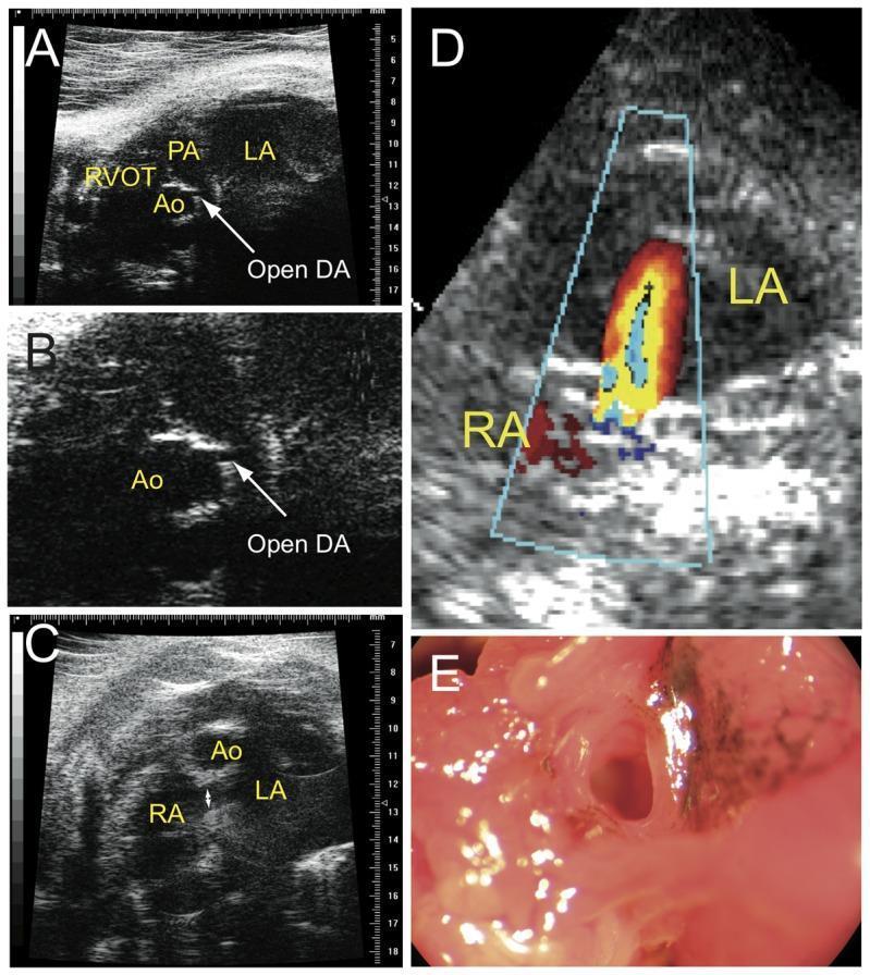
A multi-panel medical diagnostic and clinical series demonstrating congenital cardiovascular anomalies in a ctnnb1Δex3 model. Panels A and B are ultrasound images illustrating a Patent Ductus Arteriosus (PDA), with labels identifying the Left Atrium (LA), Aorta (Ao), Right Ventricle Outflow Tract (RVOT), and Pulmonary Artery (PA). An arrow points to the 'Open DA' (Ductus Arteriosus). Panel C provides an ultrasound view of the atrial septum with labels for the Right Atrium (RA), LA, and Ao, showing a patent foramen ovale. Panel D utilizes Color Doppler imaging to show a pathological right-to-left shunt of blood flow through the foramen ovale. Panel E is a clinical photograph of a dissected heart confirming the presence of an open foramen ovale, notable for the presence of dark pigmented cells (melanocytes) around the aperture. The collection demonstrates how failure of fetal cardiovascular shunts to close postnatally leads to abnormal circulation and chamber dilation.
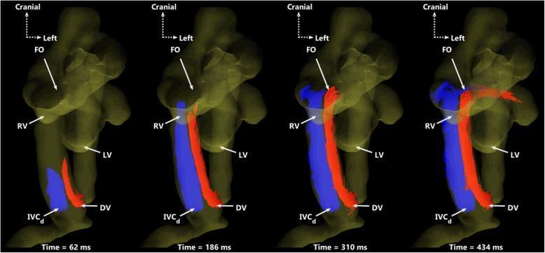
This diagnostic image series utilizes 4D flow MRI-derived particle tracing to illustrate fetal circulatory dynamics in a ventral view. The visualization demonstrates the preferential streaming of blood from two distinct venous sources over one cardiac cycle (62 ms, 186 ms, 310 ms, and 434 ms). Red particles represent blood from the ductus venosus (DV), while blue particles represent blood from the distal inferior vena cava (IVCd). The series highlights the maintenance of separate flow streams within the proximal inferior vena cava. The red DV stream is directed preferentially through the foramen ovale (FO) toward the left ventricle (LV) to supply oxygenated blood to the systemic circulation. Concurrently, the blue IVCd stream is routed primarily toward the right ventricle (RV) for delivery to the main pulmonary artery. Anatomical landmarks labeled include the ductus venosus (DV), distal inferior vena cava (IVCd), foramen ovale (FO), right ventricle (RV), and left ventricle (LV). This pedagogical tool illustrates the physiological shunting essential for fetal development and the minimal mixing of nutrient-rich and nutrient-poor blood streams.
wound healing phases stages diagram
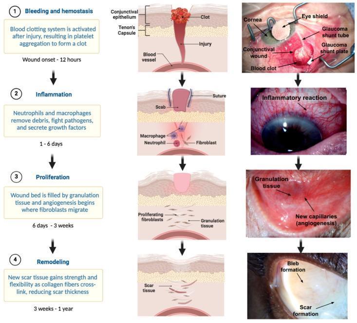
This infographic and clinical photograph series illustrates the four stages of ocular wound healing following glaucoma filtration surgery (e.g., shunt implantation). The diagram is organized into four horizontal rows representing the chronological phases: (1) Bleeding and Hemostasis (0–12 hours), showing platelet aggregation and a visible blood clot at the conjunctival wound site alongside surgical elements like the glaucoma shunt tube and plate. (2) Inflammation (1–6 days), characterized by neutrophil and macrophage recruitment and a clinical appearance of diffuse episcleral/conjunctival injection (redness). (3) Proliferation (6 days–3 weeks), showing fibroblast migration, collagen deposition, and angiogenesis (new capillary formation) clinical visible as fleshy granulation tissue. (4) Remodeling (3 weeks–1 year), depicting scar tissue maturation and the formation of a functional surgical bleb. The figure combines anatomical cross-sections of the conjunctiva and Tenon's capsule with real-world clinical images to demonstrate the physiological transition from acute injury to long-term surgical outcome in ophthalmology.
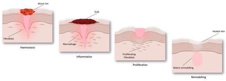
An anatomical pathophysiology diagram illustrating the four sequential stages of cutaneous wound healing: haemostasis, inflammation, proliferation, and remodeling. The illustration depicts a cross-section of the skin layers (epidermis and dermis). During 'Haemostasis', a red blood clot forms on the surface above a funnel-shaped injury, with fibroblasts visible in the surrounding dermis. In the 'Inflammation' stage, a dark-colored scab replaces the initial clot, and macrophages are identified in the underlying tissue. The 'Proliferation' stage demonstrates the formation of pinkish granulation tissue filling the wound gap, accompanied by 'proliferating fibroblasts' migrating into the site. Finally, the 'Remodeling' stage shows 'healed skin' with a restored epidermal surface and an underlying zone of 'matrix remodeling' replacing the previous injury site. The diagram serves as an educational summary of tissue repair mechanisms and cellular involvement at different phases of recovery.
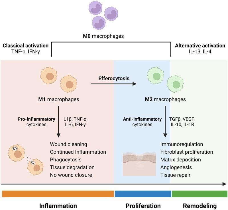
A pathophysiology diagram illustrating macrophage polarization during the stages of wound healing. The process begins with M0 macrophages (undifferentiated) which undergo 'Classical activation' via TNF-α and IFN-γ to become M1 macrophages. This pro-inflammatory phenotype predominates during the 'Inflammation' phase, secreting cytokines (IL-1β, TNF-α, IL-6, IFN-γ) and performing phagocytosis, tissue degradation, and wound cleaning. A transition via efferocytosis leads to 'Alternative activation' stimulated by IL-13 and IL-4, resulting in M2 macrophages. The M2 phenotype characterizes the 'Proliferation' and 'Remodeling' phases, producing anti-inflammatory and growth factors (TGFβ, VEGF, IL-10, IL-1R). These actions promote immunoregulation, fibroblast proliferation, matrix deposition, and angiogenesis, ultimately leading to successful tissue repair. The diagram uses color-coded backgrounds (orange for inflammation, blue for proliferation, green for remodeling) to link cellular phenotypes to clinical wound progression.
body temperature regulation thermoregulation hyperthermia heat stroke
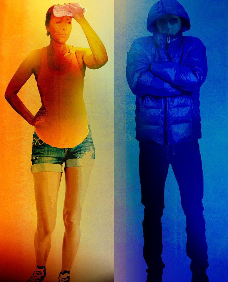
This educational comparison infographic illustrates the physiological impact of environmental temperature extremes on human health. The image is split vertically to contrast hyperthermia and hypothermia risks. The left panel, rendered in warm orange and yellow tones, depicts a figure showing signs of heat-related distress; the individual is lightly dressed in a tank top and shorts, holding a water bottle to their forehead to demonstrate cooling behaviors associated with heat exhaustion or heat stroke. The right panel, rendered in cool blue tones, shows a figure in a heavy hooded puffer jacket with arms crossed, symbolizing the body's attempt to conserve core heat during cold exposure. The visual serves as a conceptual aid for public health and environmental medicine, highlighting the physical and behavioral adaptations necessitated by temperature fluctuations. It is intended for intermediate medical or public health curricula to discuss climate-related health risks, thermoregulation, and the epidemiological relationship between seasonal temperature changes and mortality.
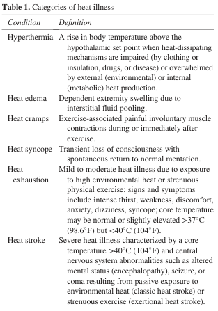
Table 1. Categories of heat illness <table><thead><tr><th>Condition</th><th>Definition</th></tr></thead><tbody><tr><td>Hyperthermia</td><td>A rise in body temperature above the hypothalamic set point when heat-dissipating mechanisms are impaired (by clothing or insulation, drugs, or disease) or overwhelmed by external (environmental) or internal (metabolic) heat production.</td></tr><tr><td>Heat edema</td><td>Dependent extremity swelling due to interstitial fluid pooling.</td></tr><tr><td>Heat cramps</td><td>Exercise-associated painful involuntary muscle contractions during or immediately after exercise.</td></tr><tr><td>Heat syncope</td><td>Transient loss of consciousness with spontaneous return to normal mentation.</td></tr><tr><td>Heat exhaustion</td><td>Mild to moderate heat illness due to exposure to high environmental heat or strenuous physical exercise; signs and symptoms include intense thirst, weakness, discomfort, anxiety, dizziness, syncope; core temperature may be normal or slightly elevated >37°C (98.6°F) but <40°C (104°F).</td></tr><tr><td>Heat stroke</td><td>Severe heat illness characterized by a core temperature >40°C (104°F) and central nervous system abnormalities such as altered mental status (encephalopathy), seizure, or coma resulting from passive exposure to environmental heat (classic heat stroke) or strenuous exercise (exertional heat stroke).</td></tr></tbody></table>
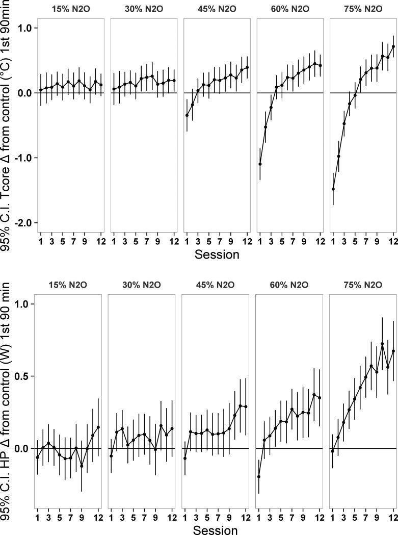
This physiological chart presents comparative data from a preclinical study examining the effects of nitrous oxide (N2O) on thermoregulation and metabolism. The data is organized into a matrix of line graphs across five N2O concentrations (15%, 30%, 45%, 60%, and 75%) over twelve 3-hour exposure sessions. The top row displays the 95% Confidence Interval (C.I.) for the change in core temperature (Tcore Δ in °C) relative to a control group during the first 90 minutes. At lower concentrations (15-30%), Tcore remains near baseline. At higher concentrations (≥45%), an initial hypothermic response (negative Δ) in early sessions transitions to progressive hyperthermia (positive Δ) by session 12, indicating thermal adaptation. The bottom row illustrates the change in heat production (HP Δ in Watts). Mirroring the temperature trends, higher N2O concentrations (60-75%) show a significant, dose-dependent increase in heat production over successive sessions. The charts highlight the pathophysiology of drug-induced thermoregulatory shifts and the development of tolerance or sensitization in metabolic responses to anesthetic gases.
female external genitalia anatomy diagram vulva hymen
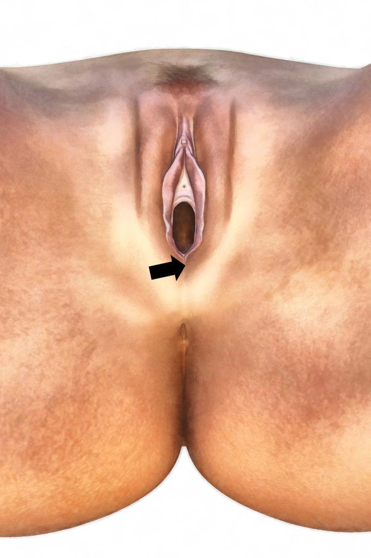
This medical anatomical diagram depicts the female external genitalia (vulva) and perineal region from a frontal perspective. Key anatomical landmarks include the labia majora, labia minora, clitoral hood, urethral meatus, and the vaginal introitus. A black arrow specifically identifies the posterior commissure of the vagina, located at the junction where the vaginal mucosa transitions into the dermal skin of the perineum. This site is clinically relevant as the target area for pulse oximetry (SpO2) measurements mentioned in the context, involving the underlying bulbospongiosus muscle. The illustration highlights the natural contours, skin texture, and spatial relationships between the vulvar structures and the anus within the perineal field. It serves as an educational reference for identifying clinical measurement sites and understanding female pelvic anatomy for gynecological or physiological studies.
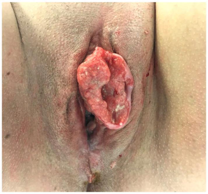
Clinical photograph of the female external genitalia showing a large, exophytic squamous cell carcinoma (SCC) of the vulva. The lesion is a centrally located, irregularly shaped mass measuring approximately 55 mm x 30 mm, originating from the left labia and extending to the midline. The tumor exhibits classic malignant morphology, including a cauliflower-like or verrucous texture with friable, erythematous tissue and areas of pale keratinization. Central ulceration and serosanguinous exudate are visible, suggesting tissue necrosis. The mass significantly distorts the local anatomy of the labia majora and minora. The surrounding perilesional skin shows signs of secondary inflammation and possible satellite nodules or lichenoid changes. This image serves as a clinical example of advanced vulvar malignancy, illustrating the need for surgical intervention such as radical local excision and assessment of inguinofemoral lymph nodes.
Clinical photograph of the female external genitalia showing the vulva on the 7th post-operative day following defibulation and excision of a vulvar inclusion cyst. The image demonstrates a healing surgical site along the labia. The wound edges are well-approximated and epithelizing, showing a characteristic transition in color from light-pink scar tissue at the superior aspect to more erythematous healing tissue at the inferior pole. Residual suture tracks or marks are visible along the linear incision line. The surrounding skin and pubic hair appear normal for the post-operative stage, with no signs of active purulent discharge or dehiscence. This visual serves as an educational example of secondary wound healing and successful surgical restoration of the vulvar anatomy (defibulation) in the context of treating complications from female genital mutilation or inclusive cysts.
autonomic nervous system sympathetic parasympathetic diagram
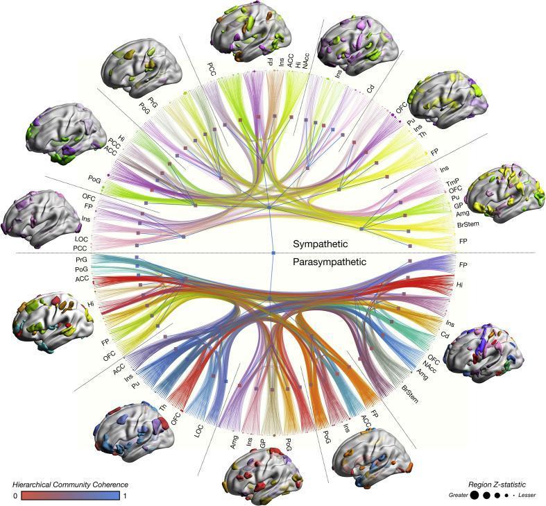
This multimodal anatomical and functional diagram illustrates the human 'autonomic connectome,' detailing the neural infrastructure of sympathetic and parasympathetic regulation. The central element is a circular network graph partitioned into two hemispheres: the upper represents the sympathetic nervous system and the lower the parasympathetic nervous system. Surrounding the center are colorful lines (edges) representing network-based statistics from functional MRI, gray matter morphometry, and white matter tractography. Edge width indicates connection effect size. The outer perimeter contains nodes corresponding to specific brain regions, such as the anterior cingulate cortex (ACC), insula (Ins), amygdala (Amg), and brainstem (BrStem). Node size is proportional to the Z-statistic importance of the region. Surrounding the central connectome are several 3D cortical surface reconstructions displaying color-coded parcellations that visualize hierarchical community structures. A color scale at the bottom indicates hierarchical community coherence (ranging from red to blue), while a size legend defines the regional Z-statistics. This educational visual explains the high-dimensional integration of disparate cortical and subcortical structures involved in autonomic homeostasis and sympathovagal balance.
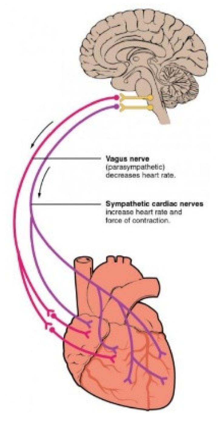
This medical illustration depicts the Autonomic Nervous System (ANS) pathways connecting the brainstem to the heart. A sagittal section of the human brain shows the medulla oblongata as the origin for both parasympathetic and sympathetic innervation. The Vagus nerve (parasympathetic), color-coded in purple, is shown descending from the brainstem to the heart, where it branches to innervate the atria and ventricles; text annotations indicate its role in decreasing heart rate. The sympathetic cardiac nerves, color-coded in red, originate from the same region and follow a parallel descending path, branching across the cardiac tissue; text labels specify their role in increasing both heart rate and the force of contraction. The diagram illustrates the functional antagonism between the two branches of the ANS in cardiac regulation, highlighting the primary innervation sites at the sinoatrial node, atrioventricular node, and the myocardium.
fracture types classification transverse oblique comminuted
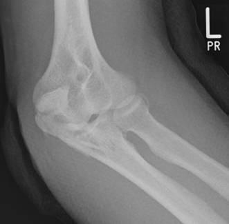
A diagnostic radiograph (X-ray) of the left elbow in an oblique view, demonstrating a comminuted fracture of the proximal ulna involving the olecranon process. The image reveals at least two distinct fracture lines, resulting in a multi-fragmentary pattern. A primary transverse fracture line extends across the olecranon, accompanied by distraction and displacement of the proximal fragment. A second fracture line indicates the presence of an intermediate fragment, which compromises the articular surface of the trochlear notch. The fracture pattern suggests a loss of structural integrity of the elbow's primary bony stabilizer against posterior displacement. Key anatomical landmarks visible include the distal humerus, the coronoid process of the ulna, and the radial head. This imaging illustrates the difficulty in detecting specific intermediate 'key' fragments using conventional biplanar radiography alone, highlighting the clinical relevance of orthopedic assessment for joint congruity and surgical planning.
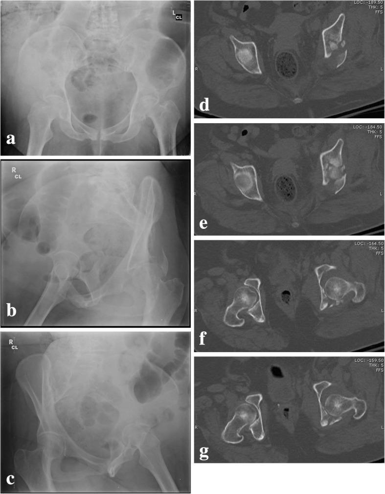
This composite figure presents pre-operative diagnostic imaging of a complex, comminuted transtectal transverse acetabular fracture with posterior wall involvement in a human patient. Panels (a–c) consist of conventional pelvic radiographs: (a) Anteroposterior view showing disruption of the ilioischial and iliopubic lines; (b) Obturator oblique view highlighting the anterior column and posterior wall; and (c) Iliac oblique view visualizing the posterior column and iliac wing. These images demonstrate significant disruption of the normal acetabular architecture and altered joint congruency. Panels (d–g) display axial Computed Tomography (CT) scans at different levels, providing a cross-sectional perspective of the fracture morphology. The CT slices clearly reveal the comminution of the acetabular roof, fracture lines extending through the quadrilateral surface, and displacement of the posterior wall fragments. The imaging collectively illustrates a T-shaped fracture pattern with high-energy articular impaction, serving as a teaching case for the Letournel classification and the assessment of femoral head stability (roof-arc measurements) in orthopedic trauma surgery.
pubic symphysis age estimation forensic anthropology
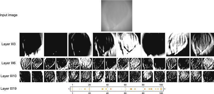
This technical diagram illustrates the internal feature extraction process of a convolutional neural network (CNN) used for forensic age estimation based on 3D scans of the human pubic symphysis. The image follows the evaluation of a 72-year-old individual through various processing stages. The 'Input image' shows a grayscale 2D projection of the 3D surface scan, appearing as a blurry anatomical outline. Subsequent rows visualize internal network layers: Layer #3 shows initial filters focusing on high-contrast edges and basic morphology; Layers #6 and #10 display multiple feature maps that refine complex textures and vertical ridge patterns characteristic of age-related skeletal degeneration. The final row, Layer #19, depicts a one-dimensional feature vector represented as a linear scale (1–100) with colored activation points, indicating the condensed high-level data used for the final age prediction (estimated here as 71.6 years). This visualization demonstrates how AI identifies subtle morphological changes in skeletal remains that may be difficult for the human eye to quantify, providing a specialized tool for forensic anthropology and clinical aging research.
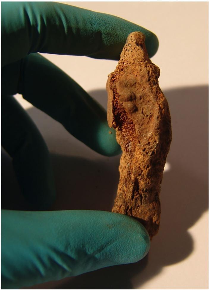
A clinical photograph of a specimen showing the symphyseal face of a left pubic bone fragment, held by a gloved hand. This osteological specimen is used for forensic age estimation based on morphological changes of the pubic symphysis. The visual features include well-marked ridges and furrows across the symphyseal surface, along with the early formation of a rim on both the ventral and dorsal margins. These specific age-related metamorphic changes are consistent with Stage II of the Acsádi and Nemeskéry classification system, typically indicating an adult individual in early to mid-middle age. The bone shows signs of weathering and mineralization changes common in exhumed skeletal remains. The image serves as an educational example of forensic anthropology and skeletal biology methods for determining biological profile in archaeological or forensic contexts.
fat embolism microscopy histology lung
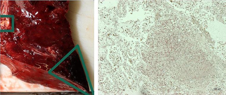
This composite educational image illustrates the pathological findings of a lung abscess and infarction through gross inspection and histology. The left panel shows a clinical photograph of a gross lung specimen. The tissue exhibits diffuse congestion and a dark red, hemorrhagic appearance. A green square highlights a localized lung abscess characterized by a paler, necrotic, and irregular surface texture. A green triangle identifies a wedge-shaped, dark, consolidated area consistent with a lung infarction, likely secondary to septic embolism. The right panel displays a corresponding histological section at 50x magnification using Chloroacetate Esterase (CAE) staining. The microscopy reveals a dense septic focus with heavy neutrophilic infiltration and disorganized cellular architecture, characteristic of acute inflammatory response and tissue liquefaction. A 100-micrometer scale bar is provided for reference. This material is significant for pathology and forensic medicine in demonstrating the pulmonary complications of systemic sepsis or infective endocarditis.
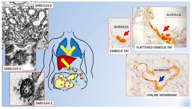
A multi-panel medical infographic and diagnostic montage illustrating the proposed pathophysiology of fat embolism in COVID-19. On the left, transmission electron microscopy (TEM) images show spherical SARS-CoV-2 virions with characteristic surface spike projections and dense internal nucleocapsids. A central anatomical diagram depicts the pathway of inflammatory factors and fat moving from visceral adipose tissue (labeled with hyperinflammation) through the venous system to the heart and lungs. On the right, histopathological images show alveolar structures. Two panels demonstrate pulmonary fat embolism with brightly stained orange/red fat globules and flattened embolic masses within the alveolar spaces. A third panel displays an alveolus with a distinct hyaline membrane, characterized by a smooth, translucent lining along the alveolar wall, typical of diffuse alveolar damage. This composite visualizes the clinical link between visceral obesity, viral infection, and sudden bilateral pneumonia.
Fetal circulation diagram showing: umbilical vein carrying oxygenated blood from placenta, ductus venosus bypassing liver to IVC, foramen ovale shunting blood from right atrium to left atrium, ductus arteriosus shunting blood from pulmonary artery to descending aorta, umbilical arteries returning blood to placenta. Label all structures clearly. Show oxygenated blood in red, deoxygenated in blue, mixed in purple. Medical textbook style diagram.
Flowchart showing changes at birth in fetal circulation: First event - Lung expansion causes drop in pulmonary vascular resistance -> increased pulmonary blood flow -> raised left atrial pressure -> foramen ovale closes functionally. Second event - cord clamping stops umbilical flow -> ductus venosus closes -> becomes ligamentum venosum. Third event - increased oxygen tension -> ductus arteriosus constricts -> closes -> becomes ligamentum arteriosum. Fourth event - umbilical arteries constrict -> become medial umbilical ligaments. Clean medical flowchart style with boxes and arrows.
Medical diagram showing wound healing phases in sequence: Phase 1 Hemostasis (0-hours): platelet plug, fibrin clot, vasoconstriction. Phase 2 Inflammation (1-4 days): neutrophils, macrophages, debridement, cytokines. Phase 3 Proliferation (4 days - 3 weeks): fibroblasts, collagen synthesis, angiogenesis, granulation tissue, epithelialization. Phase 4 Remodeling (3 weeks - 2 years): collagen crosslinking, tensile strength increase, scar maturation. Timeline bar at top showing progression. Clean educational medical illustration style.
Medical diagram showing body temperature regulation by hypothalamus. Central box labeled Hypothalamus (Thermostat). Inputs from peripheral thermoreceptors (skin) and central thermoreceptors. Two pathways: Heat loss mechanisms (sweating, vasodilation, radiation, convection, evaporation) and Heat conservation/production mechanisms (vasoconstriction, piloerection, shivering, thermogenesis). Normal core temperature 36.5-37.5 degrees C. Show positive and negative feedback loops. Clinical boxes showing heat stroke (above 40C) and hypothermia (below 35C). Educational flowchart style.
Error calling model 'gemini-3-pro-image' (RESOURCE_EXHAUSTED): 429 RESOURCE_EXHAUSTED. {'error': {'code': 429, 'message': 'Resource has been exhausted (e.g. check quota).', 'status': 'RESOURCE_EXHAUSTED'}}
We encountered an error while processing your request. Please try again or reach out to us for help.
Q.1 Describe the mechanism of regulation of body temperature, with special reference to effects of extreme heat. (20Marks) Q.2 Describe briefly the "Autonomic Nervous System". Discuss the pathophysiology of sudden death initiated through Autonomic Nervous System. Q.3 Describe anatomy of female external genitals. How is a victim of rape examined and reported? (20 Marks) (20 Marks) (40 Marks) Q.4 Write short notes on a) Age changes at pubic symphysis. b) Ewing's postulate. c) Traumatic fat emboli. d) Medicolegal aspects of violence against women. Describe in detail 'fetal circulation' changes occurring during and after birth and its medicolegal significance. (20 Marks) Q.2 Discuss biochemical parameters which help in determination of time since death. (20 Marks) Q.3 Discuss the process of wound healing and factors influencing it. Add a note on its significance in medicolegal practice. (20 Marks) Q.4 Write short notes on a) Role of histopathology in forensic medicine. b) Types and medico-legal importance of fractures. c) Post-mortem radiology: Importance and limitations. d) Toxicokinetics and toxicodynamics: Definitions and forensic relevance. Solve the answers , diagrams, images and flow charts where necessary
body temperature regulation hypothalamus thermoregulation diagram
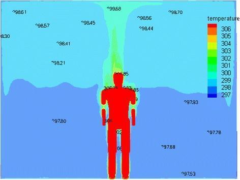
This diagnostic visualization is an educational pathophysiology diagram illustrating human thermoregulation and heat distribution within a controlled environment. The image features a human manikin model (1.75m, 75kg) serving as a proxy for a human patient to demonstrate heat exchange principles. A color-coded thermal gradient scale, ranging from 297 K (blue) to 306 K (red), maps the temperature distribution. The highest temperatures (305–306 K) are concentrated on the manikin's surface, particularly the head and torso, representing metabolic heat production. A visible thermal plume—an upward-moving column of warmer air (green to yellow)—rises directly from the head toward the ceiling, illustrating convective heat loss. Numerical annotations (e.g., 298.61, 297.53) denote ambient room temperatures, revealing a significant gradient between the 'body' surface and the surrounding environment. This content is used in medical engineering and clinical ergonomics to study thermal comfort, hospital room ventilation, and the ASHRAE standard for indoor environments. The diagram highlights the clinical significance of body-to-air heat transfer and the importance of airflow optimization for maintaining patient thermal homeostasis.
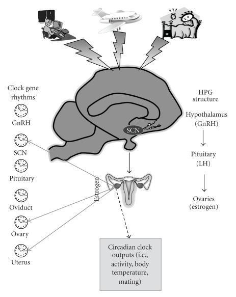
This pathophysiology diagram illustrates the integration of the hypothalamus-pituitary-gonadal (HPG) axis with circadian rhythm regulation in the female reproductive system. A sagittal section of the brain highlights the suprachiasmatic nucleus (SCN) and its relationship with GnRH neurons in the hypothalamus. The diagram outlines the hormonal cascade: Hypothalamus (GnRH) stimulates the Pituitary (LH), which acts on the Ovaries to produce Estrogen. External circadian disruptors—shift work, jet lag, and sleep deprivation—are shown influencing the SCN via lightning bolt symbols. Educational icons represent 'Clock gene rhythms' present in the GnRH neurons, SCN, Pituitary, Oviduct, Ovary, and Uterus, emphasizing peripheral molecular clocks. Solid arrows denote estrogen's feedback effects on reproductive tissues and the brain, while a dashed arrow indicates influence over circadian outputs like body temperature and activity levels. This visual serves as a summary of how environmental factors and internal clocks synchronize reproductive endocrinology.
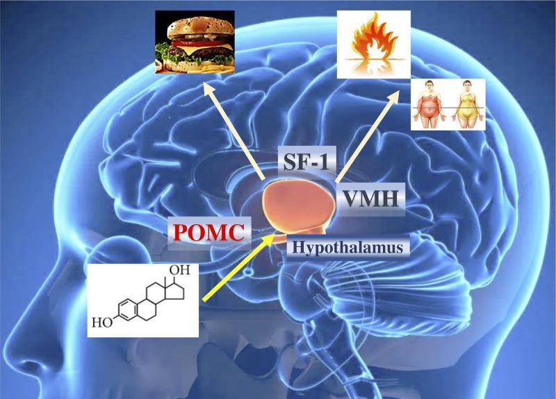
A medical illustration depicting the pathophysiology of metabolic regulation in the human brain, specifically focusing on the hypothalamus. The diagram features a translucent lateral view of the brain with the hypothalamus highlighted in orange. Key neuronal populations and regions are labeled, including POMC (pro-opiomelanocortin) and the VMH (ventromedial hypothalamus) containing SF-1 (steroidogenic factor 1) neurons. Symbolic overlays illustrate the functional outcomes of hypothalamic signaling: a hamburger icon represents food intake regulation via POMC neurons, while flame and body transformation icons signify energy expenditure and body composition changes mediated by SF-1 and VMH pathways. An estrogen (estradiol) chemical structure is shown with an arrow pointing toward the hypothalamus, indicating the hormonal influence of estrogens on these metabolic centers. This infographic demonstrates the role of estrogen receptors in central obesity control and caloric balance, serving as an educational resource for endocrinology and neurobiology.
fetal circulation changes at birth ductus arteriosus foramen ovale diagram
This composite figure presents a detailed fetal echocardiographic evaluation at 35 weeks of gestation. Panels A and B (four-chamber views) demonstrate significant chamber disproportion, with a dilated right ventricle (RV) compared to the left ventricle (LV). The flap of the foramen ovale (FFo) appears relatively fixed and exhibits tense bulging throughout both systole (A) and diastole (B). Panel C (M-mode) confirms impaired RV contractility, showing a reduced shortening fraction compared to the LV. Panel D (three-vessel and trachea view) reveals a hypoplastic aortic arch (AoA) in comparison to the larger ductus arteriosus (DA). Panels E and F utilize color Doppler imaging to illustrate abnormal hemodynamics at the interatrial septum, characterized by turbulent blood flow (marked with asterisks) and restricted passage across the foramen ovale. These findings are clinically significant for identifying secondary cardiac changes due to fetal anemia and altered preload conditions, emphasizing the assessment of the foramen ovale and ventricular function in high-risk pregnancies.
A multi-panel medical diagnostic and clinical series demonstrating congenital cardiovascular anomalies in a ctnnb1Δex3 model. Panels A and B are ultrasound images illustrating a Patent Ductus Arteriosus (PDA), with labels identifying the Left Atrium (LA), Aorta (Ao), Right Ventricle Outflow Tract (RVOT), and Pulmonary Artery (PA). An arrow points to the 'Open DA' (Ductus Arteriosus). Panel C provides an ultrasound view of the atrial septum with labels for the Right Atrium (RA), LA, and Ao, showing a patent foramen ovale. Panel D utilizes Color Doppler imaging to show a pathological right-to-left shunt of blood flow through the foramen ovale. Panel E is a clinical photograph of a dissected heart confirming the presence of an open foramen ovale, notable for the presence of dark pigmented cells (melanocytes) around the aperture. The collection demonstrates how failure of fetal cardiovascular shunts to close postnatally leads to abnormal circulation and chamber dilation.
This diagnostic image series utilizes 4D flow MRI-derived particle tracing to illustrate fetal circulatory dynamics in a ventral view. The visualization demonstrates the preferential streaming of blood from two distinct venous sources over one cardiac cycle (62 ms, 186 ms, 310 ms, and 434 ms). Red particles represent blood from the ductus venosus (DV), while blue particles represent blood from the distal inferior vena cava (IVCd). The series highlights the maintenance of separate flow streams within the proximal inferior vena cava. The red DV stream is directed preferentially through the foramen ovale (FO) toward the left ventricle (LV) to supply oxygenated blood to the systemic circulation. Concurrently, the blue IVCd stream is routed primarily toward the right ventricle (RV) for delivery to the main pulmonary artery. Anatomical landmarks labeled include the ductus venosus (DV), distal inferior vena cava (IVCd), foramen ovale (FO), right ventricle (RV), and left ventricle (LV). This pedagogical tool illustrates the physiological shunting essential for fetal development and the minimal mixing of nutrient-rich and nutrient-poor blood streams.
wound healing stages hemostasis inflammation proliferation remodeling
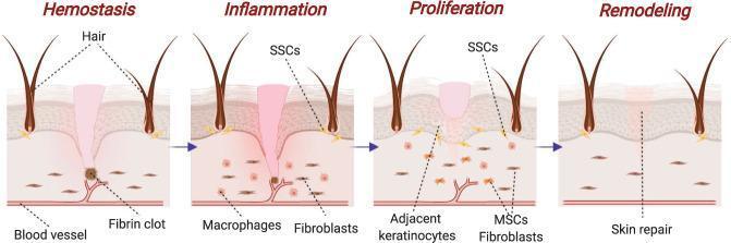
A pathophysiology diagram illustrating the four chronological stages of skin wound healing: Hemostasis, Inflammation, Proliferation, and Remodeling. The visual depicts a cross-section of the integumentary system including the epidermis, dermis, hair follicles, and vasculature. 1) Hemostasis: Shows a fibrin clot formation at the site of vascular injury to stop bleeding. 2) Inflammation: Characterized by the presence of macrophages for phagocytosis and fibroblasts beginning to migrate into the wound site; skin stem cells (SSCs) are indicated near the hair follicles. 3) Proliferation: Displays the migration and activity of adjacent keratinocytes, mesenchymal stem cells (MSCs), and fibroblasts to form a provisional extracellular matrix. 4) Remodeling: Demonstrates the final 'skin repair' phase where tissue integrity is restored, collagen fibers are realigned, and the wound site is closed. This diagram serves as an educational tool for medical students to understand the cellular and structural progression of cutaneous tissue repair.
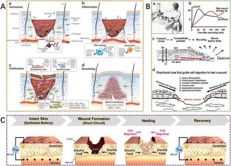
This composite educational illustration details the physiological and bioelectrical mechanisms of human wound healing. Panel A illustrates the four classical stages of repair: Hemostasis (fibrin clot and platelet aggregation), Inflammation (neutrophil and macrophage recruitment via cytokines like IL-1 and TNF̑́), Proliferation (re-epithelialization by keratinocytes, angiogenesis, and fibroblast activity), and Remodeling (matrix reorganization and eschar shedding). Panel B examines the bioelectric response, showing endogenous electric field (EF) distribution and current flow at the wound edge, which acts as a directional cue for cell migration. Panel C presents a four-phase model of the skin as an 'epithelial battery.' It contrasts intact skin, which maintains a transepithelial potential (VTEP) of approximately 60 mV, with wound formation ('short circuit' state at 0 mV), followed by the restoration of bioelectric polarity during the healing and recovery phases. The graphic emphasizes the intersection of molecular signaling and electrophysiology in dermatology and regenerative medicine.
female external genitalia vulva anatomy labeled diagram
This medical anatomical diagram depicts the female external genitalia (vulva) and perineal region from a frontal perspective. Key anatomical landmarks include the labia majora, labia minora, clitoral hood, urethral meatus, and the vaginal introitus. A black arrow specifically identifies the posterior commissure of the vagina, located at the junction where the vaginal mucosa transitions into the dermal skin of the perineum. This site is clinically relevant as the target area for pulse oximetry (SpO2) measurements mentioned in the context, involving the underlying bulbospongiosus muscle. The illustration highlights the natural contours, skin texture, and spatial relationships between the vulvar structures and the anus within the perineal field. It serves as an educational reference for identifying clinical measurement sites and understanding female pelvic anatomy for gynecological or physiological studies.
Clinical photograph of the female external genitalia showing a large, exophytic squamous cell carcinoma (SCC) of the vulva. The lesion is a centrally located, irregularly shaped mass measuring approximately 55 mm x 30 mm, originating from the left labia and extending to the midline. The tumor exhibits classic malignant morphology, including a cauliflower-like or verrucous texture with friable, erythematous tissue and areas of pale keratinization. Central ulceration and serosanguinous exudate are visible, suggesting tissue necrosis. The mass significantly distorts the local anatomy of the labia majora and minora. The surrounding perilesional skin shows signs of secondary inflammation and possible satellite nodules or lichenoid changes. This image serves as a clinical example of advanced vulvar malignancy, illustrating the need for surgical intervention such as radical local excision and assessment of inguinofemoral lymph nodes.
traumatic fat embolism syndrome pathophysiology
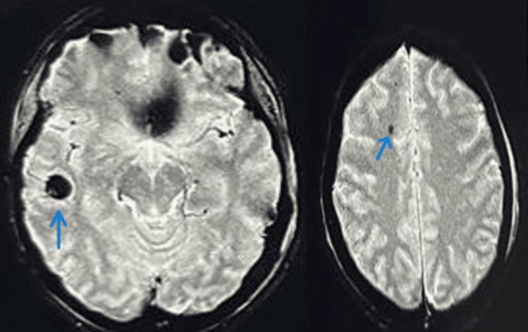
This diagnostic imaging set consists of two axial MRI brain slices demonstrating cerebral fat embolism syndrome. The images show bilateral, multifocal lesions characterized by 'starfield pattern' hypointensities. Blue arrows highlight specific small, circular-to-ovoid hypointense (dark) foci within the cerebral hemispheres, particularly visible in the right temporal lobe and the left frontal subcortical white matter. These lesions represent microhemorrhages or localized infarcts secondary to fat emboli following traumatic fractures. The anatomical structures, including the cerebral gyri, sulci, and ventricular system, are otherwise well-preserved without evidence of midline shift, mass effect, or significant herniation. The MRI findings illustrate the characteristic neurological manifestation of systemic fat embolism, emphasizing the diagnostic value of magnetic resonance imaging in patients presenting with altered mental status or neurological deficits post-trauma. The specialty is neuroradiology, suitable for intermediate to advanced medical education regarding traumatic brain injury complications.
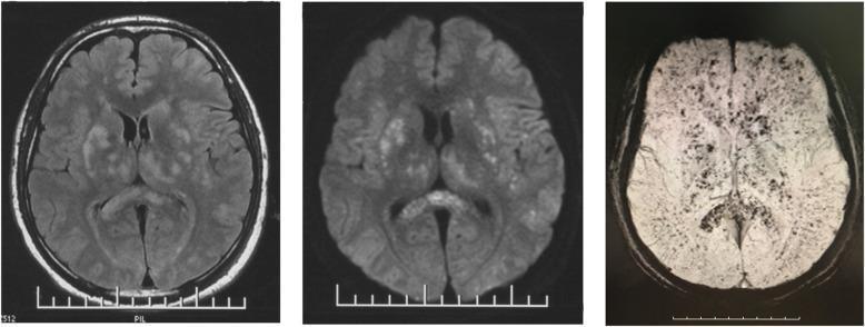
This set of axial brain MRI sequences (FLAIR, DWI, and SWI) demonstrates classic neuroimaging findings of cerebral fat embolism (CFE) syndrome following traumatic orthopedic injury. The Fluid-Attenuated Inversion Recovery (FLAIR) and Diffusion-Weighted Imaging (DWI) scans reveal numerous, small, hyperintense foci scattered throughout the brain parenchyma. This 'starfield' pattern is prominently distributed within the periventricular and subcortical white matter, basal ganglia, and deep white matter. The Susceptibility-Weighted Imaging (SWI) sequence complements these findings by showing multiple punctate hypointense spots in a similar distribution, representing microhemorrhages or susceptibility artifacts. These imaging features illustrate the diffuse embolic nature of fat particles entering the cerebral circulation, leading to multiple micro-infarcts and hemorrhagic components. The content is an essential teaching resource for distinguishing CFE from diffuse axonal injury (DAI) in the context of post-traumatic consciousness disorders and systemic clinical signs like fever and hypoxia.
types of fractures transverse spiral greenstick pathological forensic
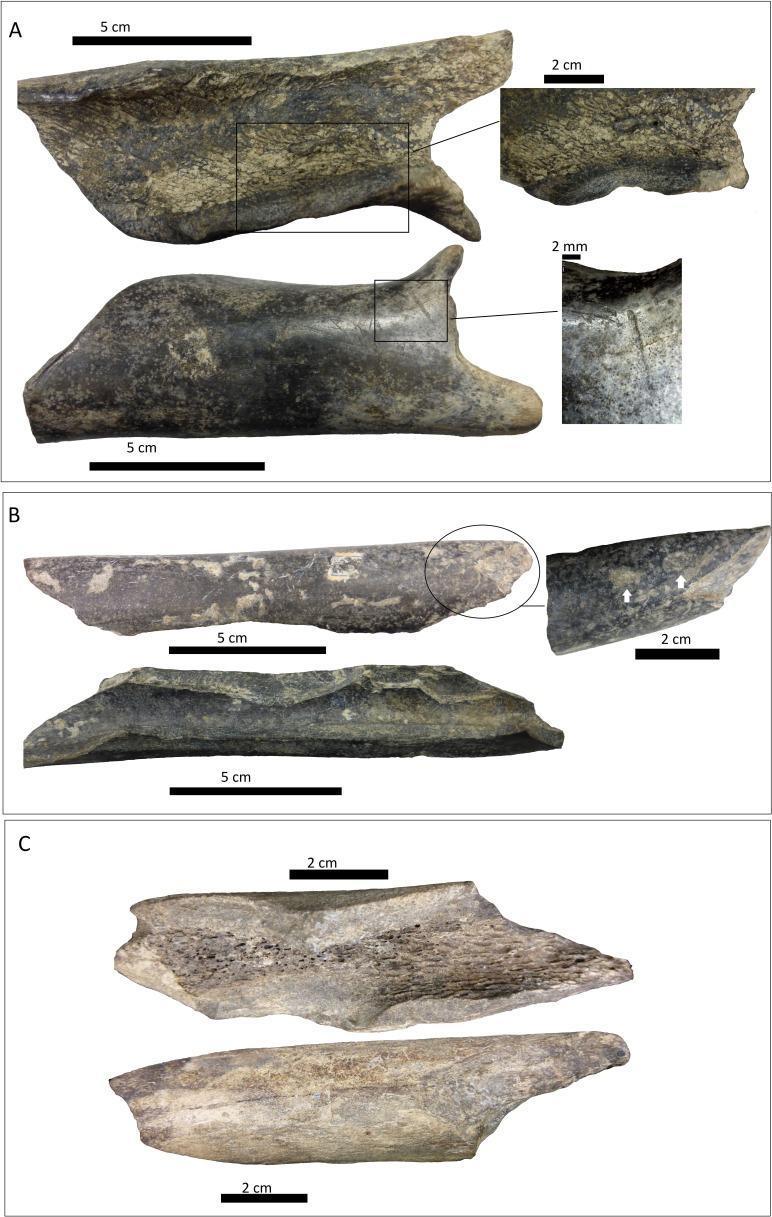
This forensic and pathological diagnostic image displays three distinct sets of bone shaft specimens (A, B, and C) illustrating various fracture patterns and surface modifications. All three specimens exhibit classic spiral fractures, characterized by smooth, twisting break surfaces indicative of torsion or high-energy dynamic forces applied to fresh bone. Panel A shows a large cortical fragment with magnified views highlighting scoring marks on the cortical face and a distinct internal notch. Panel B displays a longitudinal bone shaft with focal light pitting on the cortical surface (indicated by white arrows) and visible notches on the medullar face. Panel C depicts a more uniform bone fragment with a clearly visible trabecular (cancellous) bone structure alongside the spiral fracture edge. Educational focus is on forensic taphonomy, identifying ante-mortem or peri-mortem traumatic patterns versus post-mortem modifications. Key visual indicators include pitting, scoring, and notched depressions which help differentiate between mechanical trauma and biological interventions, such as those caused by carnivore scavenging.
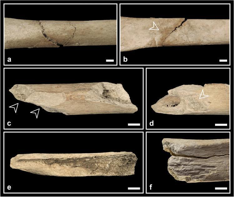
This composite of clinical photographs illustrates various perimortem fracture patterns in human skeletal specimens, specifically the tibia and femur, used for forensic and anthropological analysis. Panels a and b display a classic butterfly fracture on a tibial shaft from anterior and posterior perspectives; a transverse fracture line is visible, with a secondary incomplete fracture line branching off posteriorly (indicated by an arrowhead). Panels c and d show a femur fragment exhibiting spiral fractures and cortical bone spalling (black arrowheads), with panel d highlighting adherent sediments (white arrowhead) within the spalled region. Panel e demonstrates a bone flake with an oval profile, characterized by smooth surfaces and an acute proximal fracture angle, typical of fresh bone breakage. Panel f depicts a tibia specimen showing 'peeling' and a roughened, exfoliated cortical surface. These features are critical for distinguishing between perimortem trauma (occurring at or near the time of death) and postmortem damage, providing insights into injury mechanisms or ritualistic bone processing.
postmortem radiology CT scan virtopsy autopsy imaging
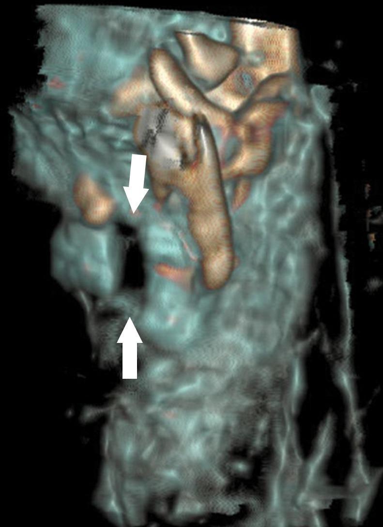
This diagnostic image is a three-dimensional (3D) volume-rendered reconstruction from a postmortem CT scan of the cervical region. The visualization focuses on the laryngeal skeleton, specifically the cricoid cartilage and its surrounding structures. High-density osseous and calcified cartilaginous tissues are rendered in tan/gold tones, while lower-density soft tissues appear in translucent blue-grey. Two white arrows indicate a clear, displaced bilateral fracture of the cricoid cartilage, characterized by a visible gap and loss of anatomical continuity in the ring-like structure. The reconstruction demonstrates the clinical utility of postmortem imaging in forensic pathology for identifying deep neck injuries, such as those resulting from blunt force or manual strangulation, which may be difficult to isolate during physical autopsy without causing iatrogenic manipulation. This material is suitable for advanced educational use in forensic radiology, trauma imaging, and laryngeal anatomy.
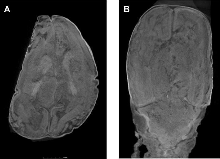
High-resolution micro-focus computed tomography (micro-CT) scans of a 13-week-gestation fetal brain, highlighting the challenges of postmortem imaging in cases of maceration and autolysis. Figure A displays an axial section at the level of the basal ganglia, while Figure B shows a coronal section through the posterior fossa and occipital lobes. Both images demonstrate significant intracranial tissue degradation. Key pathological features include a loss of the normal gray-white matter differentiation and a lack of distinct cerebral sulcation, which appears disrupted or poorly defined. The imaging resolution (35 μm) allows for detailed assessment of the internal architecture; however, the parenchymal changes shown are characteristic of postmortem autolysis, which can render fetal neuroimaging nondiagnostic for structural malformations. This content is relevant for pediatric radiology, fetal pathology, and researchers evaluating noninvasive autopsy techniques using micro-CT.
pubic symphysis age changes morphology phases Todd
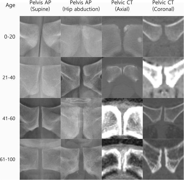
This diagnostic comparison chart illustrates the morphological changes of the pubic symphysis across four age groups (0-20, 21-40, 41-60, and 61-100 years) using four imaging modalities: Pelvis AP (supine), Pelvis AP (hip abduction), Pelvic CT (axial), and Pelvic CT (coronal). The 4x4 grid demonstrates a clear progression of joint space alterations and bony remodeling. In the younger cohorts (0-40 years), the symphysis shows smooth articular surfaces and relatively narrow, uniform joint spaces. With advancing age (41-100 years), there is a visible trend toward increased joint width, cortical thickening, and sclerosis. In the 61-100 year group, particularly in the CT axial and coronal views, significant degenerative features are evident, including irregular joint margins, subchondral sclerosis, and osteophyte formation. This visual sequence is designed for orthopedic and radiological education to assist in identifying normal age-related changes versus pathological degeneration of the pubic symphysis joint.
This technical diagram illustrates the internal feature extraction process of a convolutional neural network (CNN) used for forensic age estimation based on 3D scans of the human pubic symphysis. The image follows the evaluation of a 72-year-old individual through various processing stages. The 'Input image' shows a grayscale 2D projection of the 3D surface scan, appearing as a blurry anatomical outline. Subsequent rows visualize internal network layers: Layer #3 shows initial filters focusing on high-contrast edges and basic morphology; Layers #6 and #10 display multiple feature maps that refine complex textures and vertical ridge patterns characteristic of age-related skeletal degeneration. The final row, Layer #19, depicts a one-dimensional feature vector represented as a linear scale (1–100) with colored activation points, indicating the condensed high-level data used for the final age prediction (estimated here as 71.6 years). This visualization demonstrates how AI identifies subtle morphological changes in skeletal remains that may be difficult for the human eye to quantify, providing a specialized tool for forensic anthropology and clinical aging research.
PRAVARA INSTITUTE OF MEDICAL SCIENCES
POST GRADUATE DEGREE IN FORENSIC MEDICINE (M.D.)
PAPER - II (IInd Term Ending Examination)
Q.1 Describe the mechanism of regulation of body temperature, with special reference to effects of extreme heat. (20 Marks)
INTRODUCTION
THERMORECEPTORS (Heat Sensors)
- Located in the skin - warm receptors (C fibers) and cold receptors (A-delta fibers)
- Detect external environmental temperature changes
- Send afferent signals via spinothalamic tracts to hypothalamus
- Located in the anterior hypothalamus / preoptic area
- Respond to temperature of blood perfusing the hypothalamus
- Most sensitive - detect changes as small as 0.1°C
HYPOTHALAMIC THERMOREGULATION CENTER
┌─────────────────────────────────────────────────────────────┐
│ HYPOTHALAMUS (PREOPTIC AREA) │
│ │
│ ANTERIOR HYPOTHALAMUS POSTERIOR HYPOTHALAMUS │
│ (Heat Dissipation Center) (Heat Conservation Center) │
│ - activated by HEAT - activated by COLD │
│ - vasodilation - vasoconstriction │
│ - sweating - shivering │
│ - behavioral changes - piloerection │
└─────────────────────────────────────────────────────────────┘
MECHANISMS OF HEAT PRODUCTION
| Mechanism | Details |
|---|---|
| Basal Metabolic Rate (BMR) | Cellular metabolism generates ~60-70 kcal/hr at rest |
| Muscular Activity | Exercise raises heat production 10-20 fold |
| Shivering Thermogenesis | Involuntary rhythmic muscle contractions - increases heat production 2-5x |
| Non-shivering Thermogenesis | Brown adipose tissue (BAT) - uncoupling protein (UCP-1) converts ATP to heat |
| Specific Dynamic Action | Food digestion raises metabolic rate 10-30% |
| Hormonal | Thyroxine and epinephrine increase metabolic rate |
MECHANISMS OF HEAT LOSS
HEAT LOSS MECHANISMS
│
┌────────┼────────────┬──────────────┐
▼ ▼ ▼ ▼
RADIATION CONDUCTION CONVECTION EVAPORATION
(60%) (3%) (12%) (25%)
│ │ │ │
Infra-red Direct Moving Sweating
electromagnetic contact air/water (500mL/day)
waves with surface currents + insensible
(water 25x removes heat loss
better than air) (lungs/skin)
FLOWCHART: THERMOREGULATORY RESPONSE TO HEAT
RISE IN BODY TEMPERATURE
│
▼
ANTERIOR HYPOTHALAMUS ACTIVATED
│
┌────┴──────────────────┐
▼ ▼
CUTANEOUS SWEAT GLANDS
VASODILATION ACTIVATED
│ │
Increased blood flow Sweating → Evaporative
to skin surface cooling
│
Heat dissipated by
radiation/convection
│
▼
┌────┴────┐
▼ ▼
Heat loss Behavioral
increases changes
(heat loss (seek shade,
>> heat remove clothing,
produced) drink fluids)
│
▼
CORE TEMPERATURE RETURNS TO NORMAL (37°C)
│
▼
NEGATIVE FEEDBACK → ANTERIOR HYPOTHALAMUS INHIBITED
EFFECTS OF EXTREME HEAT
A. Heat Cramps
- Painful muscle cramps due to salt depletion from excessive sweating
- Core temperature normal
- Treatment: oral saline, rest
B. Heat Exhaustion
- Core temperature 37-40°C
- Profuse sweating, weakness, dizziness, nausea, headache, syncope
- Mechanism: dehydration + peripheral vasodilation → reduced cardiac output → reduced cerebral perfusion
- CNS function intact
- Treatment: remove from heat, IV fluids, cooling
C. Heat Stroke (Most severe - LIFE THREATENING)
- Core temperature >40°C with CNS dysfunction (confusion, seizures, coma)
- Two types:
- Classic heat stroke - elderly, sedentary, during heat waves; sweating may be absent
- Exertional heat stroke - young, active individuals; profuse sweating present
EXTREME HEAT / EXERCISE
│
▼
Core temperature >40°C
│
▼
Failure of thermoregulatory mechanisms
│
┌────┴──────────────────────────────────┐
▼ ▼ ▼
CNS dysfunction Protein Cardiovascular
(encephalopathy, denaturation collapse
seizures, coma) │ (circulatory
│ Enzyme failure shock)
▼ │
Brain edema Multi-organ
failure
│
┌──────────┼──────────┐
▼ ▼ ▼
Renal DIC Hepatic
failure (Disseminated failure
(ATN) Intravascular
Coagulation)
| System | Effect |
|---|---|
| CNS | Encephalopathy, seizures, cerebellar damage |
| CVS | Tachycardia, hypotension, arrhythmias, myocardial infarction |
| Kidneys | Acute tubular necrosis (rhabdomyolysis-induced) |
| Liver | Centrilobular necrosis (temperature-sensitive hepatocytes) |
| Blood | DIC - fibrinogen consumed, thrombocytopenia |
| Muscles | Rhabdomyolysis → myoglobinuria |
| Lungs | ARDS, pulmonary hemorrhage |
MEDICOLEGAL / FORENSIC SIGNIFICANCE
- Heat stroke deaths - autopsy may reveal cerebral edema, petechial hemorrhages in brain, myocardial degeneration, centrilobular hepatic necrosis
- Occupational heat deaths - construction workers, military personnel - compensable deaths
- Neglect cases - infants/elderly left in hot cars/rooms - criminal negligence
- Heat as precipitating factor - in pre-existing cardiac/cerebrovascular disease
- Histological evidence - protein denaturation, coagulative necrosis in multiple organs
- Hyperpyrexia - fever from infection must be distinguished from true hyperthermia at autopsy
Q.2 Describe briefly the "Autonomic Nervous System." Discuss the pathophysiology of sudden death initiated through the Autonomic Nervous System. (20 Marks)
AUTONOMIC NERVOUS SYSTEM - OVERVIEW
ANATOMY OF THE ANS
A. Sympathetic Nervous System (SNS)
- Origin: Thoracolumbar outflow - T1 to L2/L3 spinal segments
- Preganglionic fibers: Short, myelinated - synapse in paravertebral ganglia (sympathetic chain) or prevertebral ganglia
- Postganglionic fibers: Long, unmyelinated - innervate target organs
- Neurotransmitter: Preganglionic = Acetylcholine (nicotinic receptor); Postganglionic = Norepinephrine (adrenergic receptors α and β)
- Exception: Sweat glands and adrenal medulla use acetylcholine
- Adrenal medulla = modified sympathetic ganglion → releases epinephrine (80%) + norepinephrine (20%) directly into blood
- Effect: "Fight or Flight" - tachycardia, bronchodilation, pupil dilation, vasoconstriction, decreased GI motility, glycogenolysis
B. Parasympathetic Nervous System (PNS)
- Origin: Craniosacral outflow - CN III, VII, IX, X (Vagus) and S2-S4
- Preganglionic fibers: Long, myelinated - synapse in terminal ganglia near or within target organs
- Postganglionic fibers: Very short
- Neurotransmitter: Both pre- and postganglionic = Acetylcholine (muscarinic receptors at target)
- Vagus nerve (CN X) provides parasympathetic supply to heart, lungs, most abdominal viscera
- Effect: "Rest and Digest" - bradycardia, bronchoconstriction, pupil constriction, increased GI motility, glandular secretion
Comparison Table
| Feature | Sympathetic | Parasympathetic |
|---|---|---|
| Origin | T1-L2 (Thoracolumbar) | CN III, VII, IX, X; S2-S4 (Craniosacral) |
| Preganglionic | Short | Long |
| Postganglionic | Long | Short |
| Ganglia location | Paravertebral/Prevertebral | Near/within target organ |
| Preganglionic NT | Acetylcholine (nicotinic) | Acetylcholine (nicotinic) |
| Postganglionic NT | Norepinephrine | Acetylcholine (muscarinic) |
| Heart rate | Increases (β1) | Decreases |
| Blood pressure | Increases | Decreases |
| Pupils | Dilates | Constricts |
| Bronchi | Dilates | Constricts |
CARDIAC INNERVATION (Key for Sudden Death)
- Sympathetic: Cardiac nerves from T1-T5 stellate ganglia → SA node, AV node, ventricular myocardium → ↑ HR, ↑ contractility, ↑ conduction velocity
- Parasympathetic: Vagus nerve branches → SA node, AV node → ↓ HR, ↓ AV conduction (vagal tone is dominant at rest)
PATHOPHYSIOLOGY OF SUDDEN DEATH THROUGH ANS
MECHANISM 1: Vasovagal (Neurocardiogenic) Syncope → Cardiac Arrest
TRIGGER (Emotional shock, pain, fear, micturition, defecation,
neck pressure, cold water immersion, sight of blood)
│
▼
Excessive PARASYMPATHETIC (Vagal) Activation
│
┌────┴──────────────────────────────────┐
▼ ▼
Profound BRADYCARDIA VASODILATION
(sinus arrest / asystole) (arteriolar)
│ │
┌────┴───────────────────────────────── ┘
▼
SEVERE HYPOTENSION
│
▼
CEREBRAL ISCHEMIA → LOSS OF CONSCIOUSNESS
│
▼
If bradycardia/asystole prolonged → SUDDEN DEATH
- Death from fright ("voodoo death") - extreme vagal activation from intense fear
- Carotid sinus syncope - pressure on neck (strangling, tight collar) triggers exaggerated vagal response
- "Diving reflex" - sudden immersion of face in cold water → profound bradycardia
- Commotio cordis - blunt impact to precordium triggering fatal VF through ANS
MECHANISM 2: Sympathetic Surge → Fatal Arrhythmia
INTENSE STRESS / CATECHOLAMINE SURGE
(Physical exertion, emotional trauma, cocaine/amphetamines)
│
▼
Massive Sympathetic Activation
│
▼
↑ Epinephrine / Norepinephrine
│
┌────┴──────────────────────────────────┐
▼ ▼
↑ Heart rate + ↑ Myocardial
↑ Contractility Oxygen Demand
│ │
Shortened Ischemia (esp. in
refractory period coronary artery disease)
│ │
└────────────────┬─────────────────┘
▼
VENTRICULAR FIBRILLATION (VF)
│
▼
SUDDEN DEATH
MECHANISM 3: Stellate Ganglion Imbalance and Long QT
- Asymmetric sympathetic stimulation → QT prolongation → Torsades de Pointes → VF
- Jervell-Lange-Nielsen syndrome - congenital LQTS with deafness - sudden death in young
- Romano-Ward syndrome - congenital LQTS - triggered by startle/exercise
MECHANISM 4: Baroreflex Failure
- Bilateral carotid body tumors or neck dissection → loss of baroreflex
- Uncontrolled hypertensive crises → cerebral hemorrhage or cardiac failure
MECHANISM 5: Reflex Inhibition of Vital Centers
- Blow to epigastrium (solar plexus) → vagal surge → cardiac arrest
- This is the basis of "solar plexus syndrome" - seen in boxing/assault fatalities
- Laryngeal stimulation (e.g., drowning, choking) → cardiac arrest via vagal reflexes
FORENSIC SIGNIFICANCE
- Sudden unexpected deaths - autopsy may show no anatomical cause → ANS mechanism suspected
- "Fright death" - no morphological lesion found; history of extreme emotional shock
- Neck pressure deaths - carotid sinus stimulation → vagal cardiac arrest (relevant in manual strangulation)
- Sudden infant death syndrome (SIDS) - ANS immaturity with exaggerated vagal response implicated
- Drug-related sudden deaths - cocaine → sympathetic storm; organophosphate poisoning → cholinergic crisis
- Histological examination of SA/AV nodes and cardiac conduction system is essential at autopsy
Q.3 Describe anatomy of female external genitals. How is a victim of rape examined and reported? (20 Marks)
PART A: ANATOMY OF FEMALE EXTERNAL GENITALIA (VULVA)
VULVA
├── Mons Pubis
├── Labia Majora
├── Labia Minora
├── Clitoris
│ ├── Prepuce (hood)
│ ├── Glans
│ └── Frenulum
├── Vestibule
│ ├── Urethral Meatus
│ ├── Vaginal Orifice (Introitus)
│ ├── Hymen
│ ├── Bartholin's Glands (greater vestibular glands)
│ └── Skene's Glands (lesser vestibular/paraurethral)
├── Fourchette (Posterior commissure / Frenulum labiorum)
└── Perineum
| Structure | Description | Forensic Importance |
|---|---|---|
| Mons Pubis | Fatty prominence over pubic symphysis, covered by pubic hair after puberty | Pubic hair comparison in rape |
| Labia Majora | Two large folds of skin with fat; outer surface hairy, inner smooth; contains sebaceous and sweat glands | Site of bruising, abrasions in rape |
| Labia Minora | Two thin inner folds, hairless, rich in sebaceous glands; meet anteriorly to form clitoral hood and posteriorly at fourchette | Commonly bruised/lacerated in rape |
| Clitoris | Erectile organ, ~2-3cm; analogous to penis; highly sensitive; composed of corpora cavernosa | Contusions indicate violent coitus |
| Vestibule | Space enclosed by labia minora; contains urethral opening and vaginal introitus | Key examination area |
| Hymen | Thin fold of mucous membrane at vaginal entrance; variable shapes - annular, crescentic, fimbriated, septate | Central to rape examination in virgins |
| Bartholin's glands | Paired, pea-sized, at 4 and 8 o'clock positions at vaginal opening; provide lubrication during coitus | Bartholin's abscess if infected |
| Fourchette | Posterior junction of labia minora; very frequently torn in rape (first structure to tear) | Fresh tear = recent forceful penetration |
| Vaginal orifice | Variable size; depends on age, parity, and use of tampons | Dilatation assessed |
HYMEN TYPES
│
├── Annular (circular) - most common
├── Crescentic (posterior horseshoe shape)
├── Fimbriated / Denticular (frilled margins)
├── Septate (divides introitus - fibrous band)
├── Cribriform (multiple small openings)
├── Imperforate (no opening - requires surgery)
└── Parous introitus (after childbirth - remnants = carunculae myrtiformes)
PART B: EXAMINATION OF A RAPE VICTIM
GENERAL PRINCIPLES
- Examination should be done as soon as possible (ideally within 72 hours)
- Must be performed by a registered medical practitioner (preferably female doctor for female victims)
- Requires written consent from patient (or guardian if minor)
- Two doctors and a female nurse should be present
- Police may request examination under Section 164A CrPC (amended by POCSO/BNSS)
- Examination should be systematic, complete, and documented
- Maintain chain of evidence for all samples collected
EXAMINATION PROCEDURE
- Date, time, place of alleged assault
- Nature of act (vaginal, anal, oral)
- Relationship to accused
- Last voluntary coitus (if applicable)
- Last menstrual period
- Menstrual/obstetric history
- Whether the victim has bathed, changed clothes, urinated, defecated since assault
- General health, previous surgical history
- Age assessment (pubertal status - Tanner staging)
- Height, weight, built
- Mental state, signs of intoxication/sedation
- Injuries all over body:
- Defense injuries - on hands, forearms (blocking)
- Restraint marks - wrists, ankles
- Bite marks - identify shape
- Bruises, lacerations on face, neck, shoulders, thighs, breasts
- Nail scrapings for DNA (accused's skin cells)
- Oral swab if oral assault alleged
- Signs of drug/alcohol intoxication
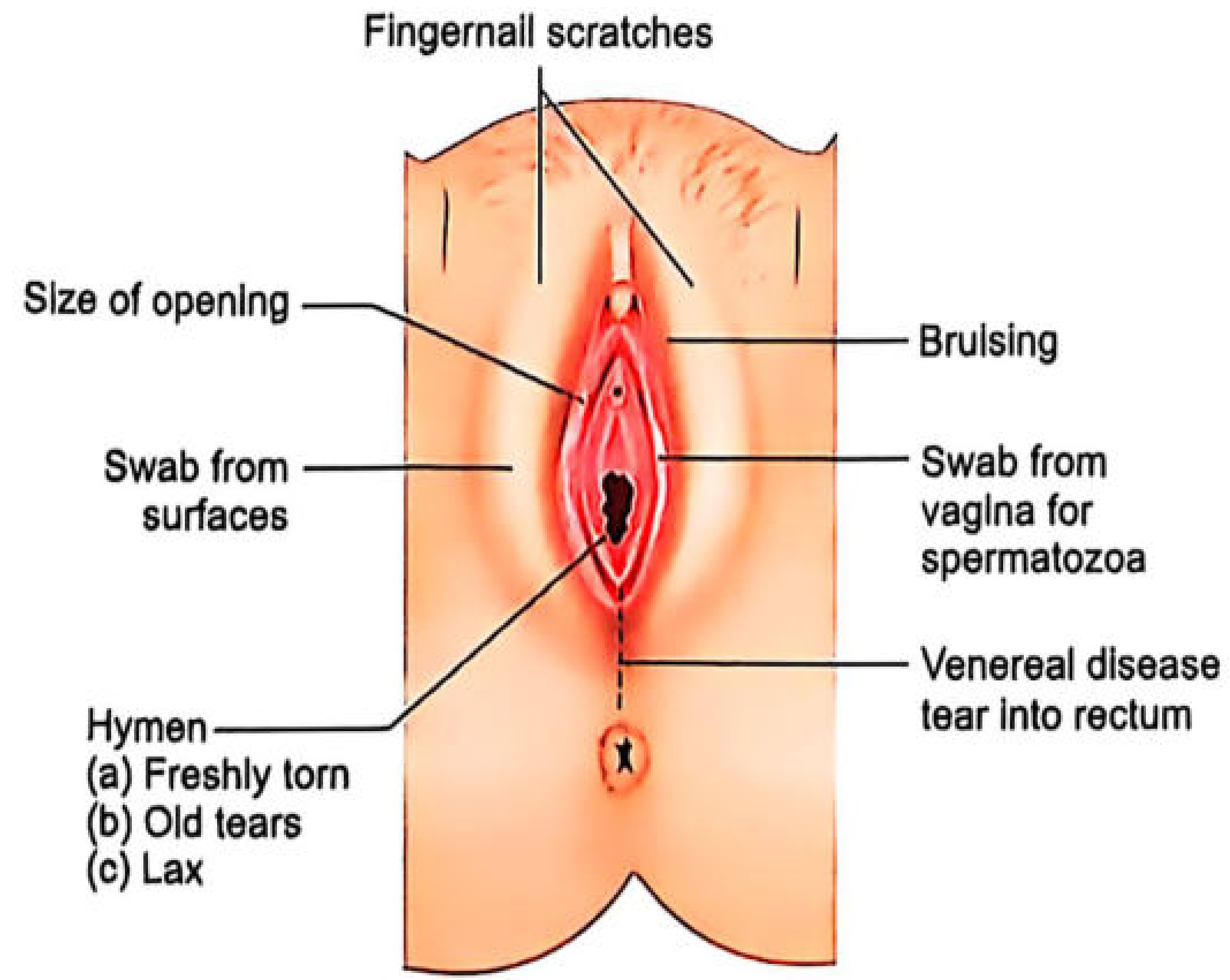
- Pubic hair - examine for matted blood/semen; comb and collect loose hairs; cut matted hair close to skin
- Thighs, labia majora, labia minora, perineum - bruises, abrasions, lacerations
- Fourchette - fresh tears (raw edges, bleeding) vs old tears (healed, smooth edges)
- Hymen examination:
- Labia separated by gentle labial traction
- Examine shape, integrity
- Fresh tears: red, raw, bleeding edges - present in recent penetration
- Old tears: healed, smooth, with healed scar tissue
- Note position (clock face - e.g., 6 o'clock tear)
- Note whether tear extends to base (hymenal margin)
- Important: hymen may be intact even after rape in experienced females or if elasticity is high
- Vagina: speculum examination (if patient consents; not in children without anesthesia)
- Note tears, bruising, discharge
- Cervix: injuries, discharge, STI signs
- Anus/rectum: if anal assault alleged - laxity, tears, bruising
| Sample | Method | Purpose |
|---|---|---|
| High vaginal swab | Sterile swab | Spermatozoa, acid phosphatase, DNA |
| Low vaginal swab | Sterile swab | Spermatozoa |
| Cervical swab | Speculum + swab | DNA of accused |
| Anal swab | If indicated | For anal rape |
| Oral swab | If indicated | For oral rape |
| Pubic hair (combed) | Comb into paper | Foreign hair comparison |
| Nail scrapings | Wooden stick | Accused's DNA |
| Blood sample | Venipuncture | DNA, alcohol, drugs, STI serology |
| Urine | Midstream | Drug/alcohol screen |
| Cervical/vaginal smear | Glass slide | Sperm motility (fresh sperm = recent rape) |
| Victim's clothing | Bag separately | Semen stains, trace evidence |
FINDINGS AND THEIR SIGNIFICANCE
- Spermatozoa in vagina (motile within 12 hours; immotile up to 72 hours; heads persist longer)
- Acid phosphatase in high concentration in vaginal swab
- Fresh hymenal tear with raw bleeding edges
- Injuries to labia, fourchette, perineum
- Y-STR DNA profile matching accused
- No hymenal tear in experienced/parous women
- No spermatozoa (if accused is azoospermic, used condom, or delay in examination)
- No injuries if victim was intoxicated, compliant under threat, or victim is experienced
MEDICOLEGAL REPORT (MLR) CONTENTS
- Name, age, address of victim
- Date, time, place of examination
- Consent details
- History as told by victim (verbatim, in quotes)
- Examination findings - general and local
- Samples collected and sent
- Opinion on:
- Age (puberty attained?)
- Evidence of recent coitus
- Evidence of forcible intercourse
- Whether injuries consistent with rape
- Doctor's signature, designation, registration number
- Report sealed and sent to police / court
Important legal point: Under IPC Section 375 (now BNS Section 63), rape is defined broadly and includes penetration of vagina, urethra, anus, mouth. The medical officer must provide objective findings only - the final determination of rape is legal, not medical.
Q.4 Write Short Notes on (40 Marks)
(a) Age Changes at Pubic Symphysis (10 Marks)
| Phase | Age (Years) | Features |
|---|---|---|
| I | 18-19 | Horizontal ridges and grooves (billowing), no dorsal plateau |
| II | 20-21 | Ridges beginning to fill; partial dorsal plateau forming |
| III | 22-24 | Ridges/grooves filling; dorsal plateau more complete |
| IV | 25-26 | Ridges/grooves almost absent; complete dorsal plateau; no ventral bevel |
| V | 27-30 | Smooth surface; ventral bevel forming; slight lipping begins |
| VI | 30-35 | Upper extremity defined; ossific nodules may appear; ventral rampart forming |
| VII | 35-39 | Ventral rampart complete; smooth face; no erratic ossification |
| VIII | 39-44 | Oval outline; slightly granular texture; moderate lipping |
| IX | 45-50 | Face more irregular; lipping; erosion of bone margins begins |
| X | >50 | Irregular face; erratic ossification; severe erosion; breakdown of margins |
| Phase | Age Range (Males) | Features |
|---|---|---|
| I | 15-23 | Billowing surface, ridges, no defined margins |
| II | 19-34 | Ridges fill in; plateau developing |
| III | 21-46 | Plateau complete; ventral bevel beginning |
| IV | 23-57 | Oval face; complete rim formation; some breakdown |
| V | 27-66 | Marked deterioration; large ossific nodules |
| VI | 34-86+ | Advanced breakdown; crenulated margins; deep erosion |
- Age estimation in unidentified skeletal remains (disaster victims, mass graves)
- Corroborating/contesting claimed age of an accused
- Limitation: Age ranges overlap; sex-specific and population-specific standards needed; degenerative disease affects accuracy
(b) Ewing's Postulate (10 Marks)
EWING'S POSTULATE - ALL CONDITIONS MUST BE MET
│
├── 1. AUTHENTICITY OF INJURY
│ The alleged trauma must be authentic, definite, and
│ of sufficient severity to cause tissue damage
│
├── 2. TISSUE INVOLVED
│ The part of the body where cancer develops must be
│ the same area where the trauma occurred
│
├── 3. HISTOLOGICAL IDENTITY
│ The tissue involved must be of the correct type
│ that is known to develop the specific cancer
│ (e.g., trauma to epithelial tissue → carcinoma)
│
├── 4. ABSENCE OF PRE-EXISTING DISEASE
│ There must be no evidence of pre-existing cancer or
│ precancerous lesion at the site of trauma
│
├── 5. ADEQUATE LATENT PERIOD
│ A sufficient time interval between trauma and cancer
│ development must be present (months to years,
│ depending on tumor type)
│
├── 6. PATHOLOGICAL CHANGES
│ There must be pathological evidence connecting
│ the trauma to the cancer (e.g., scar carcinoma,
│ post-burn carcinoma - "Marjolin's ulcer")
│
└── 7. AUTHENTICATED PROGRESS
The development and progress of the cancer should
be authenticated by medical records from time of
injury onward
- Marjolin's ulcer - squamous cell carcinoma developing in chronic burn scar
- Post-traumatic bone sarcoma - osteosarcoma at fracture site
- Paraffinoma - carcinoma at injection site
- Used in workmen's compensation cases
- Personal injury litigation where cancer follows industrial/occupational exposure
- Must satisfy all criteria - single criterion insufficient
- Applies to malignant tumors only
(c) Traumatic Fat Embolism (10 Marks)
CAUSES
| Category | Examples |
|---|---|
| Traumatic (most common) | Long bone fractures (femur, tibia), pelvis fractures, hip replacement surgery, liposuction |
| Non-traumatic | Fatty liver, pancreatitis, sickle cell disease, decompression sickness, diabetes mellitus |
PATHOPHYSIOLOGY (Two Theories)
LONG BONE FRACTURE
│
▼
Disruption of medullary sinusoids
│
▼
Fat droplets from marrow enter torn veins
│
▼
Travel to pulmonary capillaries (>20μm - get trapped)
│
▼
PULMONARY FAT EMBOLISM → Hypoxia, ARDS
│
▼
Small fat globules (<8μm) pass through pulmonary capillaries
│
▼
SYSTEMIC FAT EMBOLISM → Brain, skin, kidneys
TRAUMA / STRESS
│
▼
↑ Catecholamines → Mobilization of free fatty acids
│
▼
Free fatty acids → agglomerate into chylomicra-like globules
│
▼
Biochemical endothelial damage
│
▼
Inflammatory cascade (IL-1, TNF-α, free radical release)
│
▼
Diffuse capillary leak, endothelial injury
GURD'S CRITERIA FOR CLINICAL DIAGNOSIS
- Petechial rash (pathognomonic - over chest, axillae, conjunctivae)
- Respiratory insufficiency (PaO2 < 60 mmHg)
- Cerebral involvement (not explained by head trauma)
- Tachycardia (>110/min)
- Pyrexia (>38°C)
- Retinal fat/petechiae
- Fat in urine (fat globules)
- Thrombocytopenia
- Anemia
- High ESR
ONSET: Usually 24-72 hours after injury
FORENSIC / PATHOLOGICAL DIAGNOSIS
| Test | Finding |
|---|---|
| Frozen sections with Oil Red O / Sudan III stain | Fat droplets in pulmonary capillaries, brain capillaries (GOLD STANDARD) |
| Cryostat sections | Preserve fat for staining (alcohol processing dissolves fat - regular H&E misses it) |
| Gross autopsy | Petechiae in white matter of brain, lungs congested and heavy |
| Masson-Fontana | Useful for adrenal fat embolism |
- FES is a cause of death in trauma victims - must be documented at autopsy
- Establish whether fracture was cause of death (fat embolism chain)
- May complicate orthopedic surgeries - medico-legal liability
- Gurd's criteria used in compensation cases
- Frozen sections mandatory at autopsy if FES suspected - regular formalin-fixed paraffin sections lose fat
- Timing: if fat emboli present with cellular reaction (neutrophils around globules), survival of at least 12-24 hours post-trauma confirmed
(d) Medicolegal Aspects of Violence Against Women (10 Marks)
CATEGORIES OF VIOLENCE
VIOLENCE AGAINST WOMEN
│
├── A. PHYSICAL VIOLENCE
│ ├── Domestic violence / Intimate partner violence
│ ├── Dowry-related violence
│ ├── Acid attacks
│ └── Female infanticide / foeticide
│
├── B. SEXUAL VIOLENCE
│ ├── Rape (marital, stranger, date rape)
│ ├── Sexual harassment (workplace - POSH Act)
│ ├── Child sexual abuse
│ └── Trafficking for sexual exploitation
│
├── C. PSYCHOLOGICAL VIOLENCE
│ ├── Emotional abuse, intimidation
│ ├── Stalking
│ └── Forced marriage
│
└── D. SOCIO-CULTURAL VIOLENCE
├── Female genital mutilation (FGM)
├── Honor killing
├── Witchcraft accusations
└── Forced abortion / sterilization
KEY LAWS IN INDIA
| Law | Provision |
|---|---|
| IPC Sec 375 / BNS Sec 63 | Rape - definition, elements, punishment (7 yrs to life) |
| IPC Sec 376 / BNS Sec 64-70 | Custodial rape, gang rape, repeat offenders (10 yrs to death) |
| IPC Sec 304B / BNS Sec 80 | Dowry death (within 7 years of marriage, death by burns/bodily injury + cruelty re: dowry) |
| IPC Sec 498A / BNS Sec 85 | Cruelty by husband/relatives |
| Dowry Prohibition Act 1961 | Prohibits giving/taking dowry |
| POCSO Act 2012 | Sexual offences against children <18 years |
| Domestic Violence Act 2005 | Civil remedy for domestic violence |
| POSH Act 2013 | Sexual harassment at workplace |
| MTP Act 1971 (amended 2021) | Medical termination of pregnancy - up to 24 weeks for rape survivors |
| IEA Sec 114A / BSA | Presumption of absence of consent in certain rape cases |
FORENSIC MEDICINE ROLE
- Thorough examination of rape/assault victim
- Collection of biological evidence (semen, blood, hair, nails)
- Documentation of all injuries with body diagrams
- Collection of swabs for DNA profiling
- Estimation of age, virginity status
- Pattern of injuries indicates nature of assault
- Hesitation cuts vs. defense injuries vs. offensive injuries
- Age of bruises (color change sequence: red → purple → blue → green → yellow → brown)
- Bite mark analysis
- Burns pattern analysis (ante-mortem vs. post-mortem burns)
- Kerosene/petrol detection in viscera and clothing
- Suicidal vs. homicidal burning distinguished
- Toxicology for poison administration
- Determination of sex of foetus
- MTP performed beyond legal gestational age
- Documentation of extent and depth of burns
- Chemical identification from clothing/wound
- Ophthalmic and ENT injuries documented
- Accurate, objective, unbiased medical report
- Must avoid leading language
- Medical opinion vs. legal conclusion clearly separated
- Disclosure to police mandatory under CrPC Section 39 for cognizable offences
- Duty to report without waiting for police request in cognizable offences
- Y-STR for male contributor identification
- Mitochondrial DNA for maternal lineage
- Admissible evidence under DNA Technology (Use and Application) Regulation Act 2019
PAPER - I (Ist Year Ending Examination)
Q.1 Describe in detail 'Fetal Circulation,' changes occurring during and after birth and its medicolegal significance. (20 Marks)
FETAL CIRCULATION - INTRODUCTION
- Ductus venosus (bypasses liver)
- Foramen ovale (bypasses pulmonary circulation - atrial shunt)
- Ductus arteriosus (bypasses pulmonary circulation - arterial shunt)
FETAL CIRCULATION - PATHWAY
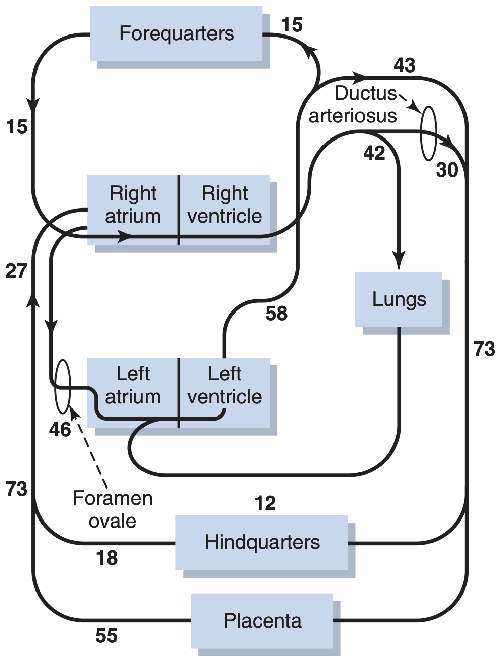
PLACENTA (O2 saturation ~80%)
│
▼ Umbilical Vein (single, carries oxygenated blood)
│
├── 50% → DUCTUS VENOSUS → IVC (bypasses liver)
│
└── 50% → Portal vein → LIVER sinusoids → Hepatic veins → IVC
↑
(mixes with deoxygenated
blood from GI tract)
│
▼
INFERIOR VENA CAVA
(mixed blood - oxygenated from ductus venosus + deoxygenated from lower body)
│
▼
RIGHT ATRIUM
│
├── ~60% → FORAMEN OVALE → LEFT ATRIUM
│ (Crista dividens directs IVC blood through FO)
│ │
│ ▼ LEFT VENTRICLE
│ │
│ ▼ ASCENDING AORTA
│ │
│ ┌────┴──────────────────┐
│ ▼ ▼
│ Coronary arteries Carotid/Subclavian
│ (Heart muscle) (Brain, Upper limbs)
│ [Well-oxygenated blood to vital organs]
│
└── ~40% + SVC blood → RIGHT VENTRICLE
│
▼
PULMONARY TRUNK
│
┌──────────┴───────────────┐
│ 10% to lungs (for lung │ 90% → DUCTUS ARTERIOSUS
│ tissue nutrition only) │ │
│ ▼
│ DESCENDING AORTA
│ │
│ ┌─────┴──────────────────────┐
│ ▼ ▼
│ Lower body/viscera 2 Umbilical Arteries
│ (less oxygenated) │
│ ▼
│ PLACENTA (for reoxygenation)
└──────────────────────────────────┘
- Liver (portal blood mixes with umbilical venous blood)
- IVC (lower body deoxygenated blood mixes)
- Right atrium (SVC blood mixes with residual IVC blood)
- Left atrium (small amount of pulmonary venous blood mixes)
- Entry of ductus arteriosus into descending aorta
- IVC blood (carrying well-oxygenated umbilical blood) is directed by the crista dividens through the foramen ovale into the left atrium → left ventricle → ascending aorta → coronary arteries and carotid arteries
- This ensures the brain and heart receive the most oxygenated blood
CHANGES AT BIRTH
BIRTH → CORD CLAMPED + FIRST BREATH
│
┌──────┴──────────────────────────────────────────┐
│ │
▼ ▼
CORD CLAMPED LUNGS EXPAND
(Placental flow stops) │
│ Pulmonary vessels
│ no longer compressed
│ │
▼ ↓ Pulmonary Vascular
↑ Systemic Vascular Resistance (5-fold)
Resistance (2-fold) │
│ ↑ Pulmonary blood flow
│ │
▼ ↑ Left Atrial Pressure
↑ Aortic pressure │
↑ Left Atrial Pressure ──────────┘
│
┌──────────────────────┼─────────────────────┐
▼ ▼ ▼
FORAMEN OVALE DUCTUS ARTERIOSUS DUCTUS VENOSUS
CLOSES CLOSES CLOSES
│ │ │
LA pressure > RA ↑ PaO2 causes Cord clamping
pressure → valve smooth muscle stops flow
pushed shut contraction │
(functional closure (functional within Fibrosis over
= immediate) 1-8 days) 2-3 months
Anatomic closure: Anatomic closure │
within 3 months (→ Lig. arteriosum) →Ligamentum
→ OVAL FOSSA by 12 weeks venosum
| Fetal Structure | Function in Fetus | Postnatal Closure | Adult Remnant |
|---|---|---|---|
| Ductus venosus | Bypasses liver (umbilical vein → IVC) | Functional: cord clamping; Anatomic: within 3-7 days | Ligamentum venosum |
| Foramen ovale | Right-to-left interatrial shunt | Functional: at birth (LA pressure > RA); Anatomic: within 3 months | Fossa ovalis (limbus = border) |
| Ductus arteriosus | Right-to-left shunt (PA → Aorta) | Functional: within 1-8 days (oxygen-induced); Anatomic: within 12 weeks | Ligamentum arteriosum |
| Umbilical vein | Carries oxygenated blood from placenta | Obliterates at birth | Ligamentum teres hepatis (round ligament of liver) |
| Umbilical arteries | Carry deoxygenated blood to placenta | Obliterate after cord clamping | Medial umbilical ligaments (proximal portions = superior vesical arteries) |
MEDICOLEGAL SIGNIFICANCE
1. Live Birth vs Stillbirth Determination
- Hydrostatic test (Docimasia pulmonum): Based on whether lungs have expanded
- If fetus breathed → lungs float in water (positive test = live birth)
- Lungs that have never expanded sink
- Important in infanticide cases
- Ductus arteriosus / Foramen ovale status at autopsy helps confirm live birth
2. Patent Ductus Arteriosus (PDA)
- If ductus arteriosus fails to close → left-to-right shunt (reversed from fetal direction)
- Aorta (high pressure) → Pulmonary artery (low pressure)
- Continuous machinery murmur
- Over time → Eisenmenger syndrome (pulmonary hypertension reverses shunt)
- Medicolegal: Life insurance disputes, flight fitness, military medical boards
3. Patent Foramen Ovale (PFO)
- Occurs in ~25-30% of adults
- Can allow paradoxical embolism (clot from venous system → crosses to arterial → stroke)
- Associated with cryptogenic stroke in young - medicolegal in workplace injury/compensation
- Scuba diving risk - decompression bubbles can pass PFO → arterial gas embolism
4. Infanticide Investigations
- Age at death (perinatally) can be assessed by whether the circulatory changes have taken place
- Fetal hemoglobin (HbF) vs. adult hemoglobin (HbA) - forensic hematology
- Ductus arteriosus histology: muscular (functional) vs. fibrosed (anatomic closure) helps establish age at death
5. Medical Negligence
- Failure to diagnose/treat congenital heart defects arising from failed circulatory changes
- Respiratory distress syndrome vs. cyanotic heart disease differential
6. Gestational Age Estimation
- Umbilical cord characteristics, placental weight, fetal circulation maturity indicators
Q.2 Discuss biochemical parameters which help in determination of time since death. (20 Marks)
INTRODUCTION
BIOCHEMICAL CHANGES AFTER DEATH
A. VITREOUS HUMOR BIOCHEMISTRY (Most Reliable)
- After death, K+ leaks from retinal cells into the vitreous
- Linear relationship between vitreous K+ and PMI:
- Formula (Sturner, 1963): PMI (hours) = 7.14 × [K+] - 39.1
- K+ rises approximately 0.17 mEq/L per hour (various studies: 0.14-0.19 mEq/L/hr)
- Normal vitreous K+ = 5-15 mEq/L
- At K+ > 15 mEq/L = death has occurred
- Useful range: 6-100 hours
- Limitation: Temperature-dependent; eye disease; sampling technique affects accuracy
- Decrease after death (moves intracellularly)
- Less reliable than K+ but useful in combination
- Decreases after death (consumed by glycolysis)
- Low vitreous glucose + high ketones = antemortem diabetic ketoacidosis
- If elevated = antemortem renal failure
- Relatively stable postmortem → forensic utility
- Product of adenosine degradation (ATP → ADP → AMP → adenosine → hypoxanthine)
- Rises linearly in first 24-36 hours
- Used to supplement K+ estimation
B. CEREBROSPINAL FLUID (CSF) BIOCHEMISTRY
| Parameter | Change After Death | Utility |
|---|---|---|
| K+ | Rises rapidly | PMI estimation (less reliable than vitreous) |
| Glucose | Falls rapidly | |
| Amino acids | Rise | |
| Glutamate | Rises significantly after 6 hours | |
| Lactate | Rises |
C. SYNOVIAL FLUID / PERICARDIAL FLUID
- Less studied but electrolyte changes similar to vitreous
- Used when vitreous not available
D. BLOOD BIOCHEMISTRY
| Parameter | Postmortem Change | Forensic Application |
|---|---|---|
| Postmortem glucose | Falls (consumed by RBCs and bacteria) | Low → starvation, DM |
| Lactic acid | Rises | Suggests perimortem physical exertion |
| Postmortem K+ (serum) | Unreliable due to hemolysis | Not used for PMI |
| Acetylcholinesterase | Reduced in organophosphate poisoning | Diagnosis |
| Troponin I, T | Elevated | Perimortem myocardial damage |
| Tryptase | Elevated in anaphylaxis | Mast cell degranulation |
| Cortisol | Elevated in stress deaths | |
| Alcohol (ethanol) | Relatively stable; slight postmortem rise from fermentation | BAC determination |
| COHb (carboxyhemoglobin) | Stable | CO poisoning diagnosis |
| MetHb | Stable | Nitrite/aniline poisoning |
E. MUSCLE BIOCHEMISTRY (Postmortem Biochemical Changes)
IMMEDIATELY AFTER DEATH
│
▼
ATP stores depleted (aerobic metabolism stops)
│
▼
Anaerobic glycolysis → Lactic acid accumulates
│
▼
Muscle pH drops (7.4 → 6.0)
│
▼
ATP depletion → Myosin-Actin cannot separate
│
▼
RIGOR MORTIS (6-12 hours onset; complete 12-24 hrs)
│
▼
Protease activity → Troponin I, Actin degradation
│
▼
RIGOR RESOLVES (24-48 hours)
F. BIOCHEMISTRY OF PUTREFACTION (Later PMI)
| Marker | Change | PMI Range |
|---|---|---|
| Bacterial amines (putrescine, cadaverine) | Rise | >24 hours |
| Volatile fatty acids (VFA) | Rise | Days |
| Indole, skatole | Rise | Days |
| Hydrogen sulfide | Rise | Days to weeks |
| Ammonia | Rise | >48 hours |
FLOWCHART: APPROACH TO PMI BY BIOCHEMICAL METHODS
BODY FOUND - ESTIMATE PMI
│
┌────┴──────────────────────────┐
│ │
▼ ▼
< 100 HOURS > 100 HOURS
│ │
▼ ▼
Vitreous K+ Putrefactive markers
(Sturner formula) (VFA, amines)
│
├── Supplement with:
│ - Vitreous hypoxanthine
│ - CSF biochemistry
│ - COHb if fire/CO suspected
│
▼
Cross-check with:
- Classical signs (rigor, livor, algor mortis)
- Stomach contents digestion state
- Entomological evidence
- Ambient temperature data
LIMITATIONS OF BIOCHEMICAL PMI ESTIMATION
- Temperature dependence - all chemical reactions accelerate with heat
- Sampling technique - vitreous must be obtained from central vitreous chamber
- Individual variation - antemortem disease alters baseline
- Putrefaction - bacteria produce/consume biochemicals, invalidating results
- No single parameter is absolute - combination approach needed
- Wide confidence intervals - biochemistry gives range, not precise time
- Refrigeration - slows biochemical changes dramatically
MEDICOLEGAL APPLICATIONS
- Determine whether victim was alive during an event (alibi corroboration/refutation)
- Distinguish antemortem vs. perimortem vs. postmortem injuries
- Verify time of death in suspicious circumstances
- Insurance fraud cases (timing of death relative to policy)
- Order of death in disaster/multiple casualties
Q.3 Discuss the process of wound healing and factors influencing it. Add a note on its significance in medicolegal practice. (20 Marks)
INTRODUCTION
TYPES OF WOUND HEALING
| Type | Description | Example |
|---|---|---|
| Primary intention | Clean wound edges approximated → heals with minimal scar | Surgical incision, sutured wound |
| Secondary intention | Wound edges cannot be approximated; heals from base upward with granulation tissue | Infected wound, large tissue defect |
| Tertiary intention (Delayed primary) | Wound initially left open, then closed later (after 4-5 days) | Contaminated traumatic wound |
PHASES OF WOUND HEALING
PHASE 1: HEMOSTASIS (Immediate - Minutes to Hours)
INJURY → Vessel Disruption
│
▼
VASOCONSTRICTION (Thromboxane A2, Serotonin)
[Brief initial response]
│
▼
PLATELET ADHESION AND AGGREGATION
(vWF bridges collagen to GPIb on platelets)
│
▼
COAGULATION CASCADE ACTIVATION
(Tissue factor → Thrombin → Fibrin)
│
▼
STABLE FIBRIN CLOT (platelet plug + fibrin mesh)
│
▼
Clot = scaffold + reservoir for growth factors
(PDGF, TGF-β, EGF released from platelet α-granules)
PHASE 2: INFLAMMATION (1-4 Days)
- Mast cell degranulation → histamine, serotonin → vasodilation + increased permeability
- Neutrophil recruitment (first cells - peak 24-48 hrs): phagocytosis, bacterial killing (oxidative burst), debridement
- Classic signs: Calor, Dolor, Rubor, Tumor (heat, pain, redness, swelling)
- Macrophages arrive (replace neutrophils as dominant cell by day 3-4)
- Macrophages: phagocytose debris, dead neutrophils; orchestrate healing via cytokines (IL-1, TNF-α, PDGF, TGF-β, VEGF)
- Lymphocytes arrive (day 5-7): regulate fibroblast activity, ECM remodeling
PHASE 3: PROLIFERATION (Day 4 to ~3 Weeks)
- Fibroblasts migrate from wound edges (stimulated by PDGF, TGF-β)
- Day 5-7: fibroblast proliferation + collagen synthesis begins
- Type III collagen (immature) initially; later replaced by Type I
- Collagen content peaks at 3 weeks
- New capillary sprouts form from existing vessels (VEGF-driven)
- Granulation tissue = new capillaries + fibroblasts + loose collagen
- Pink, vascular, friable appearance of granulation tissue
- Keratinocytes at wound edge proliferate and migrate over wound surface
- Complete within 24-48 hours for small wounds; days for larger wounds
- Creates protective epidermal barrier
PHASE 4: REMODELING / MATURATION (3 Weeks to 2 Years)
- Type III collagen replaced by Type I collagen (stronger)
- Collagen fibers reorganize along lines of tension
- Cross-linking increases (lysyl oxidase enzyme) → tensile strength increases
- Blood vessels regress → scar becomes avascular and paler
- Maximum tensile strength achieved = only 80% of original skin strength
- Scar gradually flattens and fades
TIMELINE SUMMARY TABLE (Forensic Age Estimation)
| Time After Injury | Histological Findings | Gross Appearance |
|---|---|---|
| Minutes-Hours | Hemorrhage, edema, fibrin | Red, fresh, bleeding |
| 12-24 hours | PMN (neutrophils) dominate | Red, swollen |
| 2-3 days | PMN declining; macrophages arriving | Scab forming |
| Day 3-5 | Macrophages dominant; early fibroblasts; new vessels | Granulation tissue beginning |
| Day 5-7 | Active fibroplasia; angiogenesis; early collagen | Granulation tissue obvious |
| Day 7-14 | Abundant fibroblasts; Type III collagen; re-epithelialization | Wound contracting |
| Day 14-21 | Fibrosis; decreased cellularity; collagen maturing | Pink scar |
| Weeks-Months | Type I collagen; scar remodeling | White/pale scar |
FACTORS INFLUENCING WOUND HEALING
| Factor | Effect |
|---|---|
| Infection | Delays healing - prolongs inflammation, destroys tissue |
| Blood supply | Poor perfusion (arteriosclerosis) → impaired oxygen/nutrient delivery |
| Foreign body | Perpetuates inflammation, forms sinus tracks |
| Wound size | Larger wounds heal slower |
| Radiation injury | Damages vascular endothelium, reduces fibroblast proliferation |
| Movement | Excess movement at wound site → poor healing |
| Haematoma | Acts as medium for infection; separates wound edges |
| Wound edges apposition | Good apposition → primary intention; poor → secondary |
| Factor | Effect |
|---|---|
| Nutrition | Protein deficiency → poor collagen synthesis; Vitamin C deficiency → collagen cross-linking fails (scurvy) |
| Diabetes mellitus | Impaired leukocyte function, microangiopathy, peripheral neuropathy → poor healing |
| Corticosteroids | Suppress inflammation, inhibit fibroblast proliferation, decrease collagen synthesis |
| Age | Elderly - reduced cell proliferation, decreased growth factors |
| Anemia | Reduced oxygen delivery → impaired healing |
| Immunodeficiency | Susceptibility to infection → chronic non-healing wounds |
| Drugs | NSAIDs, chemotherapy delay healing |
| Obesity | Poor blood supply to fat; increased wound tension |
| Smoking | Vasoconstriction; carboxyhemoglobin reduces tissue oxygenation |
| Hypothyroidism | Reduced metabolic rate; poor fibroblast activity |
| Uremia | Impairs leukocyte function |
ABNORMAL WOUND HEALING
| Complication | Description |
|---|---|
| Keloid | Scar extends beyond original wound margins; contains excess Type III collagen; more in dark skin; common on face, sternum, earlobes |
| Hypertrophic scar | Remains within wound margins; raised, red; may regress spontaneously |
| Wound dehiscence | Reopening of a previously closed wound (usually day 5-8 post-op) |
| Chronic non-healing ulcer | Fails to progress through healing stages; e.g., venous ulcer, diabetic foot |
| Contracture | Excessive scar contraction → functional limitation (esp. near joints) |
| Marjolin's ulcer | Malignant transformation (SCC) in chronic scar (decades later) |
MEDICOLEGAL SIGNIFICANCE OF WOUND HEALING
- The age of a wound can be estimated from histological examination
- Determines whether injury was:
- Antemortem (before death) - shows vital reaction (bleeding, inflammation, healing)
- Perimortem (at or around time of death)
- Postmortem (after death) - no vital reaction; edges dry, yellowish, shrunken
- This is critical in homicide investigations - distinguishes weapon injuries from postmortem mutilation
- Histological dating allows court to assess whether injury timeline matches accused's alibi
- E.g., neutrophil infiltration indicates injury occurred within last 24 hours
- Granulation tissue formation confirms injury was at least 4-5 days old
- Extent of healing indicates how long victim survived after injury
- Present of organization of hemothorax → survived several days after thoracic injury
- Incised wounds (clean edges) → sharp weapon
- Lacerated wounds (irregular, bridged by tissue) → blunt weapon
- Healing pattern may reveal weapon type
- Wounds on forearms/hands in various stages of healing → victim had time to defend
- Self-inflicted (hesitation cuts): parallel, superficial, accessible sites (wrist, forearm)
- Inflicted: irregular, in inaccessible areas, deeper
- Old scars provide evidence of previous assault
- Scar dating can corroborate history of domestic violence
- Sepsis from infected wound after assault → wound recipient (accused) liable for death even if original injury was non-fatal
Q.4 Write Short Notes on (40 Marks)
(a) Role of Histopathology in Forensic Medicine (10 Marks)
APPLICATIONS
- Confirms myocardial infarction (coagulative necrosis, neutrophil infiltration, contraction bands)
- Pulmonary embolism (organized thrombus in pulmonary artery)
- Subarachnoid hemorrhage microscopic confirmation
- Sudden cardiac death - conduction system histology
- Conduction system examination (SA node, AV node, Bundle of His) - mandatory in sudden unexplained cardiac deaths
- Age of wounds: neutrophils (hours), macrophages (days), fibrosis (weeks) - forensic wound dating
- Age of bruises: hemoglobin breakdown products (hemosiderin, biliverdin)
- Age of subarachnoid hemorrhage: early (hours) vs. organized (days)
- Antemortem: hemorrhage, edema, inflammatory cells, fibrin, epithelial changes
- Postmortem: no inflammation, no fibrin, no epithelial reaction, no vital response
| Condition | Histological Finding |
|---|---|
| Fat embolism | Fat globules in pulmonary/cerebral capillaries (Oil Red O on frozen sections) |
| Air embolism | Vacuoles in pulmonary capillaries |
| Drowning | Pulmonary alveolar edema; emphysema aquosum; diatoms in organs (liver, bone marrow) |
| Electrocution | Skin - nuclear streaming ("lightning trees"), vacuolization at wound edges |
| Burns | Coagulation necrosis; vascular changes indicating antemortem vs postmortem burns |
| Carbon monoxide poisoning | Cherry-red discoloration; cherry-red globules in capillaries |
| Drug deaths | Pulmonary edema (opioids); myocarditis (cocaine); granulomas (IV drug use) |
| Child abuse | Retinal hemorrhages; subdural hematoma of different ages; "corner fractures" |
| SIDS | Intrathoracic petechiae; pulmonary alveolar macrophages; brainstem gliosis |
- Bone histology (osteon counting) - Kerley's method
- Dental cementum annulations
- Cartilage mineralization
- Spermatozoa in vaginal/anal/oral swabs - cytological examination
- Vaginal smear - sperm motility, morphology
- Racial characteristics from skin histology
- Blood group antigens on tissues
- Hair histology for racial identification
- Hair sectioning for drug timeline
- Nail sections for metal poisoning
- Liver histology for steatosis (alcohol)
- Confirm diagnosis, standard of care, cause of death
- Whether appropriate treatment was given
- Brain, heart, lungs, liver, kidneys, adrenals, spleen routinely
- Specific organ blocks based on clinical context
- Fixed in 10% neutral buffered formalin
- Frozen sections for fat, carbon, enzyme histochemistry
(b) Types and Medico-Legal Importance of Fractures (10 Marks)
CLASSIFICATION BY MECHANISM (Forensic Types)
| Type | Description | Forensic Significance |
|---|---|---|
| Simple/Closed | Bone broken, overlying skin intact | Blunt force injury |
| Compound/Open | Bone communicates with external environment via skin wound | Higher risk of infection; complication of assault |
| Comminuted | Bone shattered into multiple fragments | High-velocity impact; severe blunt force |
| Transverse | Break perpendicular to long axis | Direct blow/bending force |
| Oblique | Break at angle | Bending + compressive force |
| Spiral | Spiraling along bone axis | Twisting force - common in child abuse (non-accidental injury) |
| Greenstick | Incomplete break - one cortex intact | Children (elastic bone) - bending force |
| Impacted | Bone ends driven into each other | Compressive force; falls from height |
| Pathological | Through diseased bone (tumor, osteoporosis) | Minimal force; distinguish from trauma |
| Stress/Fatigue | Repeated small stresses | Occupational; athletes; military |
| Avulsion | Fragment pulled off by tendon/ligament | Sudden violent muscle contraction |
| Depressed (skull) | Fragment pushed inward | Blunt force to skull - weapon analysis |
MEDICOLEGAL IMPORTANCE
- Skull fractures:
- Pond fracture: Depressed, circular/oval - from rounded blunt weapon
- Ring fracture (Basal): Around foramen magnum - falls on feet, buttocks → concentric force
- Hinge fracture: Transverse fracture across skull base - against lateral force
- Comminuted: Multiple fragments - severe, repeated blows
- Contrecoup fracture: At side opposite to impact - severe impact
- Spiral fractures in infants/toddlers who cannot walk → child abuse (non-accidental injury)
- Multiple fractures of different ages → repeated abuse
- Fractures inconsistent with stated mechanism of injury
- Fresh: sharp edges, hemorrhage (hours)
- Days: callus formation begins (periosteal thickening)
- Weeks: soft callus visible on X-ray
- Months: hard callus, bridging
- Old: remodeled, cortex re-established
- Multiple fractures at different stages of healing
- Metaphyseal corner fractures (bucket-handle)
- Rib fractures posteriorly
- In absence of metabolic bone disease → strong evidence of child abuse
- Fractures of forearm (radius/ulna) - blocking blows = Parry fracture (defensive fracture)
- Evidence victim attempted self-defense
- Fracture pattern reveals direction of impact
- Bumper fracture (fracture of fibula/tibia at bumper level) indicates pedestrian struck from specific direction
- Reconstruction of events from fracture analysis
- Fat embolism from long bone fractures
- Hypovolemic shock (femur fracture = 1-2L blood loss)
- Pneumothorax/hemothorax from rib fractures
- Spinal cord injury from vertebral fractures → liability of negligence if undetected
- Entrance wound: punched-in margin, internal beveling of skull
- Exit wound: external beveling, larger defect
- Intermediate range - cruciate fracture pattern
(c) Post-Mortem Radiology: Importance and Limitations (10 Marks)
MODALITIES USED
| Modality | Application |
|---|---|
| Plain X-rays | Fractures, bullets/foreign bodies, air embolism, pneumothorax, bone age |
| CT scan (PMCT) | Most widely used PM imaging - injuries, hemorrhage, gas patterns, foreign bodies |
| MRI (PMMRI) | Soft tissue injuries, brain lesions, spinal cord |
| Fluoroscopy | Angiography of vessels postmortem |
| PM Angiography | Coronary arteries, circle of Willis assessment |
| Ultrasound | Limited postmortem use |
| Micro-CT | Small specimens, pediatric/fetal cases |
IMPORTANCE / ADVANTAGES
- Creates a permanent visual record of injuries in situ
- CT provides 3D reconstruction of injury patterns
- Bullets, pellets, bomb fragments, medical devices (pacemakers, stents)
- Location, trajectory of projectiles without cutting → important in criminal cases
- All fractures including small ones detected
- 3D reconstruction of skull fractures for weapon analysis
- Spinal fractures, rib fractures
- Fracture dating on CT
- Air/gas embolism in vessels (air in cardiac chambers, great vessels)
- Pneumothorax, pneumomediastinum
- Decomposition gas distribution
- Subdural, epidural, subarachnoid hemorrhage
- Hemothorax, hemoperitoneum
- Retroperitoneal hemorrhage
- Required in certain religions that prohibit conventional autopsy
- Can identify cause of death without organ dissection
- Useful for mummified or decomposed remains
- Dental radiographs for dental identification
- Bone age estimation
- Previous healed fractures, implants, prostheses match to medical records
- Pacemaker, artificial valves, joint prostheses
- Assess function, position, integrity
- Whole-body PM X-ray ("babygram") detects multiple fractures
- Increased brain density on CT → certain poisonings (cyanide)
- Mass casualty events: rapid triage imaging of all bodies
LIMITATIONS
- Cannot detect histological changes (vital reaction, cellular infiltration)
- Cannot collect biological samples (vitreous, blood, urine, bile) for toxicology
- Cannot smell decomposition gases or odors (CO, hydrogen cyanide)
- Cannot detect color changes (CO poisoning - cherry red)
- Gas decomposition mimics air embolism
- Livor mortis can simulate hemorrhage
- Postmortem redistribution of blood mimics hemothorax
- CT poor at distinguishing bruise from normal muscle
- Mucosal injuries not detectable
- Petechial hemorrhages too small for resolution
- No imaging correlate for most toxins
- CT scanners not available in all mortuaries
- Significant expense
- Requires trained forensic radiologist
- Coronary artery lesions require PM angiography
- Micro-thrombi not detectable
- Severely decomposed bodies lose diagnostic value rapidly
(d) Toxicokinetics and Toxicodynamics: Definitions and Forensic Relevance (10 Marks)
DEFINITIONS
"What the body does to the poison" The study of the time course of absorption, distribution, metabolism, and elimination (ADME) of toxic substances in the body.
"What the poison does to the body" The study of the biochemical and physiological effects of toxic substances on the body and their mechanisms of action.
TOXICOKINETICS - ADME MODEL
TOXIC SUBSTANCE ENTERS BODY
│
▼
┌────────────────────────────────────────────────────────────────┐
│ A - ABSORPTION │
│ Routes: Oral, Inhalation, Dermal, IV, IM, Rectal, Sublingual │
│ Factors: pH, lipid solubility, molecular size, formulation │
│ 1st pass effect (oral): gut wall + liver metabolism before │
│ reaching systemic circulation │
└─────────────────────────┬──────────────────────────────────────┘
│
▼
┌────────────────────────────────────────────────────────────────┐
│ D - DISTRIBUTION │
│ Volume of Distribution (Vd) = Dose / Blood concentration │
│ High Vd = distributes widely into tissues (fat, muscle) │
│ Crosses BBB: lipid soluble, non-ionized molecules │
│ Protein binding: reduces free drug available for toxicity │
│ Redistribution after death = POSTMORTEM REDISTRIBUTION │
│ (drugs move from high-concentration tissues to blood) │
└─────────────────────────┬──────────────────────────────────────┘
│
▼
┌────────────────────────────────────────────────────────────────┐
│ M - METABOLISM │
│ Primarily in LIVER (CYP450 system) │
│ Phase I: Oxidation, reduction, hydrolysis → reactive │
│ Phase II: Conjugation → water-soluble metabolites │
│ Bioactivation: prodrugs → active toxic metabolites │
│ (e.g., paracetamol → NAPQI → hepatotoxic) │
│ Genetic polymorphisms → CYP2D6 poor/ultra metabolizers │
└─────────────────────────┬──────────────────────────────────────┘
│
▼
┌────────────────────────────────────────────────────────────────┐
│ E - ELIMINATION │
│ Half-life (t½) = time for plasma concentration to halve │
│ Routes: Renal (urine), Biliary (feces), Pulmonary (exhaled) │
│ Entero-hepatic circulation (EHC) prolongs effect │
│ First-order kinetics: constant fraction eliminated per unit │
│ time (most drugs) │
│ Zero-order kinetics: constant amount eliminated (alcohol, │
│ aspirin at toxic doses) │
└────────────────────────────────────────────────────────────────┘
TOXICODYNAMICS - MECHANISMS OF TOXICITY
| Mechanism | Example |
|---|---|
| Receptor binding | Organophosphates → AChE inhibition → cholinergic crisis; Opioids → mu receptor agonism → respiratory depression |
| Ion channel blockade | Local anesthetics → Na+ channel block → cardiac arrest; Tetrodotoxin → Na+ channel |
| Enzyme inhibition | Cyanide → cytochrome c oxidase → cellular hypoxia; CO → hemoglobin |
| Mitochondrial toxicity | Cyanide, DNP (dinitrophenol) → uncoupling of oxidative phosphorylation |
| DNA damage | Cyclophosphamide, benzene → alkylation → mutagenesis |
| Oxidative stress | Paraquat → free radical generation → lung damage |
| Immunological mechanisms | Penicillin → hapten → anaphylaxis |
| Direct cellular toxicity | Caustics → protein denaturation + coagulative necrosis |
- LD50: Lethal dose for 50% of test population - comparative toxicity measure
- Therapeutic index (TI): LD50 / ED50 - narrow TI = dangerous drug (digoxin, lithium)
- Threshold dose: Minimum dose to produce effect
- NOAEL: No observed adverse effect level
FORENSIC RELEVANCE
- Toxicokinetic data + blood/tissue levels allow calculation of amount ingested
- Distinguish therapeutic, toxic, and lethal concentrations
- Absorption pattern reveals route:
- IV → rapid peak, no first-pass → very high early blood levels
- Oral → delayed peak, first-pass effect → metabolites in liver
- Inhalation → rapid onset
- Dermal → slow absorption, low peak
- After death, drugs diffuse from high concentration tissues (liver, lungs) into blood
- Can falsely elevate blood concentrations
- Affects: tricyclics, digoxin, chlorpromazine, morphine, propranolol
- Solution: use peripheral blood (femoral vein) not central blood for PM toxicology
- PMR is a major confounding factor in forensic toxicology
- Gastric contents + absorption kinetics → approximate time of ingestion
- Stomach lavage contents vs. blood levels
- Relevant in poisoning investigations
- Chronic drug users have higher tolerance → "normal" lethal dose may not kill them
- Relevant in accidental death vs. homicide with drugs
- CYP450 inhibitors (e.g., erythromycin) may raise levels of co-administered drug
- Relevant in medical negligence cases
- Poor metabolizers may die from "normal" doses
- Ultra-rapid metabolizers may require higher therapeutic doses
- CYP2D6, CYP2C19 variants - exculpatory/explanatory evidence
- Ethanol is metabolized at ~10-15 mg/dL/hour (regardless of concentration)
- Back-calculation (retrograde extrapolation) of BAC at time of driving possible
- Medically/legally significant in drunk driving fatalities
- Specific organ damage patterns indicate specific poison:
- Hepatic necrosis (paracetamol)
- Pulmonary fibrosis (paraquat)
- Yellow liver atrophy (phosphorus)
- Garlic smell + peripheral neuropathy (arsenic)
COMPLETE COVERAGE SUMMARY
PAPER II - IInd Term (All 100 Marks Answered)
| Q | Topic | Marks | Coverage |
|---|---|---|---|
| Q1 | Temperature Regulation + Extreme Heat | 20 | Thermoreceptors, hypothalamus (anterior/posterior), heat production/loss (RESC), heat cramps/exhaustion/stroke, multi-organ failure, forensic PM findings |
| Q2 | Autonomic Nervous System + Sudden Death | 20 | SNS/PNS anatomy, neurotransmitters, comparison table, 5 mechanisms of ANS-mediated sudden death (vasovagal, catecholamine surge, LQTS, baroreflex failure, reflex inhibition) |
| Q3 | Female External Genitalia + Rape Examination | 20 | Full anatomy of vulva with hymen types, lithotomy examination, site diagram (from Reddy's FMT), sample collection table, report structure, legal points (BNS Sec 63) |
| Q4a | Age Changes at Pubic Symphysis | 10 | Todd's 10-phase system, Suchey-Brooks 6-phase system with tables |
| Q4b | Ewing's Postulate | 10 | All 7 criteria in flowchart, clinical examples, forensic applications |
| Q4c | Traumatic Fat Embolism | 10 | Mechanical + biochemical theories, Gurd's criteria, Oil Red O frozen section, medicolegal significance |
| Q4d | Medicolegal Aspects of Violence Against Women | 10 | Classification, key Indian laws (BNS/POCSO/DV Act), forensic medicine role in each category |
PAPER I - Ist Year (All 100 Marks Answered)
| Q | Topic | Marks | Coverage |
|---|---|---|---|
| Q1 | Fetal Circulation + Changes at Birth + Medicolegal | 20 | Complete pathway with mixing points, flow diagram (Guyton's), changes at birth flowchart, fate of all 5 fetal shunts, PDA/PFO medicolegal significance, infanticide |
| Q2 | Biochemical Parameters for Time Since Death | 20 | Vitreous K+ (Sturner formula), hypoxanthine, CSF, blood biochemistry, putrefactive markers, limitations |
| Q3 | Wound Healing + Factors + Medicolegal | 20 | All 4 phases with cellular timeline table, local vs systemic factors, abnormal healing, forensic wound dating, vital reaction |
| Q4a | Role of Histopathology in Forensic Medicine | 10 | 9 application areas with specific stains and findings |
| Q4b | Types and Medicolegal Importance of Fractures | 10 | 12 fracture types, bumper fracture, spiral fracture in abuse, skull fracture patterns |
| Q4c | Postmortem Radiology | 10 | CT/MRI/X-ray applications, 11 importance points, 7 limitations |
| Q4d | Toxicokinetics and Toxicodynamics | 10 | ADME flowchart, dose-response, postmortem redistribution, forensic applications including back-calculation |
Q.1 Describe the mechanism of regulation of body temperature, with special reference to effects of extreme heat. Q.2 Describe briefly the "Autonomic Nervous System". Discuss the pathophysiology of sudden death initiated through Autonomic Nervous System. Q.3 Describe anatomy of female external genitals. How is a victim of rape examined and reported? Q.4 Write short notes on a)Age changes at pubic symphysis. b)Ewing's postulate. c)Traumatic fat emboli. d) Medicolegal aspects of violence against women. Q.6 Describe in detail 'fetal circulation' changes occurring during and after birth and its medicolegal significance. Q.7 Discuss biochemical parameters which help in determination of time since death. Q.8 Discuss the process of wound healing and factors influencing it. Add a note on its significance in medicolegal practice. Q.9 Write short notes on a) Role of histopathology in forensic medicine. b) Types and medico-legal importance of fractures. c) Post-mortem radiology: Importance and limitations. d) Toxicokinetics and toxicodynamics: Definitions and forensic relevance. Solve the answers , diagrams, images and flow charts where necessary
PRAVARA INSTITUTE OF MEDICAL SCIENCES
POST GRADUATE DEGREE IN FORENSIC MEDICINE (M.D.)
Complete Model Answers - Both Papers
PAPER II
Q.1 Mechanism of Regulation of Body Temperature with Special Reference to Effects of Extreme Heat (20 Marks)
INTRODUCTION
THERMORECEPTORS
- Warm receptors (C fibers) and cold receptors (Aδ fibers) in skin
- Send signals via spinothalamic tracts to hypothalamus
- Located in preoptic area of anterior hypothalamus
- Detect blood temperature changes as small as 0.1°C
- Most sensitive and dominant
HYPOTHALAMIC THERMOSTAT
┌──────────────────────────────────────────────────────────┐
│ HYPOTHALAMUS (PREOPTIC AREA) │
│ │
│ ANTERIOR HYPOTHALAMUS POSTERIOR HYPOTHALAMUS │
│ (Heat Dissipation Center) (Heat Conservation Center) │
│ - Activated by HEAT - Activated by COLD │
│ - Sweating - Shivering │
│ - Vasodilation - Vasoconstriction │
│ - Behavioral cooling - Piloerection │
└──────────────────────────────────────────────────────────┘
HEAT PRODUCTION MECHANISMS
| Mechanism | Details |
|---|---|
| Basal Metabolic Rate | Ongoing cellular metabolism ~60-70 kcal/hr at rest |
| Muscular activity | Exercise raises heat production 10-20 fold |
| Shivering thermogenesis | Involuntary rhythmic contractions - raises heat 2-5x |
| Non-shivering thermogenesis | Brown adipose tissue (UCP-1) - uncouples ATP to heat; important in neonates |
| Specific dynamic action | Food digestion raises metabolic rate 10-30% |
| Hormonal | Thyroxine (long-term), Epinephrine (acute) increase metabolic rate |
HEAT LOSS MECHANISMS
ROUTES OF HEAT LOSS
│
┌──────────┼──────────────┬──────────────┐
▼ ▼ ▼ ▼
RADIATION CONDUCTION CONVECTION EVAPORATION
~60% ~3% ~12% ~25%
│ │ │ │
Infrared Direct contact Moving air Sweating +
EMR waves with surface currents insensible loss
(water = 25x carry heat (lungs + skin)
better than away
air)
- Sweat glands (2-3 million) can produce up to 1-2 liters/hour under maximal heat stress
- Controlled by cholinergic sympathetic fibers
THERMOREGULATORY FLOWCHART: RESPONSE TO HEAT
RISE IN BODY TEMPERATURE (e.g., exercise, hot environment)
│
▼
ANTERIOR HYPOTHALAMUS ACTIVATED
│
┌──────────┴──────────────┐
▼ ▼
CUTANEOUS VASODILATION SWEAT GLANDS ACTIVATED
(via sympathetic withdrawal (cholinergic sympathetics)
and active vasodilator fibers) │
│ Sweating → Evaporative cooling
Blood shunted to skin │
surface → radiant/ ▼
convective heat loss ┌────────────────────────┐
│ │ Heat loss > Heat gain? │
│ └────────┬───────────────┘
│ │ YES
└─────────────────────────────┼──────────────────┐
▼ ▼
Temp normalizes Behavioral response
│ (seek shade, remove
▼ clothing, drink water)
NEGATIVE FEEDBACK
Anterior hypothalamus
inhibited
EFFECTS OF EXTREME HEAT
A. HEAT CRAMPS
- Painful muscle cramps (calves, abdomen) during or after exercise
- Cause: salt depletion from excessive sweating with hypotonic fluid replacement
- Core temperature: normal
- Treatment: oral saline + rest
B. HEAT SYNCOPE
- Transient loss of consciousness
- Cause: peripheral vasodilation → venous pooling → ↓ cardiac output → cerebral hypoperfusion
- Core temperature: slightly elevated
- Treatment: lie flat, cool environment, oral fluids
C. HEAT EXHAUSTION
- Core temperature: 37°C - 40°C
- Features: profuse sweating, weakness, dizziness, nausea, headache, tachycardia, hypotension
- CNS function: intact (key distinction from heat stroke)
- Pathophysiology: dehydration + vasodilation → ↓ cardiac output → ↓ cerebral perfusion
- Treatment: remove from heat, cool environment, IV normal saline
D. HEAT STROKE - MOST SEVERE (MEDICAL EMERGENCY)
| Feature | Classic Heat Stroke | Exertional Heat Stroke |
|---|---|---|
| Population | Elderly, infants, chronic illness | Young athletes, military |
| Setting | Passive heat exposure (heat waves) | Strenuous physical exertion |
| Sweating | Often absent (anhidrosis) | Profuse sweating usually present |
| Onset | Gradual (days) | Rapid (hours) |
CORE TEMPERATURE >40°C
(Heat dissipation mechanisms overwhelmed)
│
▼
PROTEIN DENATURATION + ENZYME FAILURE
│
┌────────┼──────────────────────────────────────────┐
▼ ▼ ▼
CNS FAILURE CARDIOVASCULAR FAILURE DIRECT TISSUE DAMAGE
│ │ │
Encephalopathy Tachycardia ┌────────┼─────────┐
Seizures Hypotension ▼ ▼ ▼
Coma Arrhythmias LIVER KIDNEY MUSCLE
Cerebral Circulatory (centrilobular (Rhabdo-
edema collapse necrosis) myolysis)
│ │ │
Jaundice ATN/ARF Myoglobinuria
│
┌────────────┴────────────────┐
▼ ▼
DIC (Disseminated ARDS (Acute
Intravascular Respiratory
Coagulation) Distress Syndrome)
│ │
Hemorrhage Pulmonary edema
Thrombosis Hypoxia
│
▼
MULTI-ORGAN FAILURE
↓ → DEATH if untreated
| System | Effect |
|---|---|
| CNS | Encephalopathy, seizures, cerebellar damage (Purkinje cell loss), cerebral edema |
| CVS | Tachycardia, hypotension, arrhythmias, myocardial necrosis |
| Kidneys | Acute tubular necrosis from myoglobinuria + rhabdomyolysis |
| Liver | Centrilobular necrosis (hepatocytes most sensitive to heat) |
| Blood | DIC - fibrinogen consumed, platelet aggregation, thrombocytopenia |
| Muscles | Rhabdomyolysis → myoglobinuria → renal failure |
| Gut | Mucosal ischemia → bacterial translocation → sepsis |
| Lungs | ARDS, pulmonary hemorrhage |
FORENSIC / MEDICOLEGAL SIGNIFICANCE
- Deaths from heat stroke - autopsy findings: cerebral edema, petechiae in brain, myocardial degeneration, centrilobular hepatic necrosis, tubular necrosis in kidneys
- Infant deaths - left in hot cars/rooms = criminal negligence (heat-related child homicide)
- Elderly neglect - failure to provide cooling in extreme heat = culpable homicide
- Occupational heat deaths - factory workers, construction, military = compensable death under Workmen's Compensation Act
- Hyperpyrexia vs infection-related fever - must be distinguished at autopsy
- Core temperature at scene - important evidence; best measured rectal temperature at autopsy
- Histological evidence - protein denaturation, coagulative necrosis across organs on histopathology
Q.2 Autonomic Nervous System and Pathophysiology of Sudden Death Through ANS (20 Marks)
PART A: THE AUTONOMIC NERVOUS SYSTEM
- Sympathetic (SNS) - Thoracolumbar
- Parasympathetic (PNS) - Craniosacral
- Enteric (ENS) - gut nervous system
ANATOMY
A. SYMPATHETIC NERVOUS SYSTEM
- Origin: Thoracolumbar outflow - T1 to L2 spinal cord
- Preganglionic fibers: Short, myelinated
- Ganglia: Paravertebral (sympathetic chain) or prevertebral ganglia
- Postganglionic fibers: Long, unmyelinated
- Neurotransmitters:
- Preganglionic: Acetylcholine (nicotinic receptor)
- Postganglionic: Norepinephrine (adrenergic α and β receptors)
- Exception: Sweat glands and adrenal medulla → Acetylcholine
- Adrenal medulla: Modified sympathetic ganglion → releases Epinephrine (80%) + Norepinephrine (20%) into bloodstream
- Effect: "FIGHT OR FLIGHT"
- ↑ Heart rate (β1), ↑ Contractility
- Bronchodilation (β2)
- Vasoconstriction in skin/viscera (α1)
- Vasodilation in skeletal muscle
- Pupil dilation (mydriasis)
- ↓ GI motility, sphincter contraction
- Glycogenolysis, lipolysis
B. PARASYMPATHETIC NERVOUS SYSTEM
- Origin: Craniosacral outflow - CN III, VII, IX, X and S2-S4
- Preganglionic fibers: Long, myelinated
- Ganglia: Terminal ganglia (near or within target organ)
- Postganglionic fibers: Very short
- Neurotransmitter: Acetylcholine at both junctions (nicotinic preganglionic; muscarinic postganglionic)
- Vagus nerve (CN X): supplies heart, bronchi, esophagus, stomach, small intestine, ascending colon, liver, pancreas
- Effect: "REST AND DIGEST"
- ↓ Heart rate (M2 on SA node)
- ↓ AV conduction
- Bronchoconstriction
- Pupil constriction (miosis)
- ↑ GI motility, secretions
- Bladder contraction
COMPARISON TABLE
| Feature | Sympathetic | Parasympathetic |
|---|---|---|
| Origin | T1-L2 (Thoracolumbar) | CN III,VII,IX,X; S2-S4 |
| Preganglionic fiber | Short | Long |
| Postganglionic fiber | Long | Short |
| Ganglia | Paravertebral/Prevertebral | Terminal (near target) |
| Preganglionic NT | ACh (Nicotinic) | ACh (Nicotinic) |
| Postganglionic NT | Norepinephrine | Acetylcholine (Muscarinic) |
| Heart rate | ↑ Increases | ↓ Decreases |
| Blood pressure | ↑ Increases | ↓ Decreases |
| Pupils | Dilates | Constricts |
| Bronchi | Dilates | Constricts |
| GI | Decreases motility | Increases motility |
| Bladder | Relaxes detrusor | Contracts detrusor |
| Sweat glands | Activates (ACh) | No innervation |
CARDIAC INNERVATION (Key for Sudden Death)
- Sympathetic: T1-T5 → stellate ganglia → cardiac nerves → SA node, AV node, ventricular myocardium
- Effect: ↑ HR (chronotropy), ↑ Contractility (inotropy), ↑ AV conduction (dromotropy)
- Parasympathetic: Dorsal vagal nucleus, nucleus ambiguus (CN X) → SA node, AV node
- Effect: ↓ HR, ↓ AV conduction; vagal tone is dominant at rest
PART B: PATHOPHYSIOLOGY OF SUDDEN DEATH THROUGH ANS
MECHANISM 1: VASOVAGAL (NEUROCARDIOGENIC) SYNCOPE → CARDIAC ARREST
TRIGGER:
Severe pain, fright, sight of blood, micturition/defecation,
prolonged standing, carotid sinus pressure, cold water on face
│
▼
PARADOXICAL PARASYMPATHETIC SURGE
(Bezold-Jarisch reflex: underfilled ventricle
→ stimulates mechanoreceptors → vagal afferents)
│
┌─────────┴──────────────────────────────┐
▼ ▼
PROFOUND BRADYCARDIA PERIPHERAL
(Sinus arrest / Asystole) VASODILATION
(SA node firing suppressed) (arteriolar dilation)
│ │
└──────────────────┬─────────────────────┘
▼
SEVERE HYPOTENSION
│
▼
CEREBRAL HYPOPERFUSION
│
▼
LOSS OF CONSCIOUSNESS
│
If sustained:
▼
CARDIAC ARREST → DEATH
MECHANISM 2: SYMPATHETIC SURGE → FATAL VENTRICULAR ARRHYTHMIA
INTENSE STRESS / CATECHOLAMINE EXCESS
(Anger, excitement, cocaine, amphetamines,
pheochromocytoma, acute MI pain)
│
▼
MASSIVE SYMPATHETIC ACTIVATION
↑ Circulating Epinephrine + Norepinephrine
│
┌─────────┴──────────────────────────────┐
▼ ▼
↑ Heart Rate + ↑ Myocardial
↑ Contractility O₂ DEMAND
│ │
Shortened refractory Ischemia (esp. if
period → increased pre-existing CAD)
automaticity │
│ ▼
│ Catecholamine toxicity
│ (direct myocyte damage)
└──────────────┬────────────────────┘
▼
VENTRICULAR FIBRILLATION (VF)
│
▼
SUDDEN DEATH
MECHANISM 3: LONG QT SYNDROME AND STELLATE GANGLION IMBALANCE
- Left stellate ganglion predominance (vs. right) → asymmetric sympathetic stimulation
- Prolongs QT interval → Torsades de Pointes → degenerates into VF
- Congenital causes:
- Romano-Ward syndrome (autosomal dominant) - triggered by startle/exercise
- Jervell-Lange-Nielsen syndrome (autosomal recessive) - with congenital deafness
- Acquired: hypokalemia, drugs (haloperidol, amiodarone, erythromycin), hypothyroidism
MECHANISM 4: REFLEX CARDIAC ARREST - SPECIAL REFLEXES
SPECIFIC TRIGGERS VAGAL PATHWAY CARDIAC EFFECT
│
Blow to epigastrium ──────────────► Celiac plexus ──────────► Bradycardia
(Solar plexus punch) │ → Asystole
│
Blow to neck / Carotid ───────────► Carotid sinus ─────────► Bradycardia
sinus pressure baroreceptors + Hypotension
(Strangulation)
│
Sudden face immersion ────────────► Trigeminal nerve ──────► Profound
in cold water (diving │ Bradycardia
reflex) │ (can be fatal)
│
Laryngeal stimulation ────────────► Superior laryngeal ────► Cardiac arrest
(Choking, drowning) nerve (CN X) via vagal reflex
MECHANISM 5: COMMOTIO CORDIS
- Low-energy blunt impact to precordium during the vulnerable phase of cardiac cycle (10-30 ms before T-wave peak)
- Mechanically triggers ventricular fibrillation
- Seen in baseball, hockey, martial arts
- No structural cardiac injury found at autopsy
- Medicolegally significant in sports deaths and assault cases
MECHANISM 6: BAROREFLEX FAILURE
- Damage to bilateral carotid bodies (surgery, tumor, radiation)
- Loss of baroreflex → uncontrolled BP surges → hypertensive crisis → cerebral hemorrhage or cardiac failure
FORENSIC SIGNIFICANCE
- Sudden unexpected death with negative autopsy → ANS mechanism should be considered; test electrolytes, exclude long QT drugs
- Carotid sinus hypersensitivity → medicolegally important in neck compression deaths (strangulation may kill via vasovagal mechanism rather than asphyxia)
- Fright/fear deaths (voodoo death) → supported by elevated catecholamines and urinary VMA
- SIDS (Sudden Infant Death Syndrome) → ANS immaturity + exaggerated vagal response implicated
- Drug-related sudden death: Cocaine → sympathetic storm; Organophosphate → cholinergic crisis
- At autopsy: histological examination of SA node, AV node, His bundle, and coronary arteries is mandatory in sudden cardiac death
- Molecular autopsy (genetic testing) recommended if structural cause not found → may reveal inherited channelopathy (LQTS, Brugada) with public health/family implications
Q.3 Anatomy of Female External Genitals and Examination/Report in Rape (20 Marks)
PART A: ANATOMY OF FEMALE EXTERNAL GENITALIA (VULVA)
VULVA - ANATOMICAL COMPONENTS
│
├── 1. MONS PUBIS
│ Fatty pad over pubic symphysis; covered by pubic hair after puberty
│
├── 2. LABIA MAJORA
│ Two large folds; outer surface - hairy; inner - smooth
│ Contain fat, sebaceous glands, sweat glands, smooth muscle
│ Homologous to male scrotum
│
├── 3. LABIA MINORA
│ Two thin hairless inner folds; rich in sebaceous glands
│ Meet anteriorly → PREPUCE (hood) of clitoris
│ Meet anteriorly below clitoris → FRENULUM of clitoris
│ Merge posteriorly → FOURCHETTE (posterior commissure)
│
├── 4. CLITORIS
│ Erectile organ ~2-3 cm; composed of paired corpora cavernosa
│ Glans clitoris - highly sensitive, covered by prepuce
│ Homologous to penis
│
├── 5. VESTIBULE
│ Space enclosed by labia minora; contains:
│ ├── Urethral meatus (anterior - 2.5 cm behind clitoris)
│ ├── Vaginal orifice/Introitus (posterior)
│ ├── Hymen (at vaginal orifice)
│ ├── Bartholin's glands (4 and 8 o'clock positions)
│ └── Skene's glands (paraurethral)
│
├── 6. HYMEN
│ Thin fold of vascular mucous membrane at vaginal entrance
│ Types: Annular (most common), Crescentic, Fimbriated,
│ Septate, Cribriform, Imperforate
│
└── 7. FOURCHETTE (Posterior commissure)
Junction of labia minora posteriorly
Most frequently torn in rape (first structure to tear)
| Structure | Forensic Importance |
|---|---|
| Mons pubis | Pubic hair collection for comparison |
| Labia majora | Bruising, bite marks, abrasions from force |
| Labia minora | Bruising, lacerations - common in rape |
| Clitoris | Contusions suggest violent contact |
| Fourchette | Fresh tears = recent forceful penetration |
| Hymen | Key evidence in virgin rape; tear type and position noted |
| Bartholin's glands | Infection after rape (STI transmission) |
HYMEN TYPES
├── Annular - ring of tissue around all margins (MOST COMMON)
├── Crescentic - posterior horseshoe (absent anteriorly)
├── Fimbriated / Denticular - frilled/notched edges
├── Septate - divided by fibrous band (congenital)
├── Cribriform - multiple small perforations
├── Imperforate - no opening (requires surgery for menstruation)
└── Parous introitus - after childbirth; only remnants remain
(Carunculae myrtiformes = hymenal remnants after parity)
PART B: EXAMINATION OF A RAPE VICTIM
GENERAL PRINCIPLES BEFORE EXAMINATION
- Examination must be done as soon as possible (within 72 hours ideally - spermatozoa viable)
- Performed by a registered medical practitioner (female doctor preferred)
- Written informed consent mandatory (guardian/court order if minor or unconscious)
- Two doctors + female nurse should be present
- Examination under Section 164A CrPC (now under BNSS provisions)
- All findings documented systematically - medicolegal report is a legal document
- Chain of custody maintained for all samples
STEP 1: HISTORY TAKING
- Date, time, place of alleged assault
- Nature of act (vaginal/anal/oral/digital)
- Relationship to accused
- Whether bathed, changed clothing, urinated, defecated since assault
- Last voluntary coitus (if applicable)
- Last menstrual period, obstetric/gynecological history
- Alcohol/drug use
- General medical history
STEP 2: GENERAL PHYSICAL EXAMINATION
- Age assessment: Tanner staging (secondary sexual characteristics)
- General build, height, weight, nutritional status
- Mental state - signs of fear, trauma, intoxication/sedation
- Injuries all over body:
- Defense injuries: forearms, hands (dorsum), fingers - blocking blows
- Restraint marks: wrist, ankles (rope/ligature marks)
- Fingernail scratches
- Bite marks - shape, size photographed
- Bruises: face, neck, shoulders, breasts, inner thighs
- Nail scrapings from victim (accused's skin cells under nails)
- Oral swab if oral rape alleged
- Signs of drug-facilitated sexual assault (flunitrazepam, GHB)
STEP 3: GENITAL EXAMINATION
- Matted hair (blood/semen) → cut close to skin, retain
- Comb pubic hair → collect loose foreign hairs for comparison
- Bruises, abrasions, lacerations, bite marks
- Examine inner thighs carefully (adductor region)
- Separate with gentle labial traction
- Note bruises, tears, swelling
- Fresh tears: red, raw edges, bleeding → recent penetration
- Old tears: smooth, pale, healed margins
- Examine under adequate lighting (use Foley catheter balloon technique if needed)
- Fresh hymenal tear: bleeding, raw margins, redness, edema
- Old tear: healed, epithelialized, smooth edges
- Note: Clock position of tears (e.g., 6 o'clock posterior tear - most common)
- Penile penetration typically tears posteriorly (5-7 o'clock positions)
- Digital penetration: may not extend to hymenal margin
- Hymen may be intact in experienced/parous women, or if elasticity is high
- Mucosal tears, bruising, discharge
- High and low vaginal swabs
- Injuries, discharge
- Mucosal tears at 3 and 9 o'clock positions (from forced anal penetration)
- Laxity of anal sphincter
- Fissures, bruising, fecal blood
SAMPLES TO COLLECT
| Sample | Method | Purpose |
|---|---|---|
| High vaginal swab | Sterile swab | Spermatozoa, acid phosphatase, DNA |
| Low vaginal swab | Sterile swab | Sperm, DNA |
| Cervical swab | Speculum | Accused's DNA profile |
| Anal swab | If indicated | Anal rape |
| Oral swab | If indicated | Oral rape |
| Vaginal/cervical smear | On glass slide | Sperm motility (motile = within 12 hrs) |
| Pubic hair (combed) | Into paper envelope | Foreign hair comparison |
| Nail scrapings | Wooden stick | Accused's skin DNA |
| Blood | Venipuncture | DNA grouping, alcohol, drugs, STI serology |
| Urine | Midstream | Drug/alcohol screen |
| Victim's clothing | Separate bags | Semen stains, soil, fibers, trace evidence |
SIGNIFICANCE OF FINDINGS
- Motile spermatozoa (within 12 hours), immotile (up to 72 hours), heads persist longer
- Acid phosphatase (high levels - >50 KAU) in vaginal swab
- Y-STR DNA profile matching accused
- Fresh hymenal tear with raw bleeding edges
- Genital/perineal injuries inconsistent with consensual intercourse
- STI newly acquired after alleged assault
- Experienced/parous female - hymen already absent
- Azoospermic accused/condom used
- Delay in examination
- Victim bathed or douched
- Force not used (victim intimidated, drugged, intoxicated)
THE MEDICOLEGAL REPORT (MLR)
- Name, age, address of victim
- Name of police officer/authority requesting examination
- Date, time, place of examination
- Details of consent obtained
- History (verbatim in quotes - as stated by victim)
- General examination findings
- Local (genital) examination findings
- Samples collected and forwarded
- Medical opinion on:
- Puberty/age attained
- Evidence of recent sexual intercourse
- Evidence of forcible intercourse
- Injuries consistent with the alleged act
- Doctor's signature, qualification, registration number, institution
- Sealed, sent to Investigating Officer
Medico-legal point (BNS Section 63, formerly IPC 375): Rape definition includes penetration of vagina, urethra, anus, or mouth by any body part or object. The medical officer provides objective clinical findings only - the determination of whether rape occurred is a judicial function, not medical.
Two-Finger Test: The WHO, India's Supreme Court (Lillu @ Rajesh v. State of Haryana, 2013), and the Indian government have prohibited the two-finger test as it violates dignity and is unscientific.
Q.4 Short Notes
(a) Age Changes at Pubic Symphysis (10 Marks)
- Undergoes predictable degenerative changes related to mechanical loading and aging
- Well-studied; multiple validated classification systems exist
- Used in unidentified skeletal remains, disaster victim identification, corroboration of stated age in legal proceedings
TODD'S 10-PHASE SYSTEM (1920) - Classic
| Phase | Age (Years) | Key Features |
|---|---|---|
| I | 18-19 | Horizontal ridges and furrows (billowing); no dorsal plateau |
| II | 20-21 | Ridges beginning to fill in; partial dorsal plateau forming |
| III | 22-24 | Ridges nearly gone; complete dorsal plateau; beveling begins |
| IV | 25-26 | Smooth, flat face; complete dorsal plateau; no lipping |
| V | 27-30 | Ventral bevel forming; slight lipping begins |
| VI | 30-35 | Upper extremity defined; ventral rampart forming |
| VII | 35-39 | Ventral rampart complete; face granular; moderate lipping |
| VIII | 39-44 | Oval outline; lipping; slight erosion of margins |
| IX | 45-50 | Irregular surface; marked lipping; margin breakdown begins |
| X | >50 | Erratic ossification; severe erosion; complete rim breakdown |
SUCHEY-BROOKS SYSTEM (1990) - Most Widely Used
| Phase | Age Males | Age Females | Features |
|---|---|---|---|
| I | 15-23 | 15-24 | Billowing surface with ridges; no plateaus or rims |
| II | 19-34 | 19-40 | Ridges filling; plateau developing; rampart forming |
| III | 21-46 | 21-53 | Plateau complete; ventral bevel; breakdown beginning |
| IV | 23-57 | 26-70 | Oval face; complete rim; some breakdown |
| V | 27-66 | 25-83 | Advanced breakdown; large ossific nodules |
| VI | 34-86+ | 42-87 | Old appearance; deep erosion; crenulated margins |
- Age estimation in unidentified skeletal remains (crime scenes, mass graves, disaster victims)
- Estimate age in undocumented/disputed age of accused
- Corroborate/refute claimed age for POCSO proceedings
- Used with other skeletal age markers (cranial sutures, sternal rib ends, dental wear)
- Age ranges overlap considerably
- Sex-specific and population-specific differences exist
- Degenerative disease (arthritis) can accelerate changes
- Parity (childbirth) accelerates changes in female pubic symphysis
(b) Ewing's Postulate (10 Marks)
EWING'S POSTULATE - ALL 7 CRITERIA MUST BE SATISFIED:
┌─────────────────────────────────────────────────────────────────────┐
│ EWING'S POSTULATE │
│ (ALL conditions must be fulfilled) │
├─────────────────────────────────────────────────────────────────────┤
│ 1. AUTHENTICITY OF INJURY │
│ - Trauma must be proven, definite, severe enough to │
│ cause tissue damage - not trivial or imagined │
├─────────────────────────────────────────────────────────────────────┤
│ 2. TISSUE INVOLVED │
│ - The cancer must arise at the EXACT anatomical site │
│ of the trauma - not in a different location │
├─────────────────────────────────────────────────────────────────────┤
│ 3. TISSUE TYPE │
│ - The traumatized tissue must be of the correct type │
│ to give rise to the particular cancer │
│ (e.g., epithelial tissue → carcinoma; bone → osteosarcoma) │
├─────────────────────────────────────────────────────────────────────┤
│ 4. ABSENCE OF PRE-EXISTING DISEASE │
│ - No pre-existing cancer or precancerous lesion at the │
│ site prior to the injury must be demonstrated │
├─────────────────────────────────────────────────────────────────────┤
│ 5. ADEQUATE LATENT PERIOD │
│ - Sufficient time between trauma and cancer development │
│ (months to years depending on tumor type) │
│ - Too short (weeks) or too long (decades) weakens the link │
├─────────────────────────────────────────────────────────────────────┤
│ 6. PATHOLOGICAL EVIDENCE │
│ - Histopathological evidence connecting trauma to cancer │
│ e.g., scar tissue surrounding tumor; post-burn changes │
├─────────────────────────────────────────────────────────────────────┤
│ 7. AUTHENTICATED DOCUMENTATION │
│ - Medical records documenting both the injury AND the │
│ progression to cancer must be available │
└─────────────────────────────────────────────────────────────────────┘
- Marjolin's Ulcer: Squamous cell carcinoma (SCC) arising in a chronic burn scar - most accepted example
- Post-traumatic osteosarcoma: Bone sarcoma at site of old fracture
- Paraffinoma → carcinoma: Injection site carcinoma from paraffin oil injections
- Radiation carcinoma: After radiation treatment to an area
- Used in Workmen's Compensation claims (occupational cancer after workplace injury)
- Personal injury litigation - causation of cancer from accident
- Insurance disputes - whether cancer was caused by insured event
- All 7 criteria must be met - fulfilling only some is insufficient for legal attribution
- The postulate applies only to malignant tumors
(c) Traumatic Fat Embolism (10 Marks)
CAUSES
| Category | Examples |
|---|---|
| Traumatic (Most common) | Fractures of long bones (femur - most common, tibia, pelvis), hip/knee arthroplasty, liposuction, intramedullary nailing |
| Non-traumatic | Acute pancreatitis, fatty liver, sickle cell disease, decompression illness, diabetes mellitus, bone marrow transplant |
PATHOPHYSIOLOGY
LONG BONE FRACTURE (e.g., FEMUR)
│
▼
Disruption of intramedullary sinusoids
(medullary fat enters torn veins)
│
▼
Fat droplets enter venous circulation
│
▼
Travel to PULMONARY CAPILLARIES
(fat >20μm trapped → >7-8μm may pass through)
│
┌─────────┴─────────────────────┐
▼ ▼
PULMONARY FAT EMBOLISM Small globules (<8μm)
(mechanical obstruction) pass into systemic
Hypoxia, ARDS, RV strain circulation
│
SYSTEMIC FAT EMBOLISM
(Brain → encephalopathy)
(Skin → petechiae)
(Retina → Purtscher's)
TRAUMA → STRESS HORMONES (Catecholamines, Cortisol)
│
▼
Mobilization of free fatty acids from fat depots
│
▼
FFA coalesce into chylomicra-like globules
│
▼
Biochemical endothelial damage
+ Inflammatory cascade (IL-1, TNF-α, complement)
│
▼
Diffuse capillary leak → Multi-organ dysfunction
GURD'S DIAGNOSTIC CRITERIA
- Petechial rash (pathognomonic) - upper chest, axillae, conjunctivae, oral mucosa
- Respiratory insufficiency (PaO₂ < 60 mmHg on FiO₂ = 0.4)
- Cerebral involvement (not attributable to head injury or other cause)
- Tachycardia >110/min
- Pyrexia >38°C
- Retinal fat emboli / petechiae
- Fat globules in urine
- Thrombocytopenia (<150,000)
- Sudden ↓ Hematocrit
- High ESR (>71 mm/hr)
- Fat globules in sputum
FORENSIC / PATHOLOGICAL DIAGNOSIS
| Method | Finding | Notes |
|---|---|---|
| Frozen section + Oil Red O | Red fat droplets in pulmonary/cerebral capillaries | Gold Standard - MUST use frozen section; alcohol processing dissolves fat |
| Sudan III / Sudan IV | Fat staining in vessels | Also requires frozen sections |
| H&E | Vacuoles in vessel lumina | Only after osmium fixation (routine H&E misses fat) |
| Gross autopsy | Heavy, congested lungs; petechiae in white matter of brain | |
| Masson-Fontana | Adrenal fat embolism |
MEDICOLEGAL SIGNIFICANCE
- FES is a recognized cause of death after trauma/surgery - must document at autopsy
- Establishes causal chain: fracture → fat embolism → death → accused liable
- Forensic proof of survival: Cellular reaction around fat globules (neutrophil infiltration) = victim survived at least 12-24 hours post-injury
- Complications of orthopedic surgery → medical negligence claims
- Compensation in industrial/road traffic accidents
- Mandatory frozen sections at autopsy if FES suspected - routine paraffin processing loses all diagnostic fat
- Gurd's criteria used in compensation and medico-legal proceedings
(d) Medicolegal Aspects of Violence Against Women (10 Marks)
CLASSIFICATION OF VIOLENCE
VIOLENCE AGAINST WOMEN
│
├── A. PHYSICAL VIOLENCE
│ ├── Domestic violence / Intimate partner violence (IPV)
│ ├── Dowry-related assault and burning
│ ├── Acid attacks
│ ├── Female infanticide
│ └── Female foeticide (sex-selective abortion)
│
├── B. SEXUAL VIOLENCE
│ ├── Rape (stranger, marital, custodial, gang rape)
│ ├── Sexual harassment (workplace)
│ ├── Child sexual abuse
│ ├── Trafficking for sexual exploitation
│ └── Date rape (drug-facilitated)
│
├── C. PSYCHOLOGICAL VIOLENCE
│ ├── Emotional abuse, intimidation
│ ├── Stalking
│ └── Forced marriage
│
└── D. SOCIO-CULTURAL VIOLENCE
├── Female genital mutilation (FGM)
├── Honor killing
├── Witch-hunting
└── Forced sterilization / Forced abortion
KEY LEGISLATION IN INDIA
| Law | Key Provision |
|---|---|
| BNS Sec 63 (IPC 375) | Rape - broad definition including all forms of penetration; 7 yrs to life imprisonment |
| BNS Sec 64-70 (IPC 376) | Custodial rape, gang rape, repeat offenders - 10 yrs to death |
| BNS Sec 80 (IPC 304B) | Dowry death - death within 7 yrs of marriage under suspicious circumstances + prior dowry cruelty → presumption of guilt |
| BNS Sec 85 (IPC 498A) | Cruelty by husband/relatives; cognizable, non-bailable |
| Dowry Prohibition Act 1961 | Prohibits giving/taking dowry |
| POCSO Act 2012 | Sexual offences against children (<18 yrs); mandatory reporting by all |
| Protection of Women from Domestic Violence Act 2005 | Civil remedy; protection orders, residence orders |
| POSH Act 2013 | Sexual harassment at workplace; mandatory ICC |
| MTP Act 1971 (amended 2021) | Abortion up to 24 weeks for rape survivors; up to 20 weeks otherwise |
| IEA / BSA Sec 114A | Presumption of absence of consent in certain rape trials |
| DNA Technology Act 2019 | Regulates use of DNA evidence |
ROLE OF FORENSIC MEDICINE
- Thorough examination, sample collection, chain of custody
- DNA profiling linking accused to victim
- Age estimation (POCSO cases)
- Burns pattern: contact burn vs. flame burn vs. corrosive
- Antemortem burns: vital reaction (inflammatory cells, vesicles, erythema) vs. postmortem (no reaction)
- Kerosene/petrol detection in viscera and clothing by chromatography
- Suicidal burning (accessible areas - trunk, thighs) vs. homicidal (poured on victim - face, head involved)
- Toxicology: antemortem medication/poison before burning
- Extent and depth of burns (TBSA %)
- Chemical identification from clothing/wound wash
- Visual documentation with photography
- Ophthalmologic + ENT involvement documented
- Illegal sex determination at <20 weeks (violates PC-PNDT Act)
- Illegal MTP beyond legal gestation for sex selection
- PM fetal sex determination from gonads
- Age of bruises: color sequence (red→blue→green→yellow→brown) + histopathology
- Pattern of injuries in IPV: bilateral injuries, injuries in central/covered body areas, old + new injuries
- Mandatory reporting of cognizable offences to police (Section 39 CrPC / BNSS)
- POCSO: Mandatory reporting by any person with knowledge of child sexual abuse
- Failure to report POCSO offence = punishable offence
- Objective, evidence-based report
- Avoid conclusions beyond medical scope ("rape occurred" = judicial determination)
- Statement of medical findings: "injuries consistent with/not consistent with alleged sexual assault"
PAPER I
Q.6 Fetal Circulation, Changes at Birth and Medicolegal Significance (20 Marks)
INTRODUCTION
- Ductus venosus - bypasses liver
- Foramen ovale - bypasses right heart/pulmonary circulation (interatrial)
- Ductus arteriosus - bypasses pulmonary circulation (great vessels)
FETAL CIRCULATION PATHWAY
PLACENTA
(O₂ saturation ~80%, Pressure: high)
│
▼ UMBILICAL VEIN (single vessel - carries oxygenated blood)
│
├─── ~50% ──► DUCTUS VENOSUS ──► IVC
│ (bypasses liver)
│
└─── ~50% ──► Portal Vein ──► LIVER sinusoids
──► Hepatic veins ──► IVC
│
▼ INFERIOR VENA CAVA
(mixed: oxygenated + deoxygenated from lower body)
│
▼ RIGHT ATRIUM
│
├─ ~60% ──► FORAMEN OVALE ──► LEFT ATRIUM
│ (Crista dividens │
│ directs IVC blood) ▼
│ LEFT VENTRICLE
│ │
│ ASCENDING AORTA
│ │
│ ┌─────────────┴──────────────┐
│ ▼ ▼
│ Coronary arteries Carotid/Subclavian
│ [Heart muscle] arteries
│ [Brain, upper limbs]
│ ← BEST OXYGENATED BLOOD →
│
└─ ~40% + SVC blood ──► RIGHT VENTRICLE
│
PULMONARY TRUNK
│
┌─────────────┴────────────────┐
│ ~10% to LUNGS (for lung │ ~90% ──► DUCTUS ARTERIOSUS
│ tissue nutrition) │ │
│ ▼
│ DESCENDING AORTA
│ │
│ ┌────────────┴───────────────┐
│ ▼ ▼
│ Lower body/viscera UMBILICAL ARTERIES (×2)
│ (deoxygenated blood) │
│ ▼
└──────────────────────────────────── PLACENTA
(reoxygenation)
- Liver - umbilical venous blood + portal blood
- IVC - ductus venosus blood + lower body deoxygenated blood
- Right atrium - IVC blood + SVC blood (from head/arms)
- Left atrium - foramen ovale blood + small pulmonary venous return
- Descending aorta - ductus arteriosus blood + proximal aortic blood
CHANGES AT BIRTH
BIRTH → CORD CLAMPED + FIRST BREATH TAKEN
│
┌─────────┴────────────────────────────────────────────┐
│ │
▼ ▼
CORD CLAMPED LUNGS EXPAND
│ │
Placental flow stops Pulmonary vessels no longer compressed
│ + Hypoxic vasoconstriction removed
▼ │
↑ SYSTEMIC VASCULAR ↓ PULMONARY VASCULAR
RESISTANCE (×2) RESISTANCE (×5)
│ │
↑ Aortic pressure ↑ Pulmonary blood flow to lungs
↑ Left Atrial Pressure ↑ Left Atrial Pressure
│ │
└──────────────┬───────────────────────────┘
▼
LA PRESSURE > RA PRESSURE (reversal of fetal pattern)
│
┌────────────────┼──────────────────────────────┐
▼ ▼ ▼
FORAMEN OVALE DUCTUS ARTERIOSUS DUCTUS VENOSUS
CLOSES CLOSES CLOSES
│ │ │
Valve pushed ↑ PaO₂ → Cord clamping
shut from left smooth muscle stops umbilical
side (septum contraction flow
primum apposed (FUNCTIONAL: │
to septum 1-8 days) Fibrosis: weeks
secundum) │ │
│ Fibrosis: → LIGAMENTUM
FUNCTIONAL: 12 weeks VENOSUM
AT BIRTH │
│ → LIGAMENTUM
ANATOMIC: ARTERIOSUM
3 months
│
→ FOSSA OVALIS
POSTNATAL FATE OF FETAL STRUCTURES
| Fetal Structure | Fetal Function | Stimulus for Closure | Adult Remnant |
|---|---|---|---|
| Umbilical vein | Oxygenated blood from placenta to liver | Cord clamping | Ligamentum teres hepatis (round ligament of liver) |
| Ductus venosus | Bypasses liver (umbilical vein → IVC) | Cessation of umbilical flow | Ligamentum venosum |
| Foramen ovale | Right-to-left interatrial shunt | LA pressure > RA pressure | Fossa ovalis (limbus = former FO border) |
| Ductus arteriosus | Right-to-left shunt (PA → Aorta) | ↑ PaO₂; prostaglandin withdrawal | Ligamentum arteriosum |
| Umbilical arteries | Deoxygenated blood fetus → placenta | Vasoconstriction at cord clamping | Medial umbilical ligaments (proximal parts = superior vesical arteries) |
MEDICOLEGAL SIGNIFICANCE
LUNGS REMOVED AT AUTOPSY
│
▼
PLACED IN WATER
│
┌────┴────────────────────────────┐
▼ ▼
LUNGS FLOAT LUNGS SINK
│ │
Baby BREATHED Baby NEVER BREATHED
(Live birth, then died) (Stillborn OR died
before breathing)
│
▼
LIVE BIRTH CONFIRMED
(Infanticide possible)
- Critical: Putrefaction gas or artificial air inflation can cause false positive (sinking does not 100% exclude live birth)
- Histology of alveoli: expanded alveoli with stretched walls = breathing occurred
- Failure of ductus arteriosus to close → left-to-right shunt (reversed)
- Aorta (high pressure) → PA (low pressure)
- Clinical: Continuous "machinery" murmur at 2nd left intercostal space
- Over time: volume overload → pulmonary hypertension → Eisenmenger syndrome (shunt reversal)
- Medicolegal: Life insurance disputes, fitness for military/aviation, compensation claims
- Premature neonates: PDA common - indomethacin (COX inhibitor - reduces prostaglandin) closes it medically; prostaglandin E₁ keeps it open (in ductus-dependent congenital heart disease)
- Present in 25-30% of adults (incomplete anatomical fusion)
- Risk: paradoxical embolism (venous thrombus → crosses PFO → arterial system → stroke)
- Cryptogenic stroke in young - medicolegal in compensation cases
- Scuba diving risk: decompression nitrogen bubbles → PFO → arterial gas embolism → decompression illness
- Medicolegal: fitness for commercial diving, aviation, high-altitude work
- Extent of circulatory changes (ductus closure, lung expansion) indicates:
- Age at death
- How long the infant survived after birth
- Fetal hemoglobin (HbF) vs Adult hemoglobin (HbA) - indicates age/maturity
- Conduction system histology: functional vs. fibrosed ductus → establishes postnatal survival period
- Failure to diagnose/treat congenital cardiac abnormalities arising from incomplete circulatory adaptation
- Mismanagement of persistent fetal circulation (PPHN) in neonates
Q.7 Biochemical Parameters in Determination of Time Since Death (20 Marks)
INTRODUCTION
WHY BIOCHEMISTRY FOR PMI?
- Cellular aerobic metabolism stops
- Anaerobic metabolism continues briefly
- Membrane integrity fails → ion shifts begin
- Enzymatic autolysis begins
- These changes occur predictably and can be measured
A. VITREOUS HUMOR BIOCHEMISTRY (MOST RELIABLE)
- Enclosed within the globe → protected from bacterial contamination
- Protected from putrefaction longer than blood or CSF
- Easy, clean sample collection at autopsy (25-gauge needle into posterior chamber)
DEATH
│
▼
Cell membrane ATP-dependent Na⁺/K⁺ pump FAILS
│
▼
K⁺ leaks OUT of retinal cells into vitreous
│
▼
Vitreous K⁺ rises LINEARLY with PMI
│
▼
STURNER'S FORMULA (1963):
PMI (hours) = 7.14 × [K⁺ vitreous in mEq/L] - 39.1
Rate of rise: ~0.17 mEq/L/hour (range 0.14-0.19)
Normal vitreous K⁺ = 5-15 mEq/L
Useful range: 6 to 100 hours PMI
| Vitreous K⁺ (mEq/L) | Approximate PMI |
|---|---|
| 5-15 | Normal (immediate or early death) |
| 15-20 | ~6-12 hours |
| 20-25 | ~12-25 hours |
| 25-30 | ~25-40 hours |
| 30-40 | ~40-60 hours |
| >40 | >60 hours |
- Adenosine breakdown product: ATP → AMP → adenosine → inosine → hypoxanthine
- Rises linearly in first 24-36 hours
- Less affected by temperature than K⁺
- Complements K⁺ in early PMI
- Decreases after death (consumed by anaerobic glycolysis)
- Very low vitreous glucose in presence of high ketones → antemortem diabetic ketoacidosis
- Elevated values → antemortem renal failure (diagnostically valuable)
- Relatively stable postmortem → forensic utility in identifying cause of death
- Decrease slightly after death
- Less reliable than K⁺; used in combination analysis
B. BLOOD BIOCHEMISTRY
| Parameter | Postmortem Change | Forensic Application |
|---|---|---|
| Carboxyhemoglobin (COHb) | Very stable | CO poisoning diagnosis; % COHb indicates exposure duration |
| Methemoglobin | Relatively stable | Nitrite/aniline/dapsone poisoning |
| Troponin I/T | Elevated (from myocardial injury) | Confirms perimortem myocardial ischemia |
| Tryptase | Elevated in anaphylaxis | Mast cell degranulation - anaphylactic death |
| β-tryptase | Specific to mast cell degranulation | Most sensitive anaphylaxis marker |
| Glucose | Falls rapidly (RBC glycolysis + bacteria) | Low = starvation or perimortem hyperglycemia reversal |
| Lactic acid | Rises after death | High = perimortem physical exertion, circulatory failure |
| Cortisol | Elevated in stress deaths | Adrenal crisis, severe physiological stress |
| Ethanol | Slight postmortem rise from fermentation | BAC - back-calculate to time of death |
| Acetylcholinesterase | Reduced in organophosphate poisoning | Diagnosis of OP poisoning |
- Central blood (cardiac/vena cava) unreliable → use femoral vein blood for PM toxicology
- Drugs redistribute from liver/lung to central blood after death → falsely elevated central blood levels
C. CEREBROSPINAL FLUID (CSF) BIOCHEMISTRY
| Parameter | Change | Utility |
|---|---|---|
| K⁺ | Rises (slower than vitreous) | Less reliable than vitreous K⁺ |
| Glucose | Falls | |
| Amino acids (glutamate, aspartate) | Rise markedly after 6 hours | |
| Lactate | Rises |
D. MUSCLE / BIOCHEMICAL TIMELINE
DEATH
│
├── Minutes: ATP stores begin depleting
│ Aerobic metabolism ends
│
├── 0-6 hours: Anaerobic glycolysis → lactic acid → pH falls (7.4 → 6.0)
│ ATP depleted → actin-myosin remain cross-linked
│ RIGOR MORTIS begins (6-12 hours)
│
├── 12-36 hours: Full rigor; vitreous K⁺ rising linearly
│
├── 36-72 hours: Rigor resolves (proteolysis by cathepsins)
│ Putrefaction begins (warmer environments)
│
└── >72 hours: Putrefactive biochemistry predominates
E. PUTREFACTIVE BIOCHEMICAL MARKERS (Late PMI >48-72 hours)
| Marker | Change | Approximate PMI |
|---|---|---|
| Volatile fatty acids (VFA) - propionic, butyric | ↑ Rising | Days |
| Putrescine (from putrescine) | ↑ | >24 hours |
| Cadaverine (from lysine) | ↑ | >24 hours |
| Skatole, Indole | ↑ | Days |
| Ammonia | ↑ Markedly | >48 hours |
| Hydrogen sulfide | ↑ | Days to weeks |
FLOWCHART: APPROACH TO PMI USING BIOCHEMISTRY
BODY FOUND
│
├── PMI < 100 HOURS?
│ │
│ ▼
│ VITREOUS K⁺ (Sturner formula)
│ Supplement with:
│ - Vitreous hypoxanthine
│ - CSF K⁺ / amino acids
│ - Blood tryptase (anaphylaxis)
│ - Blood COHb / MetHb (poisoning)
│ - Blood troponin (cardiac cause)
│
├── PMI > 100 HOURS?
│ │
│ ▼
│ Putrefactive markers:
│ - VFA, putrescine, cadaverine
│ - Entomological evidence
│ - Soil analysis
│
└── COMBINE WITH:
- Classical signs (rigor, livor, algor)
- Stomach contents digestion state
- Ambient temperature history
- Scene factors (heating, cooling)
LIMITATIONS
- Temperature-dependent - warm environment accelerates all reactions (need correction)
- Individual variation - antemortem disease alters baseline values
- Sampling technique - vitreous must come from central chamber (not anterior chamber)
- Putrefaction - bacteria produce/consume biochemicals after >48-72 hrs → unreliable
- Wide confidence intervals - results give a range, not precise time
- Refrigeration dramatically slows all changes
- No single parameter is absolute - multiparameter approach recommended
MEDICOLEGAL APPLICATIONS
- Verify or refute alibi ("accused was 200 km away at claimed time of death")
- Establish sequence in multiple-victim deaths (insurance, inheritance claims)
- Distinguish antemortem vs. perimortem vs. postmortem injuries
- Identify timing of sexual assault relative to death
- Insurance fraud - policy taken out after actual death
Q.8 Wound Healing - Process, Factors, and Medicolegal Significance (20 Marks)
INTRODUCTION
TYPES OF WOUND HEALING
| Type | Mechanism | Scar | Example |
|---|---|---|---|
| Primary intention | Clean edges reapproximated (sutures, staples, glue) | Minimal scar | Surgical incision, clean laceration |
| Secondary intention | Wound left open; heals from base upward with granulation tissue | Larger scar | Infected wound, large tissue loss |
| Tertiary (Delayed primary) | Wound intentionally left open 4-5 days, then closed | Intermediate | Contaminated traumatic wound |
PHASES OF WOUND HEALING
PHASE 1: HEMOSTASIS (0 minutes - few hours)
INJURY → Blood vessel disruption
│
▼
VASOCONSTRICTION (immediate, brief)
(Thromboxane A₂, serotonin from platelets)
│
▼
PLATELET ADHESION
(vWF bridges subendothelial collagen to platelet GPIb)
│
▼
PLATELET ACTIVATION + AGGREGATION
(ADP, TXA₂ released → more platelets recruited)
│
▼
COAGULATION CASCADE (Extrinsic: Tissue Factor → Thrombin → Fibrin)
│
▼
STABLE FIBRIN CLOT (platelet plug + fibrin mesh + RBCs)
│
▼
Clot = scaffold for cells + reservoir for growth factors
(Platelets release: PDGF, TGF-β, EGF, VEGF from α-granules)
PHASE 2: INFLAMMATION (Day 1 - Day 4)
- Mast cell degranulation → histamine, serotonin → vasodilation + ↑ vascular permeability
- Neutrophils arrive first (peak 24-48 hrs)
- Chemotaxis via IL-8, C5a, LTB4, fMLP
- Kill bacteria (respiratory burst: O₂ → H₂O₂ → HOCl)
- Debride necrotic tissue
- Classical signs: Calor (heat), Dolor (pain), Rubor (redness), Tumor (swelling), Functio laesa (loss of function)
- Macrophages dominate (by day 3-4)
- M1 phase: phagocytose debris, dead neutrophils, bacteria
- M2 phase: orchestrate repair via PDGF, TGF-β, VEGF, IL-10
- "Master orchestrators" of wound healing
- Lymphocytes arrive (day 5-7): regulate fibroblast activity
PHASE 3: PROLIFERATION (Day 4 - 3 Weeks)
Fibroblasts migrate from wound margins
(Stimulated by: PDGF, TGF-β, FGF)
│
▼
Fibroblast proliferation (Day 5-7)
│
▼
Collagen synthesis begins
Type III collagen (immature, weak) first
│
▼
Type I collagen replaces Type III later
│
▼
Collagen content peaks at 3 weeks
- New capillaries sprout from existing vessels (VEGF, FGF-2 driven)
- Endothelial cell proliferation and migration
- Granulation tissue = new capillaries + fibroblasts + loose Type III collagen + macrophages
- Gross: pink, vascular, friable, bleeds easily
- Keratinocytes at wound margin proliferate and migrate
- Driven by: EGF, KGF, TGF-α
- Migrate beneath scab/fibrin over provisional ECM (fibronectin, vitronectin)
- Complete in 24-48 hrs (small wounds) to days (larger wounds)
- Restores protective epidermal barrier
- Myofibroblasts (fibroblasts with actin filaments) contract wound edges
- Reduces wound size by up to 40-80% in secondary intention wounds
- Driven by TGF-β
PHASE 4: REMODELING/MATURATION (3 Weeks - 2 Years)
Type III collagen → replaced by → Type I collagen
(disorganized) (organized along lines of stress)
│
▼
Collagen CROSS-LINKING by lysyl oxidase
│
▼
↑ Tensile strength (slowly)
│
▼
Blood vessels regress → scar becomes avascular + pale
│
▼
MAXIMUM tensile strength reached = only 80% of original tissue
FORENSIC TIMELINE - HISTOLOGICAL AGE ESTIMATION OF WOUNDS
| Time Post-Injury | Histological Findings | Gross Appearance |
|---|---|---|
| 0-6 hours | Hemorrhage, edema, fibrin, margination of PMNs | Red, fresh wound, bleeding |
| 12-24 hours | Neutrophil infiltration (peak); fibrin deposition | Red, swollen, warm |
| 2-3 days | Neutrophils declining; monocytes appearing; early macrophages | Scab forming |
| 3-5 days | Macrophages dominant; early fibroblast migration; capillary budding | Granulation tissue begins |
| 5-7 days | Active fibroplasia; moderate angiogenesis; Type III collagen; re-epithelialization | Pink granulation tissue |
| 7-14 days | Abundant fibroblasts; collagen maturing; reduced cellularity | Wound contracting |
| 2-3 weeks | Fibrosis; decreased vascularity; Type I replacing III | Pink/red scar |
| Months-Years | Organized collagen bundles; dense fibrous scar | Pale/white scar |
FACTORS INFLUENCING WOUND HEALING
| Factor | Mechanism of Effect |
|---|---|
| Infection | Prolongs inflammation; tissue destruction; delays collagen synthesis |
| Blood supply | Ischemia → ↓ O₂ delivery → impaired fibroblast/leukocyte function |
| Foreign body | Perpetuates inflammation; promotes sinus formation |
| Dead space/haematoma | Bacterial medium; separates wound edges; inhibits healing |
| Radiation injury | Damages vasculature; fibroblast dysfunction; obliterative endarteritis |
| Repeated trauma | Prevents maturation; prolongs inflammatory phase |
| Wound tension | Excess tension → poor apposition; dehiscence risk |
| Factor | Effect |
|---|---|
| Malnutrition/Protein deficiency | ↓ Collagen synthesis; impaired immune function |
| Vitamin C deficiency (Scurvy) | Collagen cross-linking fails (hydroxylation of proline/lysine requires Vit C + prolyl hydroxylase); wound dehiscence |
| Vitamin A deficiency | Impaired epithelialization and collagen synthesis |
| Zinc deficiency | Cofactor for collagen synthesis and cell proliferation |
| Diabetes mellitus | Impaired neutrophil function; microangiopathy; peripheral neuropathy; ↑ infection risk |
| Corticosteroids | Suppress inflammation; inhibit fibroblast proliferation; ↓ collagen synthesis |
| Age (elderly) | ↓ Cell proliferation; ↓ growth factors; ↓ vascular response |
| Anemia | ↓ Oxygen delivery → impaired healing |
| Obesity | Poor blood supply to fat; ↑ wound tension; ↑ infection risk |
| Smoking | Nicotine → vasoconstriction; COHb reduces O₂ delivery |
| Hypothyroidism | ↓ Metabolic rate; poor fibroblast activity |
| Uremia/Renal failure | Impairs leukocyte function; ↑ infection |
| Chemotherapy/Immunosuppressants | ↓ Cell proliferation; ↑ infection susceptibility |
| NSAIDs | Inhibit PG synthesis → impair hemostasis + inflammatory phase |
ABNORMAL WOUND HEALING
| Complication | Key Features | Forensic Note |
|---|---|---|
| Keloid | Extends beyond wound margins; excessive Type III collagen; recurs after excision | More common in dark skin; common over sternum, earlobes, deltoid |
| Hypertrophic scar | Raised but within margins; may regress | Distinguished from keloid by borders |
| Contracture | Excessive scar contraction → functional limitation | Common over joints, neck, axillae after burns |
| Chronic ulcer | Fails to progress (venous, diabetic, pressure) | |
| Wound dehiscence | Reopening of closed wound (Day 5-8 post-op) | Sepsis risk |
| Marjolin's ulcer | SCC in chronic burn scar (decades later) | Ewing's postulate applies |
MEDICOLEGAL SIGNIFICANCE OF WOUND HEALING
INJURY BEFORE DEATH INJURY AFTER DEATH
(ANTEMORTEM) (POSTMORTEM)
│ │
▼ ▼
VITAL REACTION PRESENT: NO VITAL REACTION:
- Active bleeding (infiltration - Dry, yellowish wound edges
of RBCs into tissue) - No fibrin deposition
- Inflammatory cells - No cellular infiltration
- Fibrin exudate - No histamine flare
- Tissue edema - No protein exudate
- Histamine flare (zone of
redness around wound)
│
▼
CONFIRMED ANTEMORTEM INJURY
- Critical in homicide to distinguish weapon injuries from postmortem mutilation
- Postmortem injuries may have blood from lividity seeping in - must NOT be confused with vital reaction
- Neutrophil infiltration = injury within last 12-24 hours
- Macrophage dominance = injury at least 2-4 days ago
- Granulation tissue = injury at least 4-5 days ago
- Fibrosis = injury at least 2-3 weeks ago
- Courts use histological dating to assess alibi credibility
- Organized hemothorax (fibrin + macrophages) = victim survived at least several days after chest trauma
- Healing bone callus at fracture = survived at least 1-2 weeks
| Wound Type | Features | Weapon |
|---|---|---|
| Incised | Clean, straight edges, no bruising at margins | Sharp edge weapon (knife, razor) |
| Lacerated | Irregular, bridged by tissue strands, bruised margins | Blunt weapon |
| Stab | Puncture, depth > length | Pointed weapon |
| Contusion | Bruising, no skin break (usually) | Blunt force |
| Feature | Self-inflicted (Suicidal) | Homicidal/Assault |
|---|---|---|
| Location | Accessible areas (wrist, forearm, neck) | Any location, including inaccessible |
| Pattern | Parallel, multiple shallow "hesitation cuts" + one deep | Irregular, varying depth |
| Clothing | Usually removed/pulled up at site | Intact (cut through clothing) |
| Defense injuries | Absent | Present (hands, forearms) |
| Distribution | Single site | Multiple sites |
- Wounds on palms, dorsum of hands, forearms from blocking blows
- Presence indicates victim was alive and conscious enough to defend
- Absence in a beating death raises suspicion of victim being unconscious/drugged
- Old white scars = previous assaults (domestic violence history)
- Multiple scars at various stages = pattern of repeated injury (battered woman)
- Scar dating = corroborates history of prior unreported assaults
- If victim dies of sepsis from an infected wound caused by assault → accused liable for death even if original injury was non-lethal
- Wound histology documents the assault-related wound + subsequent infection
Q.9 Short Notes
(a) Role of Histopathology in Forensic Medicine (10 Marks)
APPLICATIONS
| Condition | Histological Finding |
|---|---|
| Myocardial infarction | Coagulative necrosis, neutrophilic infiltration (24-48 hrs), granulation tissue (5-7 days), fibrosis (2+ weeks) |
| Pulmonary embolism | Thrombus in pulmonary artery - organized vs. fresh (dating possible) |
| Subarachnoid hemorrhage | Hemosiderin-laden macrophages, iron pigment |
| Sudden cardiac death | Conduction system histology (SA/AV node, His bundle) - structural changes |
| Meningitis | Leptomeningeal neutrophilic infiltration |
- Antemortem injury: hemorrhage (RBCs in tissue), fibrin, inflammatory cells, edema, enzymatic changes
- Postmortem injury: none of the above; collapsed vessels; no cellular response
- Neutrophils (hours), macrophages (days), fibroblasts (days), collagen (weeks), scar (months)
- Crucial in assault, child abuse, domestic violence cases
| Condition | Key Histological Finding / Stain |
|---|---|
| Fat embolism | Fat globules in pulmonary/cerebral capillaries - Oil Red O / Sudan III on FROZEN sections |
| Drowning | Emphysema aquosum; alveolar edema; diatoms in liver/bone marrow (confirmatory) |
| Carbon monoxide | Cherry-red discoloration; globules in capillaries |
| Electrocution | Nuclear streaming ("lightning trees"); vacuolization of epidermis at entry wound |
| Burns (ante vs. post) | Antemortem: coagulative necrosis + inflammatory cells + vesicle formation; Postmortem: heat artifact only |
| Drug deaths (IV) | Talc/foreign body granulomas in lung from IV drug use |
| Cocaine | Contraction band necrosis in myocardium; coronary vasospasm |
| Opioid overdose | Pulmonary edema; congested organs |
| Paraquat | Progressive pulmonary fibrosis (alveolitis → fibrosis in weeks) |
| SIDS | Intrathoracic petechiae; pulmonary alveolar macrophages; brainstem gliosis |
| Child abuse | Subdural hematoma of different ages; metaphyseal microfractures; retinal hemorrhages |
- Kerley's method: Count osteons (Haversian systems) per unit area in cortical bone
- More osteons = older individual
- Dental cementum annulations: Annual layers in dental cementum (like tree rings)
- Fracture dating from callus histology
- Spermatozoa detection in vaginal/anal/oral swabs (cytological smear)
- Sperm morphology and motility assessment
- PSA (Prostate Specific Antigen) detection in vaginal swabs
- Racial indicators from hair morphology (Negroid: round cross-section + central melanin; Caucasoid: oval; Mongoloid: round + medullary pattern)
- Blood group antigens on tissue sections
- Bone histology for sex and age
- Hair sectioning for drug timeline (1 cm ≈ 1 month of growth)
- Nail sections (arsenic, thallium - metal poisoning bands)
- Liver histology: steatosis (alcohol/obesity), centrilobular necrosis (right heart failure/CCl₄/paracetamol)
- Confirm diagnosis, treatment adequacy, causal chain
- Establish standard of care breach through tissue pathology
(b) Types and Medicolegal Importance of Fractures (10 Marks)
TYPES OF FRACTURES
| Type | Description | Mechanism | Forensic Significance |
|---|---|---|---|
| Simple (Closed) | Bone broken; overlying skin intact | Any force | Standard blunt trauma |
| Compound (Open) | Communicates with exterior through skin wound | High force or sharp trauma | Risk of infection; complication chain |
| Comminuted | Bone shattered into multiple fragments | High velocity, severe blunt force | Severe assault; MVA |
| Transverse | Perpendicular to long axis | Direct bending force | Direct blow to bone |
| Oblique | Diagonal break | Bending + compression | Falls, MVA |
| Spiral | Spiraling fracture line along shaft | Twisting (torsional) force | Child abuse (non-ambulatory infants cannot sustain twisting) |
| Greenstick | Incomplete; one cortex intact | Bending in children | Children's elastic bone; child abuse |
| Impacted | Bone ends driven into each other | Axial compression | Fall from height; landing on heels |
| Pathological | Through diseased bone (tumor, osteoporosis, Paget's) | Minimal or no trauma | Distinguish from traumatic; rule out primary disease |
| Stress/Fatigue | Repeated low-level stress accumulates | Repetitive loading | Occupational; military; athletes |
| Avulsion | Fragment torn off by tendon/ligament | Sudden violent muscle contraction | Sports injuries; indirect trauma |
| Depressed | Fragment pushed inward (skull) | Localized blunt force to skull | Weapon analysis from shape/size of depression |
SKULL FRACTURES - FORENSIC CLASSIFICATION
| Type | Features | Forensic Significance |
|---|---|---|
| Linear | Single line fracture | Moderate force; common in MVA |
| Depressed | Fragment pushed below table; circular/oval | Shape indicates weapon (hammer vs. rock) |
| Comminuted | Multiple fragments at impact site | Severe, repeated blows |
| Pond fracture | Depressed, infant; brain uninjured | Forceps delivery; child abuse |
| Ring fracture | Around foramen magnum | Fall on feet/head; vertical force transmitted up/down spine |
| Hinge fracture | Transverse across skull base | Lateral impact; MVA rollovers |
| Contre-coup fracture | On opposite side from impact | Acceleration-deceleration; can confuse scene reconstruction |
MEDICOLEGAL IMPORTANCE
- Fracture pattern reconstructs mechanism: direction, force, weapon type
- Comminuted depressed skull fracture = repeated, severe blows (homicide vs. single accident)
- Bumper fractures of tibia/fibula: level of bumper indicates victim's height at impact; direction shows vehicle movement
- Ring fractures suggest fall from height or jump
BATTERED BABY SYNDROME indicators:
│
├── SPIRAL FRACTURES in pre-ambulatory infants
│ (infants <1 yr cannot twist their own limbs)
│
├── METAPHYSEAL "corner fractures" (bucket-handle fractures)
│ from violent shaking/jerking
│
├── POSTERIOR RIB FRACTURES (from squeezing the chest)
│
├── MULTIPLE FRACTURES OF DIFFERENT AGES
│ (on X-ray: acute + healing callus + old remodeled)
│
└── FRACTURES INCONSISTENT WITH STATED MECHANISM
("baby fell off sofa" → cannot explain bilateral femur fractures)
- Ulna fracture from blocking a blow with raised forearm
- Confirms victim was alive, conscious, and defending themselves
- Important in distinguishing assault from accident
- Fresh: sharp, hemorrhagic edges (hours)
- Periosteal thickening (days)
- Soft callus on X-ray (7-14 days)
- Hard callus (4-8 weeks)
- Remodeled bone (months) → implies injury was NOT recent
- Fracture level and direction of force reconstructs collision
- Pedestrian bumper height → vehicle identification
- Occupant fractures → restraint or lack thereof
- Fat embolism from long bone fractures
- Hypovolemic shock (femur = 1-2L blood loss)
- Pneumothorax/hemothorax from rib fractures
- Spinal cord injury from vertebral fractures → paralysis
- Failure to diagnose spinal fracture at hospital = medical negligence
- Skull: entrance = internal beveling; exit = external beveling
- Range estimation from fracture morphology
(c) Post-Mortem Radiology: Importance and Limitations (10 Marks)
IMAGING MODALITIES
| Modality | Main Forensic Use |
|---|---|
| Plain X-ray | Fractures, bullets/pellets, foreign bodies, bone age, air |
| CT scan (PMCT) | Most widely used - injuries, hemorrhage, gas, foreign bodies, 3D reconstruction |
| MRI (PMMRI) | Soft tissue injuries, brain/spinal cord, cartilage |
| PM Angiography (CTA) | Coronary arteries, aorta, circle of Willis |
| Micro-CT | Small specimens, pediatric/fetal pathology |
| Dental radiographs | Identification, dental age estimation |
IMPORTANCE / ADVANTAGES
- Permanent 3D visual record of all injuries in situ
- Cannot be altered after dissection; legally robust documentation
- Bullets, pellets, bomb fragments, needles located precisely
- Trajectory mapping without cutting tissue
- Critical in criminal cases (ballistics)
- All fractures detected including hairline
- 3D skull fracture reconstruction for weapon morphology analysis
- Dating of fractures (callus formation visible)
- Air embolism: gas in cardiac chambers, major vessels
- Pneumothorax, pneumomediastinum
- Intra-organ gas from putrefaction vs. pathological gas
- Subdural, epidural, subarachnoid, intracerebral hemorrhage
- Hemothorax, hemoperitoneum, retroperitoneal hemorrhage
- Families objecting to conventional autopsy (Jewish, Muslim traditions) → PMCT offers alternative
- May satisfy legal requirements in some jurisdictions
- Dental radiograph comparison (most accurate identification method)
- Previous healed fractures, surgical implants, prostheses matched to antemortem records
- Bone age estimation
- Whole-body "babygram" X-ray detects multiple fractures
- Mass casualties: rapid imaging triage of all bodies
- Interpol DVI standards incorporate PMCT
- Pacemaker (MUST remove before MRI - but assess on CT)
- Artificial heart valves, joint prostheses - position and integrity
LIMITATIONS
- Cannot collect biological samples (vitreous, blood, bile, CSF) for toxicology/biochemistry
- Cannot perform histopathology
- Cannot detect color changes (cherry-red of CO, cyanide almond smell)
- Cannot perform microbiological cultures
- Putrefaction gas mimics air embolism
- Livor mortis fluid can simulate hemorrhage
- Postmortem blood pooling mimics hemopericardium
- Bruise vs. normal muscle: poor contrast on CT
- Mucosal injuries, petechiae: below resolution limit
- Ligamentous injuries
- No imaging correlate for most toxic substances
- Exception: some specific findings (e.g., dense brain on CT with cyanide)
- Coronary artery atherosclerosis requires PM coronary angiography for full assessment
- Micro-thrombi, small emboli not detectable
- CT scanner not in all mortuaries
- Requires trained forensic radiologist for interpretation
- PMCT + angiography = expensive
- Severely decomposed bodies rapidly lose all diagnostic value
- Gas artifact predominates
- Vital reaction, wound age, cellular changes - all require microscopy
(d) Toxicokinetics and Toxicodynamics: Definitions and Forensic Relevance (10 Marks)
DEFINITIONS
TOXICOKINETICS = "What the BODY does to the POISON" Study of the time course of absorption, distribution, metabolism, and elimination (ADME) of toxic substances.
TOXICODYNAMICS = "What the POISON does to the BODY" Study of the biochemical and physiological effects of toxic substances on living organisms and their mechanisms of action, including the dose-response relationship.
TOXICOKINETICS - THE ADME MODEL
TOXIC SUBSTANCE
│
▼ ─────────────── A: ABSORPTION ──────────────────────────────┐
│ │
│ Routes: Oral (GI), Inhalation (lungs), Dermal, │
│ IV/IM/SC (injection), Rectal, Sublingual │
│ │
│ FIRST-PASS EFFECT (oral route): │
│ Gut wall → Portal vein → LIVER metabolism │
│ → Reduces bioavailability before reaching systemic │
│ circulation (e.g., morphine 30% bioavailability oral) │
└──────────────────────────────────────────────────────────────┘
│
▼ ─────────────── D: DISTRIBUTION ──────────────────────────┐
│ │
│ Vd (Volume of Distribution) = Dose / Blood Concentration │
│ High Vd = extensive tissue distribution │
│ Blood-brain barrier crossing: lipophilic, uncharged │
│ Protein binding: reduces free (active) drug │
│ │
│ POSTMORTEM REDISTRIBUTION (PMR): │
│ After death, drugs diffuse from high-concentration │
│ organs (liver, lung) → blood │
│ → Falsely ELEVATES central blood drug concentrations │
│ → Must use PERIPHERAL BLOOD (femoral vein) at autopsy │
└────────────────────────────────────────────────────────────┘
│
▼ ─────────────── M: METABOLISM ─────────────────────────────┐
│ │
│ Primarily LIVER (Cytochrome P450 - CYP system) │
│ Phase I: Oxidation, Reduction, Hydrolysis │
│ → Creates reactive/polar metabolites │
│ Phase II: Conjugation (glucuronidation, sulfation) │
│ → Water-soluble for excretion │
│ │
│ BIOACTIVATION: prodrug → active toxic metabolite │
│ Paracetamol → NAPQI (N-acetyl-p-benzoquinone imine) │
│ → hepatotoxic at overdose (depletes glutathione) │
│ │
│ GENETIC POLYMORPHISMS (CYP2D6, CYP2C19): │
│ Poor metabolizers → toxic accumulation at "normal" doses │
│ Ultra-rapid metabolizers → sub-therapeutic levels │
└─────────────────────────────────────────────────────────────┘
│
▼ ─────────────── E: ELIMINATION ──────────────────────────┐
│ │
│ Routes: Renal (urine - most common), Biliary (feces), │
│ Pulmonary (exhaled - ethanol, volatile substances), │
│ Sweat, Saliva, Breast milk │
│ │
│ Half-life (t½) = time for plasma concentration to halve │
│ Steady-state = ~5 half-lives │
│ │
│ FIRST-ORDER KINETICS: constant fraction eliminated/time │
│ (most drugs) │
│ │
│ ZERO-ORDER KINETICS: constant AMOUNT eliminated/time │
│ (ethanol - 10-15 mg/dL/hr; aspirin at toxic doses) │
│ → FORENSIC: back-calculation (retrograde extrapolation) │
│ of BAC at time of driving │
└───────────────────────────────────────────────────────────┘
TOXICODYNAMICS - MECHANISMS OF TOXICITY
| Mechanism | Example | Clinical Effect |
|---|---|---|
| Receptor agonism | Opioids → μ receptor | Respiratory depression, coma |
| Receptor antagonism | Atropine → muscarinic block | Tachycardia, dry mouth, mydriasis |
| Enzyme inhibition | Organophosphates → AChE inhibition | Cholinergic crisis (SLUDGE: Salivation, Lacrimation, Urination, Defecation, GI distress, Emesis) |
| Mitochondrial toxicity | Cyanide → Cytochrome c oxidase block | Histotoxic hypoxia; cellular asphyxia |
| Ion channel block | Local anesthetics, tetrodotoxin → Na⁺ channel | Cardiac arrest; paralysis |
| Oxidative stress | Paraquat → free radical generation (superoxide) | Pulmonary fibrosis; multi-organ failure |
| DNA damage | Benzene, cyclophosphamide → alkylation | Carcinogenesis; bone marrow suppression |
| Direct cellular toxicity | Corrosives (H₂SO₄, NaOH) → protein denaturation | Coagulative/liquefactive necrosis |
| Membrane disruption | Detergents, solvents | Cell lysis |
| Immunological | Penicillin → hapten → IgE-mediated | Anaphylaxis |
- LD₅₀: Dose killing 50% of test animals → comparative toxicity
- ED₅₀: Effective dose in 50%
- Therapeutic Index (TI) = LD₅₀ / ED₅₀ → narrow TI = dangerous (digoxin TI ~2; penicillin TI ~100)
- Threshold dose: Minimum dose to produce any effect
- NOAEL: No Observed Adverse Effect Level
FORENSIC RELEVANCE
- Blood/tissue levels + toxicokinetic parameters → calculate total body burden
- Compare to published LD₅₀ and postmortem reference ranges
- IV route: rapid peak, no first-pass, very high early blood levels
- Oral: delayed peak, first-pass reduces levels, metabolites in liver
- Inhalation: rapid onset, primarily pulmonary metabolites
- Dermal: slowest onset, sustained levels
- Stomach contents + absorption kinetics + blood levels → estimate time of ingestion
- Gastric vs. intestinal vs. blood distribution indicates absorption phase
- Drugs in liver/lungs redistribute to central blood after death
- Can make therapeutic blood levels appear toxic
- Solution: Femoral vein blood (peripheral) is standard for PM toxicology
- PMR affects: tricyclics, digoxin, chlorpromazine, morphine, methadone, propranolol
- Zero-order elimination: 10-15 mg/dL/hour
- Retrograde extrapolation: BAC at time of driving = BAC at autopsy + (15 mg/dL × hours elapsed)
- Postmortem fermentation produces ethanol (vitreous most resistant → use vitreous ethanol)
- Applied in drink-driving fatality investigations
- Chronic heroin users tolerate doses that would kill naive individuals
- High postmortem morphine level in known user ≠ automatically homicide
- Relevant in distinguishing accidental overdose from homicide by poisoning
- CYP inhibitors (e.g., erythromycin inhibits CYP3A4) → elevated levels of co-administered drug
- Relevant in polypharmacy medical negligence cases
- CYP2D6 poor metabolizer → death from "normal" codeine dose
- Exculpatory evidence in medical negligence; explains unexpected deaths
- Pharmacogenomic testing on postmortem DNA now available
| Pattern | Suggests |
|---|---|
| Centrilobular hepatic necrosis | Paracetamol, CCl₄, chloroform |
| Yellow phosphorescence | White phosphorus |
| Pulmonary fibrosis | Paraquat |
| Peripheral neuropathy + Mees' lines | Arsenic, thallium |
| Cherry-red tissues | Carbon monoxide |
| Skin ulcers (rain-drop pigmentation) | Chronic arsenic |
| Garlic odor | Arsenic, phosphine |
| Gastric hemorrhage | Corrosives, alcohol |
Study tip for exam: Every question from these papers is interconnected - vital reaction links wound healing to time since death; ANS links thermoregulation to sudden death; fetal circulation links to neonatology and infanticide. Learn the mechanisms deeply and the applications in forensic context will follow naturally.