
I will give you question answer me in such a way that i can defend myself un front of examiner .... What are the different types of incisions for pyelolithotomy turner warwick , reed dosrsal lambotomy etc give me table name of incision and incision landmarks , what are the incision used for upper middle and lower ureter open surgery , if after renal surgery pyelo or ureterolithotomy if drain out put is high how we will manage it what are the didifferent management options what is the difference between drain and nephrostomy tube give me answer of all these questions from authentic source of urology so i can mention it in my answer
Turner Warwick pyelolithotomy incision types dorsal lumbotomy Reed incision landmarks urology
high drain output after pyelolithotomy ureterolithotomy management urine leak nephrostomy
Complete Urology Exam Answer: Renal/Ureteric Stone Surgery
1. INCISIONS FOR PYELOLITHOTOMY - Tabular Format
| Incision Name | Landmarks / Description | Key Features |
|---|---|---|
| Flank incision (Subcostal) | Below the 11th or 12th rib, along the flank; extends from lateral border of erector spinae posteriorly to the lateral edge of rectus anteriorly | Standard approach; gives good renal pelvis access; no rib resection needed in most cases |
| Supracostal incision | Placed above the 11th or 12th rib (supra = above); approaches through the bed of the rib | Better superior exposure; risk of pleural entry |
| Dorsal Lumbotomy (Dorsal approach / "Reed" incision) | Vertical or oblique incision lateral to the sacrospinalis (erector spinae) muscles, between 12th rib superiorly and iliac crest inferiorly; patient in prone position | Allows medial retraction of quadratus lumborum; incision of anterior fascicle of dorsal lumbar fascia provides entry; no major muscle cutting; quick access to posterior renal pelvis and proximal ureter |
| Turner-Warwick extended pyelolithotomy incision | Extended flank approach using a long flank or thoraco-abdominal incision for complex/staghorn stones; may involve rib resection | Provides wide exposure; used for large staghorn calculi; can be combined with Gil-Vernet pyelotomy (intrarenal approach) |
| Gil-Vernet pyelotomy (not strictly an incision but an operative approach) | Extended longitudinal incision of the renal pelvis after sinus dissection; exploits the renal sinus fat plane to expose infundibula and calyces | Minimal renal parenchymal disruption; used for large pelvis stones |
| Subcostal (12th rib) / Infracostal flank | Below the 12th rib; most commonly used for pyelolithotomy | Safest approach, avoids pleura; limited superior exposure |
| Anterior subcostal (Kocher/transverse) | Transverse subcostal incision at the level of the umbilicus or below; used when bilateral or horseshoe kidney surgery required | More abdominal exposure; less common for simple pyelolithotomy |
Key exam point (Hinman's Atlas): "A flank incision is made below either the 11th or 12th rib depending on the position of the kidney." For pyelolithotomy, the patient is positioned in the lateral decubitus (flank) position with the kidney bridge elevated and the table flexed.
Key exam point (Smith & Tanagho): "The proximal ureter may be approached with a dorsal lumbotomy. An incision lateral to the sacrospinalis muscles allows medial retraction of the quadratus lumborum. The anterior fascicle of the dorsal lumbar fascia must be incised to gain proper exposure despite the appearance of potentially opening the peritoneum."
2. INCISIONS FOR OPEN URETEROLITHOTOMY - By Level
| Location of Stone | Approach of Choice | Incision Options | Key Landmarks |
|---|---|---|---|
| Upper ureter (above iliac vessel crossing) | Extraperitoneal, retroperitoneal | - Supracostal flank (above 11th rib) - Subcostal flank (below 12th rib) - Dorsal lumbotomy (best for proximal ureter) | Between 12th rib and iliac crest laterally; patient in flank position |
| Middle ureter | Extraperitoneal anterior | - Flank incision (muscle-splitting) - Anterior abdominal muscle-splitting incision - Midline extraperitoneal | Paramedian or muscle-splitting from umbilicus level; lateral approach |
| Lower ureter (below iliac vessels, pelvic) | Extraperitoneal pelvic | - Low midline (infraumbilical) - Pfannenstiel incision - Gibson incision (oblique iliac fossa) | Gibson: curved incision from ASIS to pubic tubercle, 2 fingers above inguinal ligament |
Source (Hinman's): "Proximal ureter: a supracostal, subcostal, or flank incision provides optimal exposure. Alternatively, a lumbotomy can be used for proximal stones. Distal ureter: stones distal to the ureter crossing the iliac vessels can be approached extraperitoneally via a low midline, Pfannenstiel, or Gibson incision."
3. HIGH DRAIN OUTPUT AFTER RENAL/URETERIC SURGERY - Management
First - Confirm what the drain output contains
- Send the drain fluid for creatinine level
- If drain creatinine > serum creatinine (typically >2x) = urinary leak
Step-by-Step Management Algorithm
- Low output (slowly decreasing) = conservative management likely sufficient
- High output (>100-200 mL/day, not decreasing) = active leak, needs intervention
- Keep the drain in situ - do NOT remove it prematurely
- Ensure the Foley catheter is in place and draining freely (decompress the bladder)
- Keep the ureteral stent (if placed) patent and functioning
- Check ureteral stent position on X-ray if obstruction is suspected
| Option | Indication | Mechanism |
|---|---|---|
| Keep drain + ensure Foley catheter drainage | All cases first | Decompresses system, reduces pressure driving the leak |
| Ureteral stent (double-J) | No stent in situ, or stent displaced | Bridges the ureterotomy/pyelotomy, diverts urine internally, reduces leak pressure |
| Nephrostomy tube insertion | Obstruction above leak site, stent cannot be placed retrograde, or high-output leak not responding to stenting | Diverts urine above the leak site; reduces urine flow through the repair |
| Prolonged drain use | Controlled small leak with no sepsis | Allow fistula to close spontaneously over days-weeks |
| Radiological/CT assessment | Suspected urinoma or collection | Rule out collection requiring percutaneous drainage |
| Percutaneous drainage of urinoma | Large collection forming | Prevents sepsis, abscess |
| Return to theatre / re-exploration | Complete disruption of anastomosis, devascularised ureter, large uncontrolled leak | Surgical repair or nephrectomy in worst case |
Source (Hinman's Atlas, post-pyelolithotomy): "The Jackson-Pratt drain is removed when output is less than 50-100 mL over 24 hours." This is the threshold for safe drain removal.
Source (Hinman's Atlas, open ureterolithotomy): "The Foley catheter can be removed on postoperative day 1, and the drain can be removed 24 hours later if output is low."
Key principle to state in exam:
4. DIFFERENCE BETWEEN DRAIN AND NEPHROSTOMY TUBE
| Feature | Surgical Drain (JP drain / Robinson drain / corrugated drain) | Nephrostomy Tube |
|---|---|---|
| Definition | A passive or active tube placed in the perinephric/retroperitoneal space outside the urinary tract | A tube placed percutaneously or surgically directly into the renal collecting system (pelvis/calyx) |
| Position | Outside the urinary system, in the retroperitoneal/perinephric space | Inside the renal pelvis or collecting system |
| Primary purpose | To drain blood, lymph, and any leaked urine from the surgical wound bed; detect urine leak early | To divert urine directly from the kidney, bypassing any obstruction or disruption distally |
| How it works | Drains fluid that has collected externally around the repair | Drains urine before it reaches the ureterotomy/pyelotomy site - true proximal diversion |
| Indication | Placed routinely after pyelolithotomy/ureterolithotomy as a safety drain; post-op monitoring | Obstruction, failed retrograde stenting, high-output urine leak not controlled by stenting, sepsis from obstructed system |
| Effect on leak | Does NOT reduce the leak - it only collects fluid that has already leaked | REDUCES the leak by diverting flow away from the repair site |
| Duration | Removed when output < 50-100 mL/24h (usually day 2-5 post-op) | Kept for weeks; removed when distal passage confirmed on nephrostogram |
| Route | Placed surgically during primary operation, exits through skin near wound | Placed under fluoroscopic/ultrasound guidance percutaneously (or operatively) through renal parenchyma |
| Risk | Infection along track if kept too long; secondary infection | Bleeding (traverses parenchyma), tube displacement, encrustation |
Key exam sentence:
Quick Summary for Viva Defence
Complication of TURP
Complications of TURP - Complete Exam Answer
Classification of TURP Complications
Immediate (Intraoperative)
Early (Within 30 days)
Late (After 30 days)
1. TUR SYNDROME (Most Imp - Favourite Examiner Question)
- Resection time >90 minutes (Smith & Tanagho)
- Large gland with many open venous sinuses
- Raised intravesical pressure (irrigant bag too high)
- Hypotonic irrigants (distilled water, glycine, sorbitol)
- Capsular perforation
| Solution | Osmolality (mOsm/L) | Advantages | Disadvantages |
|---|---|---|---|
| Distilled water | 0 | Best visibility | Hemolysis, hemoglobinemia, hemoglobinuria, hyponatremia |
| Glycine 1.5% | 200 | Less TUR syndrome risk | Visual disturbance, hyperammonemia, hyperoxaluria |
| Sorbitol 3.3% | 165 | Same as glycine | Hyperglycemia, lactic acidosis, osmotic diuresis |
| Mannitol 5% | 275 | Iso-osmolar, not metabolized | Osmotic diuresis, volume expansion |
| Normal saline (bipolar TURP) | 308 | Eliminates hyponatremia | Volume overload risk still present |
- Neurological: nausea, vomiting, confusion, agitation, visual disturbances, seizures, coma
- Cardiovascular: hypertension (early), bradycardia, then hypotension
- Specific to glycine: transient blindness (glycine acts on retinal glycine receptors)
- Halt surgery if >1000 mL absorbed (males), >750 mL (females)
- Terminate surgery if >2000 mL absorbed (males), >1000-1500 mL (females)
- With saline: terminate after >2500 mL absorbed
| Severity | Serum Na | Treatment |
|---|---|---|
| Mild | >125 mEq/L | Cease irrigation, restrict free water, observe |
| Moderate | 120-125 mEq/L | IV furosemide 10-40 mg (free water diuresis) |
| Severe | <120 mEq/L | Furosemide + hypertonic 3% saline (50-200 mL slowly) |
| With seizures | Any | IV magnesium (MgSO4 - blocks NMDA receptors, counteracts glycine excitatory effects) |
| Normal osmolality but volume overload | Any | Hemodialysis preferred |
Exam pearl: "Rate of Na correction must not exceed 8-10 mEq/L per day to avoid osmotic demyelination syndrome (central pontine myelinolysis)."
Bipolar TURP: Uses normal saline as irrigant, eliminating the risk of hypo-osmolar hyponatremia. Volume overload can still occur with prolonged resection (Campbell-Walsh).
2. HEMORRHAGE
- Incidence ~8% requiring transfusion (older database studies) (Hinman's)
- Bipolar electrosurgery has reduced intraoperative hemorrhage significantly
- Causes: opening of large venous sinuses, arterial bleeders
- Occurs within first few weeks
- Sloughing of eschar covering resected vessels
- Management: Large-bore catheter (24 Fr or larger) + hand irrigation with catheter-tip syringe to evacuate clots → continuous bladder irrigation (CBI) → bleeding usually stops in 24-48 hours without return to OT (Hinman's)
3. CAPSULAR PERFORATION / EXTRAVASATION
- Extravasation of irrigating fluid into periprostatic tissue and space of Retzius
- More common with aggressive resection technique
- Clinical sign: Suprapubic tension/distension during or after TURP
- Management (Hinman's):
- 2-cm transverse suprapubic incision → Kelly clamp "popped" through fascia above pubic bone → spread 2 cm in space of Retzius → evacuates bloody fluid → leave 3/4-inch Penrose drain through fascial opening → remove next day
4. RETROGRADE EJACULATION
- Incidence: 75% (Smith & Tanagho)
- Hinman's calls it "an inevitability, not a complication" because bladder neck is deliberately resected
- Mechanism: Resection of bladder neck destroys sympathetic-mediated closure of internal sphincter during ejaculation → semen passes retrograde into bladder
- Counselling is mandatory pre-operatively
5. ERECTILE DYSFUNCTION (IMPOTENCE)
- Incidence: 5-10% (Smith & Tanagho)
- Mechanism: Thermal injury or pressure on cavernous nerves, psychological, or pre-existing vascular disease
- Usually not caused by standard TURP if resection stays proximal to verumontanum
6. URINARY INCONTINENCE
- Incidence: >1% (Smith & Tanagho)
- Two types (Hinman's):
- Detrusor overactivity (DO) / urge incontinence: Pre-existing from bladder outlet obstruction; usually resolves over weeks-months as bladder remodels after obstruction relief; may persist for several months
- Intrinsic sphincter deficiency (ISD) / stress incontinence: From overly aggressive apical resection below verumontanum, or unmasking of pre-existing sphincter weakness (esp. post-radiotherapy, post-brachytherapy)
Exam pearl: "The verumontanum is the anatomical landmark protecting the external sphincter. Resection should stop at or just proximal to the verumontanum to avoid sphincter damage."
7. URETHRAL STRICTURE / BLADDER NECK CONTRACTURE
- Incidence mentioned by Smith & Tanagho as a recognized complication
- Urethral stricture: from endoscope trauma, especially at the meatus or bulbar urethra
- Bladder neck contracture: from overly aggressive bladder neck resection, especially in small prostates where scarring/fibrosis causes later contracture
- Hinman's advises: "If the prostate is small, consider TUIP which preserves the bladder neck tissue and minimizes bladder neck contracture"
8. URINARY TRACT INFECTION / SEPSIS
- From bacteriuria pre-operatively, instrumentation, or catheter
- Prophylactic antibiotics mandatory pre-operatively
- Persistent infection post-TURP may indicate residual adenoma or abscess
9. RECTAL INJURY (Rare, Serious)
- Most common in: re-do prostatectomy, post-radiation patients where tissue planes are obliterated (Hinman's)
- Small rectal injury: Non-operative - urethral drainage + bowel rest + antibiotics
- Persistent fistula (rectourethral fistula): Diverting colostomy + fistula repair (staged approach)
10. DEEP VEIN THROMBOSIS / PULMONARY EMBOLISM
- Patient in prolonged lithotomy position
- Prevention: Sequential compression devices, early mobilization
Summary Table for Quick Viva Recall
| Complication | Incidence | Key Fact |
|---|---|---|
| Retrograde ejaculation | 75% | Inevitability, not a complication |
| TUR Syndrome | 10-15% | Hyponatremia from hypotonic irrigant |
| Hemorrhage requiring transfusion | ~8% | Bipolar TURP reduces this |
| Erectile dysfunction | 5-10% | Counsel pre-op |
| Incontinence | >1% | Protect verumontanum |
| Bladder neck contracture | <1-5% | Worse with small prostate |
| Rectal injury | Rare | Post-radiation high risk |
Key Exam Points to State Confidently
-
"Bipolar TURP uses normal saline as irrigant, eliminating hypo-osmolar TUR syndrome but volume overload risk remains." (Campbell-Walsh)
-
"Spinal anaesthesia is preferred for TURP because the awake patient can report early symptoms of TUR syndrome (nausea, confusion, visual changes)." (Hinman's + Miller's)
-
"Treatment of TUR syndrome: furosemide for free water diuresis; hypertonic 3% saline only if Na <120 mEq/L and symptomatic; magnesium for seizures when glycine is the cause."
-
"Resection must remain proximal to the verumontanum to protect the external (rhabdosphincter) sphincter and prevent permanent incontinence."
-
"Retrograde ejaculation occurs in 75% - patient must be counselled pre-operatively. It is a consequence of the procedure, not a surgical error."
Different types of drains
Types of Surgical Drains - Complete Exam Answer
Diagram from Bailey & Love (Fig 7.20)
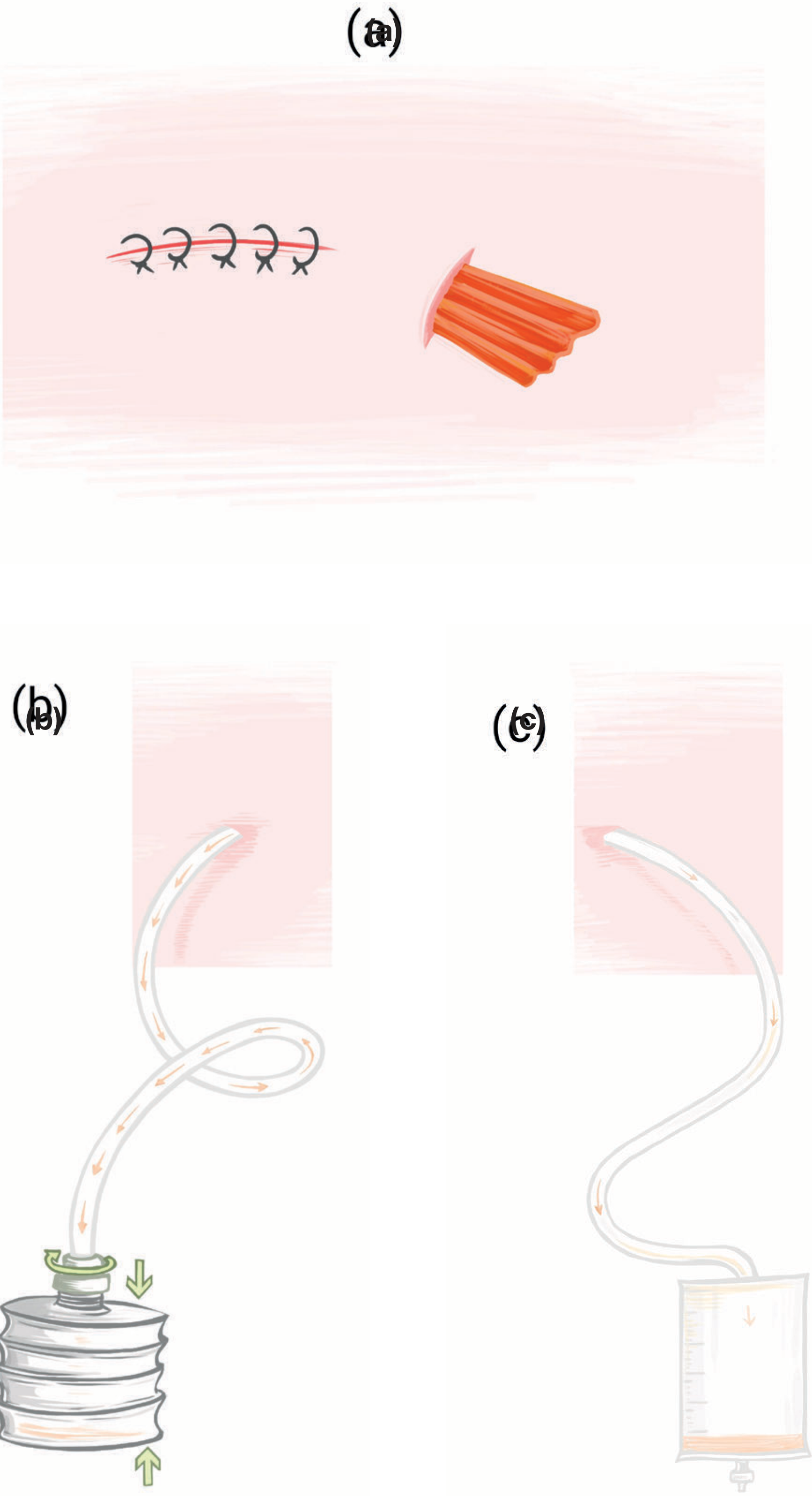
Primary Classification (Bailey & Love)
DRAINS
├── OPEN DRAINS (Passive, gravity-based)
└── CLOSED DRAINS
    ├── Active (Suction / Suctioned)
    └── Passive (Non-suctioned)
Detailed Classification Table
| Category | Type | Examples | Mechanism | Uses | Disadvantage |
|---|---|---|---|---|---|
| Open Drain | Passive, gravity | Penrose drain, Corrugated (latex) drain | Forms a channel between body cavity and external environment; fluid drains by gravity/capillary action | Debrided wounds, abscess cavities | Unsightly; frequent dressing changes; acts as conduit for bacterial colonisation (retrograde infection) |
| Closed Active (Suction) | Active, negative pressure | Jackson-Pratt (JP) drain, Redivac drain, Hemovac | Maintains negative pressure - actively suctions fluid out; obliterates dead space | Axillary dissection, ventral hernia repair, head & neck surgery, mastectomy, post-pyelolithotomy/nephrectomy | Risk of sucking viscera/omentum into tip if placed intra-abdominally; caution near vital structures |
| Closed Passive (Non-suction) | Passive, gravity/capillary | Robinson drain, urinary catheter, nasogastric tube | Uses capillary action and gravity; no negative pressure | Abdominal cavity drainage (without risk of sucking viscera), biliary, urinary | Less effective at obliterating dead space |
| Tube Drain (Specific) | Passive/active variant | T-tube (biliary), Intercostal chest drain, Nephrostomy tube | Specific anatomical tubes draining a defined hollow structure | T-tube: post-CBD exploration; Chest drain: pleural effusion/pneumothorax; Nephrostomy: renal pelvis | Site-specific complications |
| Sump Drain | Active, double-lumen | Shirley sump drain, Yeates drain | Outer lumen drains fluid; inner lumen allows air ingress, preventing suction blockage; continuous lavage possible | Pancreatic fistula, contaminated cavities | More complex; infection risk along air channel |
Individual Drains - Detailed Description
1. Penrose Drain
- Type: Open, passive
- Material: Soft, flat latex rubber tube (like a collapsed tube)
- Mechanism: Wicks fluid to the surface by capillary action + gravity
- Use: Abscess cavities, debrided infected wounds
- Named after: Charles Bingham Penrose (American gynaecologist)
- Disadvantage: Open to environment - highest infection risk; needs frequent dressing changes
2. Corrugated Drain
- Type: Open, passive
- Material: Corrugated (ribbed/ridged) rubber or plastic sheet
- Mechanism: The ridges create channels along which fluid tracks to the surface (capillary action)
- Use: Superficial wound drainage, debrided wounds, abscess beds
- Difference from Penrose: Corrugated has ridged surface creating multiple channels; Penrose is a flat tube
3. Robinson Drain
- Type: Closed, passive, non-suction
- Material: Soft silicone or PVC tube with multiple side holes
- Mechanism: Gravity and capillary action; closed system - connects to a drainage bag
- Use: Abdominal cavity drainage (prevents suction on omentum/viscera), post-cholecystectomy, pelvic drainage
- Advantage over open drains: Closed system reduces infection risk
4. Jackson-Pratt (JP) Drain
- Type: Closed, active (suction), negative pressure
- Material: Flat, perforated, grenade/bulb-shaped silicone drain connected to a collapsible bulb
- Mechanism: Bulb is compressed and re-connected - creates sustained negative pressure; actively sucks fluid
- Use: Post-pyelolithotomy, post-nephrectomy, mastectomy, liver surgery
- Key fact (Hinman's): "JP drain removed when output is less than 50-100 mL over 24 hours"
5. Redivac / Hemovac Drain
- Type: Closed, active (suction)
- Material: Perforated tube connected to a rigid vacuum bottle (Redivac) or a flat coiled drain with spring-loaded disc (Hemovac)
- Mechanism: Pre-compressed vacuum container maintains sustained negative suction
- Use: Orthopaedic surgery (hip/knee arthroplasty), head & neck, mastectomy
- Difference from JP: Larger reservoir; Hemovac has a disc that is compressed to create suction; Redivac uses a sealed glass/plastic vacuum bottle
6. Sump Drain (Shirley / Yeates)
- Type: Closed, active, double-lumen
- Material: Outer large drainage lumen + inner smaller air-inlet lumen
- Mechanism: Fluid drains out through outer lumen while air enters through inner lumen (prevents suction effect, maintains continuous flow); can also allow lavage
- Use: Pancreatic fistula, large infected cavities, high-output fistulae where irrigation is needed
- Advantage: Does not block; allows continuous lavage if needed
7. T-Tube
- Type: Closed, passive
- Material: Latex or silicone T-shaped tube
- Mechanism: Placed in common bile duct (CBD) after exploration; short arms sit inside CBD, long arm exits through abdominal wall; bile drains passively into bag
- Use: Post-CBD exploration for choledocholithiasis
- Removed when: Cholangiogram (T-tube cholangiogram) at day 10-14 shows no leak and free flow to duodenum
8. Intercostal Chest Drain (ICD)
- Type: Closed, passive (underwater seal) or active (with suction)
- Material: Firm PVC tube (24-32 Fr) inserted through intercostal space
- Mechanism: Underwater seal creates one-way valve; air/fluid exits pleural space on expiration but cannot re-enter
- Use: Pneumothorax, haemothorax, empyema, post-thoracic surgery
- Landmark: 5th intercostal space, mid-axillary line ("safe triangle")
9. Nephrostomy Tube
- Type: Closed, passive/active diversion tube
- Material: Soft malecot or pigtail catheter placed percutaneously into renal pelvis under ultrasound/fluoroscopy
- Mechanism: Diverts urine directly from kidney before it reaches obstructed or injured ureter
- Use: Ureteric obstruction, post-ureterolithotomy urine leak, pyonephrosis
Properties of an Ideal Drain (Bailey & Love)
- Be non-irritating to surrounding tissues
- Be non-reactive (inert material)
- Drain effectively without blockage
- Not act as a conduit for infection
- Be easy to remove without fragmentation
Indications for Surgical Drain (Bailey & Love)
- Anticipated collection of blood/serum/lymph (e.g. axillary dissection, hernia repair)
- Dead space obliteration
- Established abscess/collection drainage
- Controlled fistula - transforms an uncontrolled internal leak into a controlled external fistula
- Biliary or urinary leak monitoring
Arguments For and Against Routine Drain Use (Bailey & Love)
| Arguments FOR (Protagonists) | Arguments AGAINST (Detractors) |
|---|---|
| Removes blood, serous fluid, bile, pus, pancreatic secretions | Increases intra-abdominal and wound infection (retrograde bacteria) |
| Early warning signal for haemorrhage or anastomotic leak | Delays recovery and increases hospital stay |
| Provides track for long-term drainage | Increases abdominal pain |
| Decreases pulmonary function | |
| May falsely reassure the clinician (drain can block while leak continues) |
Quick Summary Table for Viva
| Drain | Open/Closed | Active/Passive | Key Feature |
|---|---|---|---|
| Penrose | Open | Passive | Flat soft latex; abscess, infected wounds |
| Corrugated | Open | Passive | Ridged; multiple channels |
| Robinson | Closed | Passive | Silicone tube; abdominal; no suction |
| Jackson-Pratt | Closed | Active (suction) | Bulb suction; post-renal surgery |
| Redivac/Hemovac | Closed | Active (suction) | Vacuum bottle; orthopaedics, mastectomy |
| Sump (Shirley/Yeates) | Closed | Active + air inlet | Double lumen; pancreatic fistula |
| T-tube | Closed | Passive | CBD after exploration |
| Chest drain (ICD) | Closed | Passive (UWS) | Pleural space; one-way valve |
| Nephrostomy | Closed | Passive | Renal pelvis; proximal urinary diversion |