attachment
attachment

Doagnise the radio finding

analyse_media
Reading File
Finding Sources
Finding Sources
Searching Images

shoulder fracture dislocation proximal humerus radiograph
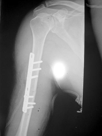
Anteroposterior (AP) radiograph of the right shoulder and humerus demonstrating post-surgical management of a complex upper limb injury. The image shows the glenohumeral joint, acromion, and clavicle. The proximal humerus is reduced within the glenoid fossa, indicating successful management of a prior anterior dislocation. Along the mid-diaphyseal region of the humerus, an orthopedic internal fixation device is visible, consisting of a metallic compression plate secured with multiple cortical screws across a transverse fracture site. The hardware provides stabilization and alignment of the humeral shaft. Key anatomical landmarks include the humeral head, scapular glenoid, and humeral diaphysis. This imaging serves as an educational example of managing ipsilateral shoulder dislocation and humeral shaft fracture using open reduction and internal fixation (ORIF).
Anteroposterior (AP) radiograph of the right shoulder and humerus demonstrating post-surgical management of a complex upper limb injury. The image shows the glenohumeral joint, acromion, and clavicle. The proximal humerus is reduced within the glenoid fossa, indicating successful management of a prior anterior dislocation. Along the mid-diaphyseal region of the humerus, an orthopedic internal fixation device is visible, consisting of a metallic compression plate secured with multiple cortical screws across a transverse fracture site. The hardware provides stabilization and alignment of the humeral shaft. Key anatomical landmarks include the humeral head, scapular glenoid, and humeral diaphysis. This imaging serves as an educational example of managing ipsilateral shoulder dislocation and humeral shaft fracture using open reduction and internal fixation (ORIF).

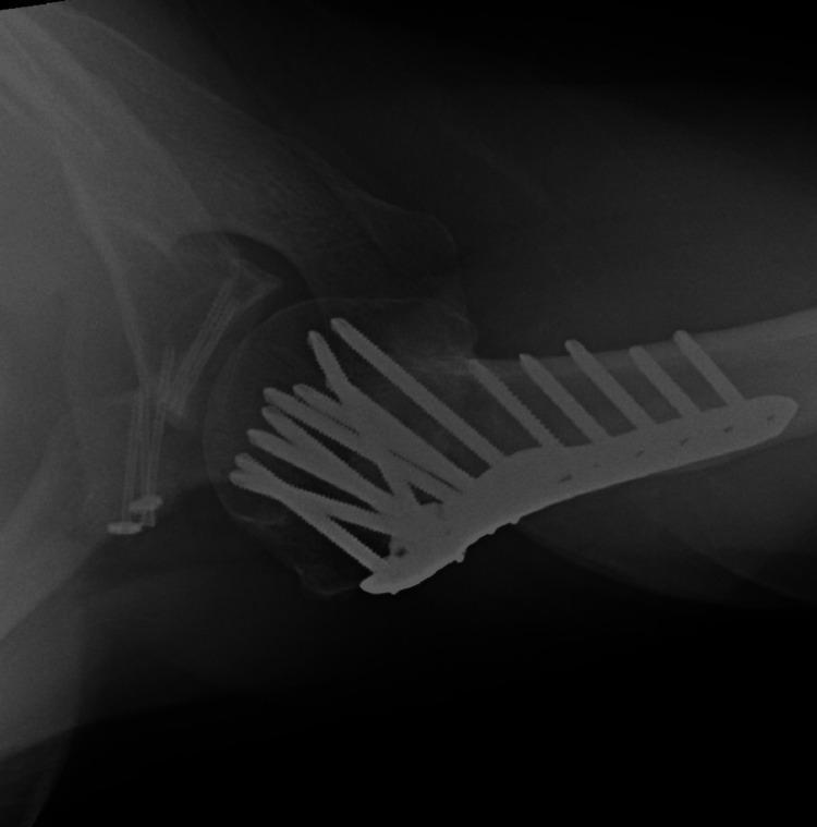
Postoperative axial radiograph of the left shoulder demonstrating complex orthopedic internal fixation for a multi-part fracture involving the proximal humerus, glenoid, and coracoid process. A large, contoured proximal humeral locking plate (PHILOS) is visible on the lateral humerus, secured with multiple divergent locking screws into the humeral head in a fan-like configuration and several bicortical screws along the humeral shaft. On the scapular side, at least two cannulated screws are visible traversing the anterior glenoid rim to stabilize an anterior glenoid fragment. Additional fixation is noted near the coracoid process with screws and washers. The image shows evidence of successful fracture union at 14 months post-surgery, with the glenohumeral joint maintaining a concentric, reduced position. This radiograph illustrates surgical management for high-energy shoulder trauma involving complex fracture-dislocation or 'floating shoulder' variants.
Postoperative axial radiograph of the left shoulder demonstrating complex orthopedic internal fixation for a multi-part fracture involving the proximal humerus, glenoid, and coracoid process. A large, contoured proximal humeral locking plate (PHILOS) is visible on the lateral humerus, secured with multiple divergent locking screws into the humeral head in a fan-like configuration and several bicortical screws along the humeral shaft. On the scapular side, at least two cannulated screws are visible traversing the anterior glenoid rim to stabilize an anterior glenoid fragment. Additional fixation is noted near the coracoid process with screws and washers. The image shows evidence of successful fracture union at 14 months post-surgery, with the glenohumeral joint maintaining a concentric, reduced position. This radiograph illustrates surgical management for high-energy shoulder trauma involving complex fracture-dislocation or 'floating shoulder' variants.

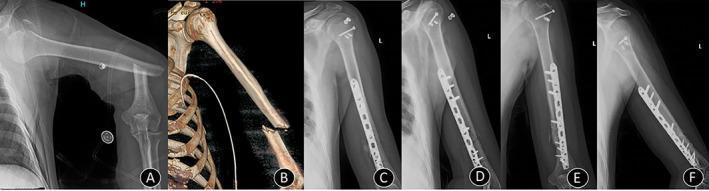
A multi-modal series of six images (A-F) documenting the clinical progression of complex orthopedic trauma. Panel A: Plain radiograph of the left shoulder and humerus showing an anterior glenohumeral dislocation associated with a fracture of the greater tuberosity and a displaced transverse fracture of the proximal-to-mid humeral shaft. Panel B: A 3D CT reconstruction demonstrating the humeral shaft fracture following successful reduction of the shoulder joint. Panels C and D: Immediate postoperative anteroposterior (AP) and lateral X-rays showing internal fixation. The shaft fracture is stabilized with a long locking compression plate (LCP) and multiple cortical/locking screws. The greater tuberosity is secured with cannulated compression screws, and radiopaque suture anchors are visible in the proximal humerus, indicating rotator cuff repair. Panels E and F: Follow-up radiographs at four months demonstrate anatomical alignment, successful maintenance of shoulder reduction, and progressive bony union (callus formation) at both fracture sites. This series serves as an educational example of managing combined shoulder dislocation-fractures through open reduction and internal fixation (ORIF).
A multi-modal series of six images (A-F) documenting the clinical progression of complex orthopedic trauma. Panel A: Plain radiograph of the left shoulder and humerus showing an anterior glenohumeral dislocation associated with a fracture of the greater tuberosity and a displaced transverse fracture of the proximal-to-mid humeral shaft. Panel B: A 3D CT reconstruction demonstrating the humeral shaft fracture following successful reduction of the shoulder joint. Panels C and D: Immediate postoperative anteroposterior (AP) and lateral X-rays showing internal fixation. The shaft fracture is stabilized with a long locking compression plate (LCP) and multiple cortical/locking screws. The greater tuberosity is secured with cannulated compression screws, and radiopaque suture anchors are visible in the proximal humerus, indicating rotator cuff repair. Panels E and F: Follow-up radiographs at four months demonstrate anatomical alignment, successful maintenance of shoulder reduction, and progressive bony union (callus formation) at both fracture sites. This series serves as an educational example of managing combined shoulder dislocation-fractures through open reduction and internal fixation (ORIF).

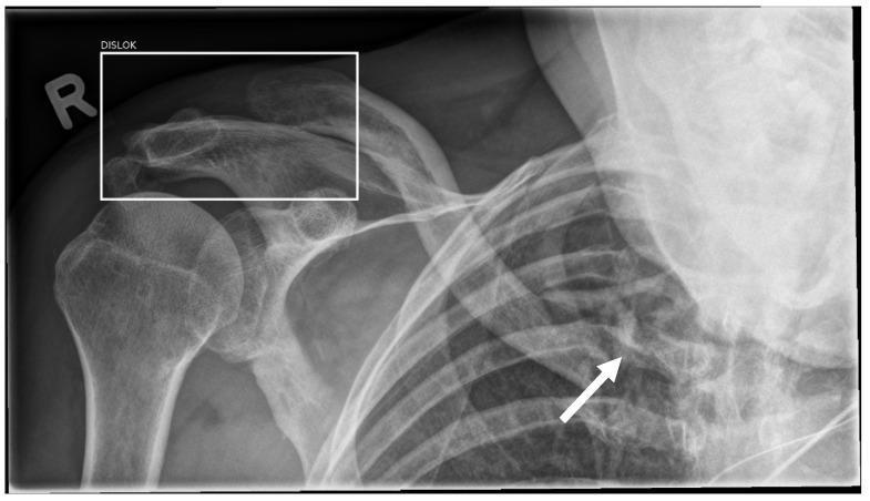
A standing anteroposterior (AP) radiograph of the right shoulder and upper thoracic region. Anatomical structures include the proximal humerus, scapula, clavicle, and the upper rib cage. The image demonstrates two distinct traumatic injuries. First, a white bounding box labeled 'DISLOK' highlights a dislocation at the acromioclavicular (AC) joint. Second, a white arrow points to a displaced fracture of the proximal clavicle near the sternoclavicular region, characterized by cortical disruption and bony overriding. The radiograph also contains an 'R' marker indicating the right side. This image is used in medical education to illustrate the clinical presentation of multiple injuries in the shoulder girdle and the utility/limitations of AI-assisted fracture and dislocation detection in a diagnostic imaging workflow.

A standing anteroposterior (AP) radiograph of the right shoulder and upper thoracic region. Anatomical structures include the proximal humerus, scapula, clavicle, and the upper rib cage. The image demonstrates two distinct traumatic injuries. First, a white bounding box labeled 'DISLOK' highlights a dislocation at the acromioclavicular (AC) joint. Second, a white arrow points to a displaced fracture of the proximal clavicle near the sternoclavicular region, characterized by cortical disruption and bony overriding. The radiograph also contains an 'R' marker indicating the right side. This image is used in medical education to illustrate the clinical presentation of multiple injuries in the shoulder girdle and the utility/limitations of AI-assisted fracture and dislocation detection in a diagnostic imaging workflow.

Radiographic Findings & Impression

View: AP radiograph of the left shoulder/proximal humerus, with visualized upper chest ("L" marker confirms left side; image also includes both lung apices/upper zones).

Findings

1. Left shoulder joint
  • The humeral head is displaced away from the glenoid fossa, sitting low and medial to its expected articulation, with loss of the normal smooth glenohumeral congruity.
  • The contour of the humeral head/greater tuberosity region is irregular, suggesting an associated fracture fragment rather than a simple dislocation.
  • The clavicle and acromion appear intact and in normal position above the joint.
Impression: Left glenohumeral fracture-dislocation (fracture of the proximal humerus, likely involving the surgical neck and/or greater tuberosity, with associated dislocation of the humeral head from the glenoid). Correlate clinically for mechanism (traumatic fall/direct blow) and consider CT for fragment number/displacement (Neer classification) prior to definitive management.
2. Bilateral lung fields
  • Diffuse, fine reticulonodular/nodular opacities scattered throughout both lungs, without a clear zonal predominance visible on this cropped view.
  • Mediastinum and trachea appear midline; no obvious large pleural effusion.
Impression: Bilateral diffuse micronodular lung pattern - differential includes:
  • Miliary tuberculosis (classic 1-3 mm random nodules) - Tintinalli's Emergency Medicine, and Grainger & Allison's Diagnostic Radiology
  • Disseminated fungal infection (e.g., coccidioidomycosis) - Murray & Nadel's Textbook of Respiratory Medicine
  • Hematogenous pulmonary metastases (random nodular pattern)
  • Less likely: pneumoconiosis or diffuse interstitial lung disease, depending on nodule size/distribution and clinical context

Overall Assessment

This film shows two distinct abnormalities: an acute/subacute left proximal humeral fracture-dislocation, and an incidental (or potentially related) diffuse bilateral micronodular lung pattern. If there is a known primary malignancy or the humeral fracture occurred with minimal trauma, a pathological fracture through a metastatic bone lesion with pulmonary metastases becomes an important differential to exclude - the fragmented, comminuted appearance at the fracture site would support this. Alternatively, if there is a history of fever, weight loss, or TB exposure, miliary tuberculosis with an unrelated traumatic shoulder injury is equally plausible.
This cannot be finalized on a single cropped AP film. Recommended next steps:
  • Clinical history: trauma mechanism, constitutional symptoms, known malignancy, TB risk factors
  • Dedicated shoulder series (AP + axillary/scapular Y view) or CT shoulder to characterize the fracture-dislocation and check for pathological features (lytic destruction, cortical breach)
  • Full PA/lateral chest radiograph or CT chest to characterize the nodular pattern's size, distribution, and zonal predominance
  • Correlation by a radiologist with the complete study, not this single cropped image
For reference, similar fracture-dislocation patterns are illustrated below:
Anterior glenohumeral dislocation with associated greater tuberosity and humeral shaft fracture
I don't have the patient's clinical history (age, trauma mechanism, symptoms, prior malignancy) - that information would substantially narrow this differential, particularly for the lung finding. If you can share that context, I can refine the impression.
This is a shared conversation. Sign in to Orris to start your own chat.