
PERICARDIAL EFFUSION CXR
pericardial effusion chest x-ray water bottle heart shadow
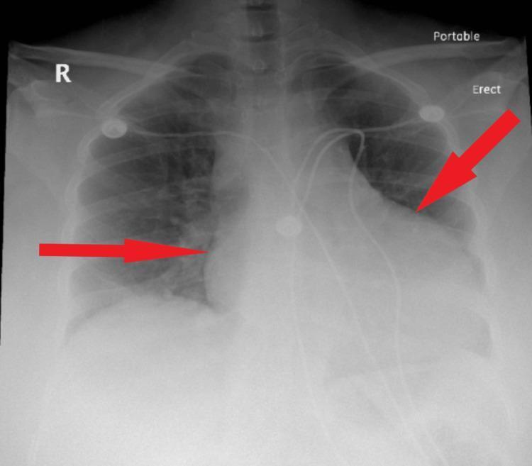
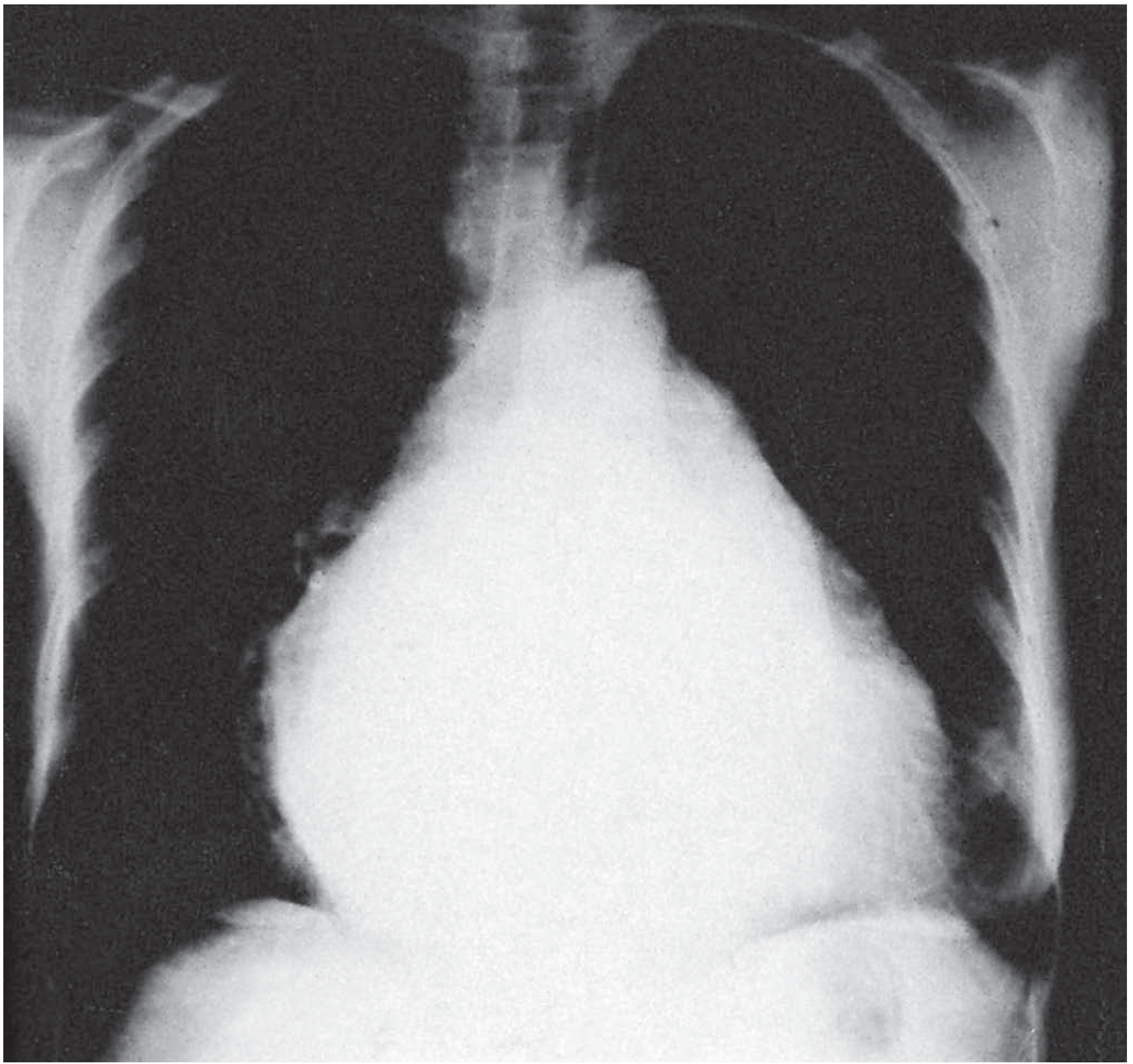
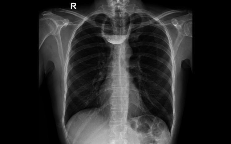
A posteroanterior (PA) view portable chest X-ray demonstrating a significantly enlarged cardiac silhouette, often described as a 'water-bottle' heart shape. Red arrows point to the widened lateral margins of the mediastinum/heart, which obscure the bilateral lung hila. Despite the massive cardiomegaly, the peripheral lung fields remain relatively clear without overt signs of pulmonary edema, vascular congestion, or consolidation. The costophrenic angles are partially visualized, and the trachea appears midline. Multiple external monitoring electrodes and wires are visible on the patient's chest. This radiographic presentation is highly characteristic of a large pericardial effusion, as the rapid accumulation of fluid expands the pericardial sac symmetrically, causing a globular enlargement of the cardiac shadow that masks underlying pulmonary structures.
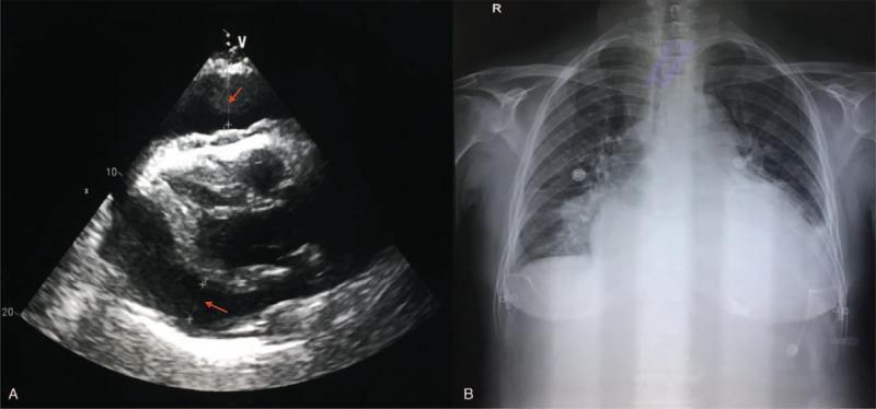
The image consists of two diagnostic panels demonstrating a large pericardial effusion. Panel (A) is an echocardiogram in a parasternal long-axis or similar view showing a significant, dark (anechoic) space surrounding the heart, representing a large pericardial effusion. Red arrows indicate the fluid layer between the epicardium and the parietal pericardium, which causes visible compression of the cardiac chambers. Panel (B) is a posterior-anterior (PA) chest X-ray showing severe cardiomegaly with a 'water-bottle' heart configuration, characterized by a globally enlarged, globular cardiac silhouette with indistinct borders. This radiographic sign is highly suggestive of massive pericardial fluid accumulation. The lungs appear relatively clear, though the bases are obscured by the enlarged cardiac shadow and the diaphragm. The combination of these images serves as a classic educational example of the diagnostic correlation between ultrasound and radiography for pericardial disease.
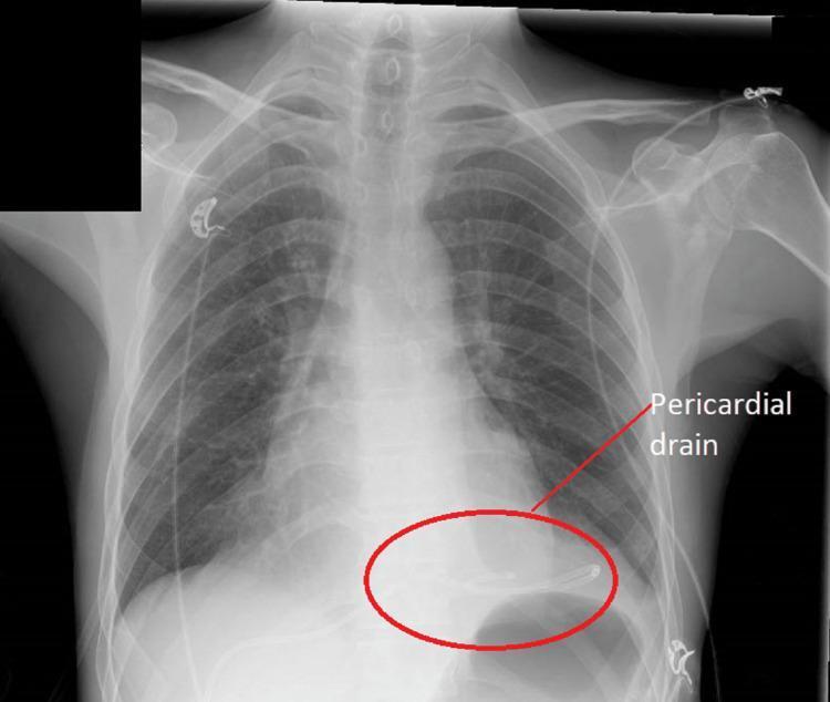
This diagnostic image is an anteroposterior (AP) chest X-ray demonstrating the post-procedural state of a patient following the drainage of a large pericardial effusion. The central focus is the cardiac shadow, which shows a significant reduction in the cardiothoracic ratio compared to typical 'water bottle' heart configurations seen in tamponade. A radiopaque pericardial drain is clearly visible and highlighted by a red oval and label in the lower left cardiac region (anatomical right side of the heart silhouette). The drain's distal pigtail or curved tip is positioned within the pericardial space to facilitate continuous drainage. The surrounding lung fields are clear, with no visible pulmonary edema, pleural effusion, or infiltrates, suggesting stable respiratory status. Several external ECG leads are visible on the chest wall. This visual serves as an educational tool for identifying the radiological signs of effective pericardiocentesis and the proper positioning of a subcostal pericardial drainage catheter.
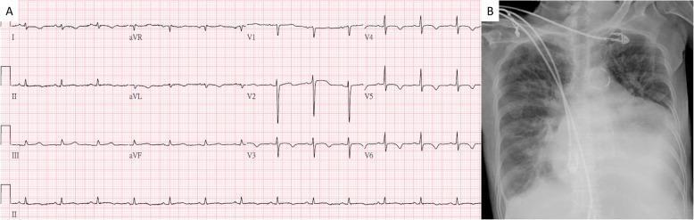
This composite diagnostic image features two panels related to a cardiovascular assessment. Panel A shows a 12-lead Electrocardiogram (ECG) demonstrating a normal sinus rhythm. A key finding is the low voltage QRS complexes globally across both limb and precordial leads, which can be clinically associated with conditions such as pericardial effusion or obesity. Panel B is a posterior-anterior (PA) chest X-ray exhibiting a significantly enlarged cardiac silhouette, resulting in a 'water bottle' heart shape often characteristic of massive pericardial effusion. There is a notable increase in the cardiothoracic ratio. The lung fields show some patchy opacities potentially suggesting mild pulmonary congestion or edema. Radiopaque medical monitoring leads are visible over the thoracic region. Together, these images illustrate the classic findings of pericardial effusion: dampened electrical activity on ECG and a globose cardiac shadow on radiography.
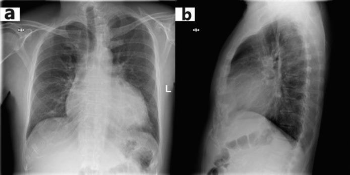
This diagnostic image displays a standard chest X-ray in two views: (a) Posteroanterior (PA) frontal view and (b) Lateral profile view. The frontal view reveals significant enlargement of the cardiac silhouette, known as cardiomegaly, with an increased cardiothoracic ratio (CTR). The heart exhibits a widened, somewhat globular shape, which is a classic radiographic sign associated with a large pericardial effusion ('water bottle heart' configuration). In the lateral view (b), the enlarged cardiac shadow occupies an increased portion of the retrosternal space, further confirming global cardiac enlargement. Despite the cardiomegaly, the lung fields appear relatively clear without overt signs of pulmonary edema or cephalization of vessels. The costophrenic angles are visible, and the diaphragm maintains a normal contour. This clinical photograph is a primary teaching example for identifying cardiomegaly and assessing the cardiothoracic ratio in the context of acute pericarditis or pericardial effusion.
"pericardial effusion" AND "chest radiography"
Pericardial Effusion on Chest X-Ray (CXR)
Key Radiographic Findings
- The classic sign is a symmetrically enlarged, flask/globular-shaped cardiac shadow with loss of the normal distinct cardiac borders (loss of the "waist" between the heart and great vessels).
- A minimum of ~200-250 mL of pericardial fluid is generally required to produce visible cardiomegaly on CXR - smaller effusions are often radiographically silent (Rosen's Emergency Medicine, p. 1231).
- Unlike heart failure, the peripheral lung fields typically remain relatively clear without pulmonary edema or vascular congestion - a useful distinguishing feature, since an acutely dilated heart from effusion behaves differently than a failing myocardium (Tintinalli's Emergency Medicine, p. 1708-1711).
- Because the pericardium can stretch, a rapidly enlarging cardiac silhouette on serial films (especially without a clear clinical explanation like new heart failure) should raise suspicion for an expanding effusion rather than chronic cardiomegaly.
- A radiolucent stripe representing the pericardial fluid separating the epicardial fat from the mediastinal/pericardial fat, best seen on lateral views. It is a specific but insensitive sign - rarely visualized in practice (Tintinalli's Emergency Medicine, p. 1708-1711).
- Widened mediastinum
- In chronic/longstanding effusions, pericardial calcification may be present (suggesting constrictive pericarditis or a chronic process)
- The trachea typically remains midline (helps distinguish from other causes of mediastinal widening)
Correlation With Echocardiography
Quick Summary Table
| Finding | Significance |
|---|---|
| Water-bottle/globular heart | Classic sign; needs ~200-250 mL+ fluid |
| Clear lung fields with cardiomegaly | Distinguishes from CHF-related cardiomegaly |
| Rapid interval enlargement on serials | Suggests expanding effusion |
| Epicardial fat pad sign (lateral view) | Specific but rarely seen |
| Normal CXR | Does NOT exclude acute effusion/tamponade |
VISUAL FOR CXR VS MS FINDINGS
mitral stenosis chest x-ray straightening left heart border double density left atrial enlargement
mitral stenosis chest x-ray left atrial enlargement cardiomegaly pulmonary congestion
Visual Comparison: CXR Findings — Pericardial Effusion vs Mitral Stenosis
Pericardial Effusion — "Water Bottle" Heart
- Globular, symmetric enlargement of the entire cardiac silhouette
- Sharp, well-defined borders (fluid smooths out normal contour undulations)
- Loss of the normal cardiac "waist" (the indentation between the right atrium/aorta and the left ventricle)
- Lung fields usually clear despite the large heart shadow
- Rapid change in size on serial films
Mitral Stenosis — "Mitral Configuration" Heart
- Straightening of the left heart border (loss of the normal concave contour, filled in by the enlarged left atrial appendage/pulmonary artery)
- Double density behind the right heart border — the enlarged left atrium's silhouette overlapping the right atrium (Goldman-Cecil Medicine, p. 839-844)
- Left atrial enlargement can splay the carina and elevate the left main bronchus
- Prominent pulmonary vasculature: upper lobe diversion (cephalization), Kerley B lines, and interstitial edema from chronic pulmonary venous hypertension
- Overall heart size may be only mildly increased despite marked left atrial enlargement (unlike the globally massive silhouette of effusion)
Side-by-Side Comparison Table
| Feature | Pericardial Effusion | Mitral Stenosis |
|---|---|---|
| Overall heart shape | Globular, symmetric "water bottle" | Normal-to-mild enlargement with straightened left border |
| Heart border | Smooth, sharp, loses normal contour landmarks | Straightened left border; "double density" at right border |
| Which chamber drives the shape | Whole pericardial sac (extracardiac fluid) | Left atrium specifically |
| Lung fields | Typically clear | Congested — cephalization, Kerley B lines, interstitial edema |
| Onset on serial films | Can change rapidly (hours-days) | Develops slowly over years (chronic rheumatic disease) |
| Confirmatory test | Echocardiography (anechoic fluid, chamber collapse) | Echocardiography (thickened/calcified mitral valve, elevated transmitral gradient, planimetry) |
| Associated ECG clue | Low-voltage QRS, electrical alternans | Notched/biphasic P waves ("P mitrale"), right axis deviation |
middle mediastinal mass d/d
middle mediastinal mass CT chest lymphadenopathy diagram mediastinal compartments
"mediastinal mass" AND differential diagnosis
Middle Mediastinal Mass — Differential Diagnosis
Differential Diagnosis by Category
1. Lymphadenopathy (most common cause)
- Malignant: lymphoma, metastatic bronchogenic carcinoma (subcarinal/paratracheal spread), metastases from extrathoracic primaries
- Infectious: tuberculosis, histoplasmosis, coccidioidomycosis
- Inflammatory/granulomatous: sarcoidosis (often with hypercalcemia, polyarthralgia, cough)
- Lymphoproliferative: Castleman disease (angiofollicular lymph node hyperplasia, HHV-8 associated, shows postcontrast enhancement on CT)
2. Foregut Duplication Cysts (most common cystic lesion here)
- Bronchogenic cyst - smooth-walled, non-enhancing, fluid-attenuation lesion; classically subcarinal or paratracheal
- Esophageal duplication cyst
- Neurenteric cyst
- Pericardial cyst - classic "spring water cyst," typically at the right cardiophrenic angle
3. Vascular Masses
- Aortic arch aneurysm or dissection
- Aberrant subclavian artery / vascular rings
- Azygos or hemiazygos vein dilation
- Superior vena cava dilation/mass effect (SVC syndrome from adjacent malignancy)
4. Esophageal Lesions
- Esophageal carcinoma
- Large hiatal hernia extending into mediastinum
- Achalasia (markedly dilated, fluid/food-filled esophagus)
- Esophageal diverticulum
5. Tracheal Masses
- Primary tracheal tumors (rare)
- Extrinsic compression from adjacent nodal disease or thyroid extension
6. Other
- Substernal/ectopic thyroid extending inferiorly (usually anterior but can extend into middle mediastinum)
- Mediastinal granuloma (fibrosing mediastinitis - presents with dyspnea, wheezing, hemoptysis)
- Morgagni hernia (technically at the cardiophrenic angle, can mimic a middle mediastinal mass on CXR)
Quick Reference Table
| Diagnosis | Key Clues | Imaging Features |
|---|---|---|
| Lymphoma | Night sweats, weight loss, extrathoracic adenopathy, high ESR/CRP | Bulky, homogeneous nodal masses, any compartment |
| Metastatic lung cancer nodes | Smoker, primary lung mass | Enlarged subcarinal/paratracheal nodes |
| TB lymphadenitis | Constitutional symptoms, exposure history | Necrotic, rim-enhancing nodes; may calcify |
| Sarcoidosis | Bilateral hilar + paratracheal adenopathy, hypercalcemia | Symmetric, non-necrotic nodes ("1-2-3 sign") |
| Bronchogenic cyst | Often incidental, may cause airway compression | Smooth, thin-walled, water-attenuation, non-enhancing; subcarinal common |
| Pericardial cyst | Asymptomatic, incidental | Right cardiophrenic angle, water density |
| Aortic aneurysm | Chest/back pain, pulsatile | Continuous with aorta, wall calcification |
| Achalasia | Dysphagia, regurgitation | Air-fluid level, dilated esophagus, "bird beak" on esophagram |
| Castleman disease | Young patient, often asymptomatic | Avidly enhancing mediastinal mass/nodes |
Diagnostic Approach
- CXR first - usually the initial study, though limited sensitivity (Grainger & Allison, p. 1398)
- Contrast-enhanced CT chest - defines location, relationship to vessels/airway, and characterizes cystic vs solid vs vascular
- EBUS-guided or CT-guided FNA/core biopsy - preferred tissue sampling for nodal/solid masses; combined FNA + core-needle biopsy reaches ~98% accuracy vs ~79% for either alone (Schwartz's Principles of Surgery, p. 2925)
- MRI - useful when CT is equivocal, especially for cystic lesions or vascular invasion assessment
- Avoid direct biopsy of suspected cysts (bronchogenic, pericardial) due to infection risk - if symptomatic, go to surgical resection instead
Predisposing esophageal ca conditions cures of smooth vs nodular septal thickening
1. Predisposing Conditions for Esophageal Carcinoma
Squamous Cell Carcinoma (SCC)
| Risk Factor | Notes |
|---|---|
| Tobacco smoking | Relative risk ~6.2-fold higher with >25 cigarettes/day; risk falls after 10 years of cessation |
| Alcohol use | Synergistic with smoking - combined use confers 10-25 fold increased risk |
| Achalasia | Stasis and chronic mucosal irritation; SCC may emerge 10-20 years after diagnosis |
| Caustic esophageal injury (lye/corrosive strictures) | Chronic inflammation and stricture-related dysplasia |
| Plummer-Vinson (Paterson-Brown Kelly) syndrome | Glossitis, cervical esophageal webs, iron-deficiency anemia |
| Tylosis palmaris et plantaris | Rare hereditary desquamating condition with near 100% lifetime SCC risk |
| Nutritional deficiency | Diets low in fruits/vegetables; deficiencies in vitamins A, B12, C, E, folate, zinc, selenium, molybdenum |
| Hot beverages / thermal injury, nitrosamine-rich foods, betel nut/paan chewing | Chronic mucosal irritation (geographic "Asian esophageal cancer belt" - Caspian region, Iran, India, China) |
| Head and neck SCC (synchronous/metachronous) | Shares smoking/alcohol risk factors - "field cancerization" |
| Radiation exposure | Prior mediastinal radiotherapy |
Adenocarcinoma
| Risk Factor | Notes |
|---|---|
| Barrett esophagus | Single most important precursor - metaplastic columnar epithelium with dysplasia risk |
| Chronic GERD / acid reflux | Drives Barrett metaplasia |
| Obesity | Increases intra-abdominal pressure and reflux, also independent metabolic risk |
| Tobacco use | Weaker association than with SCC but still contributory |
| Prior mediastinal radiotherapy | Shared with SCC |
2. Smooth vs Nodular (Irregular/Beaded) Interlobular Septal Thickening on HRCT
Smooth Septal Thickening
| Cause | Key Associated Findings |
|---|---|
| Pulmonary edema (cardiogenic/hydrostatic) | Gravity-dependent distribution, pleural effusions, cardiomegaly, may show "crazy paving" with ground-glass opacity |
| Pulmonary alveolar proteinosis | "Crazy paving" pattern (smooth septal thickening + ground-glass opacity) is classic |
| Pulmonary veno-occlusive disease (PVOD) | Smooth septal thickening is a prominent finding, plus centrilobular ground-glass nodules |
| Lymphangitic spread (early/mild cases) | Can occasionally look smooth before becoming nodular |
| Pulmonary hemorrhage syndromes | Interstitial component from blood/edema |
| Idiopathic pulmonary arteriovenous malformations related changes | Smooth septal thickening with centrilobular ground-glass nodularity described in some vascular processes |
Nodular / Irregular / Beaded Septal Thickening
| Cause | Key Associated Findings |
|---|---|
| Lymphangitic carcinomatosis | Classic cause of irregular/beaded septal thickening; often asymmetric, with hilar/mediastinal adenopathy |
| Sarcoidosis | Less common cause of nodular septal thickening; perilymphatic micronodules along septa, fissures, bronchovascular bundles; upper-lobe predominant |
| Silicosis / coal workers' pneumoconiosis | Nodular thickening with perilymphatic micronodules, may progress to conglomerate masses |
| Amyloidosis (pulmonary) | Can produce nodular/beaded septal pattern with calcification |
| Erdheim-Chester disease | Rare cause of nodular septal infiltration |
Practical Distinguishing Point
- Smooth + ground-glass + gravity-dependent → favor edema/PAP ("crazy paving")
- Nodular/beaded + hilar adenopathy + known malignancy → favor lymphangitic carcinomatosis
- Perilymphatic micronodules along septa/fissures + upper lobe → favor sarcoidosis
- A perilobular pattern (thickening that looks septal but is actually peripheral lobular infiltration) can mimic true septal thickening and is most often seen in organizing pneumonia - worth distinguishing as a mimic rather than true septal disease.
achalasia vs scleroderma barium study findings
achalasia barium swallow bird beak dilated esophagus
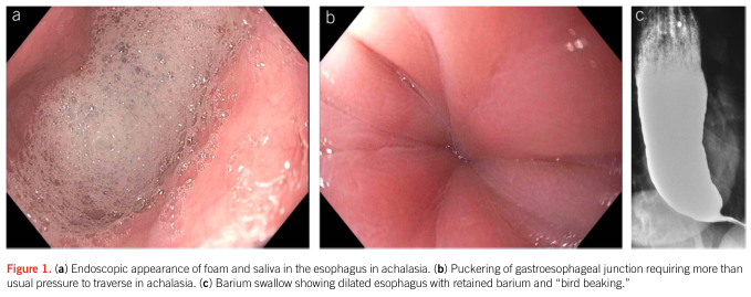
Summary : This figure presents three medical imaging views illustrating features of achalasia in the esophagus: endoscopic foam/saliva accumulation, puckering at the gastroesophageal junction, and a barium swallow radiograph showing classic "bird beak" appearance.
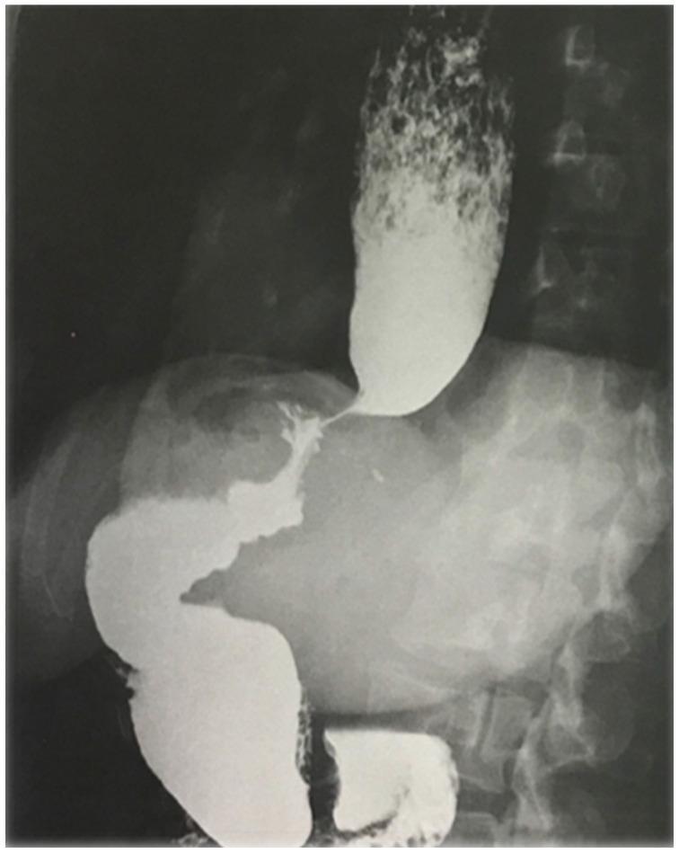
A barium swallow X-ray (fluoroscopy) of the upper gastrointestinal tract demonstrating a markedly dilated esophagus. There is a prominent accumulation and stasis of contrast medium within the esophageal lumen, with visible mottling suggestive of retained food particles. The distal esophagus exhibits a sharp, smooth tapering near the esophagogastric junction, characteristic of a 'bird's beak' deformity often associated with achalasia. Below the diaphragm, the gastric sleeve from a previous laparoscopic sleeve gastrectomy (LSG) is visible with regular contours and normal transit of the contrast bolus that has managed to pass. The image illustrates secondary achalasia or post-surgical esophageal dysmotility, highlighting the contrast between the dilated thoracic esophagus and the restricted passage through the lower esophageal sphincter.
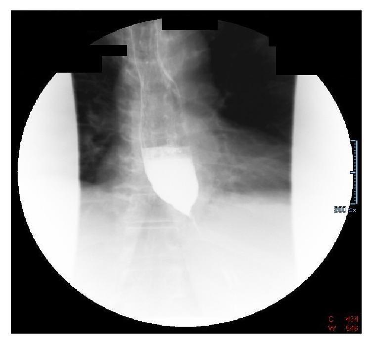
A diagnostic fluoroscopic image of a barium swallow (esophagram) demonstrating classic radiologic features of achalasia or pseudoachalasia. The image shows a markedly dilated thoracic esophagus filled with radiopaque barium contrast. There is a characteristic abrupt, symmetrical tapering of the distal esophagus at the gastroesophageal junction, creating the diagnostic 'bird's beak' appearance. Significant barium column stasis is visible proximal to the lower esophageal sphincter, indicating functional or mechanical obstruction. Minimal contrast is seen passing into the stomach, which is partially visualized in the lower quadrant. This finding is highly suggestive of impaired lower esophageal sphincter relaxation or extrinsic compression, correlating clinically with symptoms such as progressive dysphagia and weight loss.
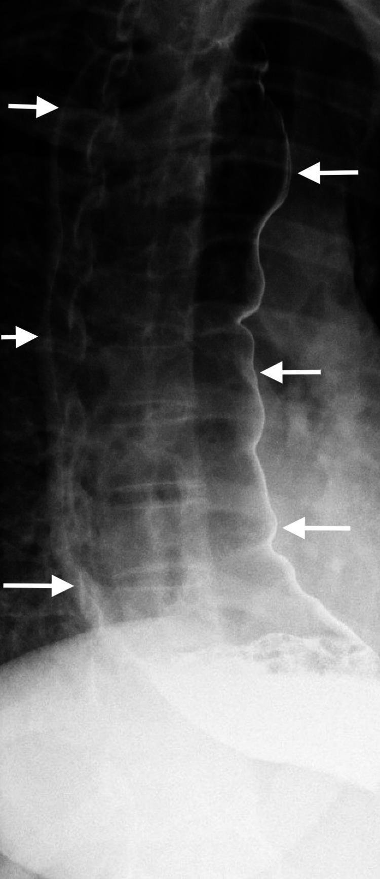
This diagnostic image is a barium swallow esophagram (X-ray radiography) illustrating end-stage achalasia. The primary finding is a massively dilated, tortuous, and elongated thoracic esophagus, referred to as a 'sigmoid esophagus' or 'S-shaped' esophagus due to its resemblance to the sigmoid colon. White arrows delineate the lateral and medial borders of the dilated lumen, which is filled with radiopaque contrast. The esophageal body shows significant loss of primary peristalsis and characteristic pooling of barium. Distally, the esophagus exhibits smooth tapering near the gastroesophageal junction (GEJ), which typically presents as the 'bird-beak' sign in this pathology, though the distal-most segment is partially obscured by the extensive pooling in the dilated lumen. This visual is characteristic of chronic, severe achalasia leading to decompensation of the esophageal musculature. It is a critical educational resource for understanding advanced gastrointestinal motility disorders and the radiological manifestations of obstructive esophageal pathology in a clinical setting.
scleroderma esophagus barium swallow dilated atonic aperistalsis
Achalasia vs Scleroderma: Barium Swallow Findings
Achalasia
- Markedly dilated proximal esophagus (megaesophagus) with pooling/stasis of barium and often retained food debris (mottled appearance)
- Smooth, symmetric, tapered narrowing of the distal esophagus at the gastroesophageal junction - the LES fails to relax
- Aperistalsis in the esophageal body
- In advanced/end-stage disease: massive dilation with tortuosity, described as a "sigmoid" or "S-shaped" esophagus
- Poor/absent passage of barium into the stomach despite prolonged standing
- Important: the tapering is smooth and symmetric, without shouldering - shouldering or an irregular/eccentric narrowing should raise suspicion for pseudoachalasia from malignancy at the GE junction
Scleroderma (Systemic Sclerosis)
- Dilated, barium-filled esophagus - due to smooth muscle atrophy/fibrosis replacing the distal two-thirds of the esophageal wall
- Aperistalsis of the distal two-thirds (which is smooth muscle) while the proximal skeletal-muscle third is often preserved
- LES is weak/incompetent (patulous), not tight - this is the opposite of achalasia. Barium refluxes freely from the stomach back into the esophagus
- Often associated with a hiatal hernia
- With chronic reflux, a distal peptic stricture can develop, causing localized narrowing with proximal dilatation - this can superficially mimic a stricture pattern but the mechanism (reflux-induced) and LES tone differ from achalasia
- Delayed esophageal clearance is most apparent when the patient is supine (gravity is the only driving force left, since peristalsis is absent); emptying may look relatively normal upright
- Barium studies are frequently combined with hand films to show sclerodactyly/calcinosis if systemic sclerosis is suspected clinically
Side-by-Side Comparison
| Feature | Achalasia | Scleroderma |
|---|---|---|
| Esophageal body | Dilated, aperistaltic (can become sigmoid/tortuous) | Dilated, aperistaltic (distal 2/3 only) |
| LES / GE junction | Narrowed, "bird-beak" taper, fails to relax | Widened, patulous, incompetent |
| Barium passage into stomach | Delayed/obstructed despite standing | Passes relatively freely; reflux occurs |
| Reflux of barium from stomach | Absent (LES too tight) | Present (LES too loose) |
| Associated finding | Retained food/barium debris, "sigmoid esophagus" in late disease | Hiatal hernia, distal peptic stricture from chronic GERD |
| Underlying pathology | Loss of myenteric (Auerbach) plexus ganglion cells | Smooth muscle atrophy/fibrosis of muscularis |
| Mimic to exclude | Pseudoachalasia (malignant infiltration at GEJ - look for shouldering/irregularity) | Peptic stricture, other causes of GERD-related stricture |
why we do upside down patient in scleroderma
scleroderma barium swallow head down position Trendelenburg gastroesophageal reflux test
Why the "Upside-Down" (Head-Down/Trendelenburg) Position Is Used in Scleroderma Barium Studies
The Core Reason: Testing LES Competence
- Increases intra-abdominal/intragastric pressure relative to the thorax, forcing barium against the gastroesophageal junction
- Demonstrates free reflux of barium from the stomach back up into the esophagus if the LES is truly incompetent - confirming the pathologic weak sphincter tone characteristic of scleroderma
- Differentiates scleroderma from achalasia - in achalasia the LES fails to relax but retains high resting tone, so tilting the patient head-down will NOT produce reflux (the tight sphincter holds). In scleroderma, the same maneuver readily provokes reflux because the sphincter has lost its barrier function. This is a practical bedside/fluoroscopic way to distinguish a "tight but non-relaxing" LES from a "weak/incompetent" LES.
- Assesses esophageal clearance without peristalsis - since scleroderma causes aperistalsis of the distal two-thirds of the esophagus, the only mechanism left to clear swallowed barium is gravity. Studying the patient recumbent/head-down reveals deficient esophageal emptying in the recumbent position - retained barium pools in the dilated, atonic esophagus because there's no peristaltic wave to push it down and gravity is working against clearance in that position. This is itself a diagnostic radiologic sign of scleroderma esophageal involvement.
Summary of What You're Looking For
| Position | Achalasia | Scleroderma |
|---|---|---|
| Upright | Delayed emptying, bird-beak taper, no reflux | May empty passively via gravity through patulous LES |
| Head-down/tilted | LES remains closed - no reflux provoked | Free reflux of barium into esophagus - confirms incompetent LES |
| Recumbent | Less specific | Deficient/delayed esophageal emptying due to aperistalsis - barium pools |
A Caveat Worth Noting
types of esophageal diverticulum
Zenker diverticulum barium swallow pharyngoesophageal
This lateral barium swallow radiograph (contrast esophagram) of the cervical and upper thoracic region demonstrates a large Zenker's diverticulum. A significant, well-defined, radiopaque, sac-like outpouching is visible originating from the posterior wall of the pharyngoesophageal junction, specifically at Killian's dehiscence above the cricopharyngeal muscle. The diverticulum is filled with barium contrast, appearing as an elongated, dependent sac approximately 7 cm in length that displaces the esophagus anteriorly. There is a clear narrowing or 'neck' at the proximal connection point where the diverticulum communicates with the esophageal lumen. The image highlights significant barium retention within the pouch, a hallmark finding of this false diverticulum. This diagnostic image is intended for medical education regarding upper gastrointestinal pathologies, specifically pharyngeal pouch disorders and causes of cervical dysphagia and regurgitation.
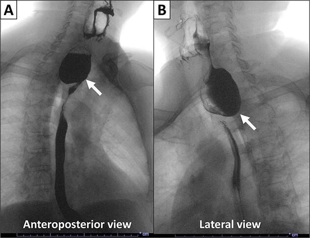
This diagnostic image is an X-ray esophagogram (barium swallow) showing a Zenker’s diverticulum in a human patient. The visual is divided into two panels: an Anteroposterior (AP) view (A) and a Lateral view (B). Both views demonstrate a large, contrast-filled, saccular outpouching measuring approximately 4 x 5 cm. In the AP view, the diverticulum appears as a prominent, rounded, radiopaque mass originating from the proximal esophagus. The Lateral view confirms the pathology is a pulsion diverticulum arising from the posterior aspect of the esophagus, specifically through Killian's triangle (the pharyngoesophageal junction). White arrows in both panels point to the relatively narrow neck of the diverticulum, measuring 1.7 cm in width. The contrast material (barium suspension) clearly opacifies the diverticular lumen and outlines the distal esophageal lumen, which appears narrowed due to the mass effect of the filled sac. Key educational concepts include the diagnosis of upper esophageal dysphagia and the typical posterior location of pharyngoesophageal diverticula.
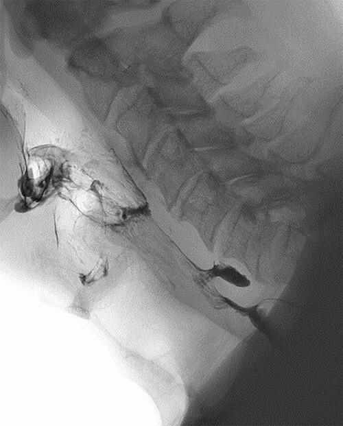
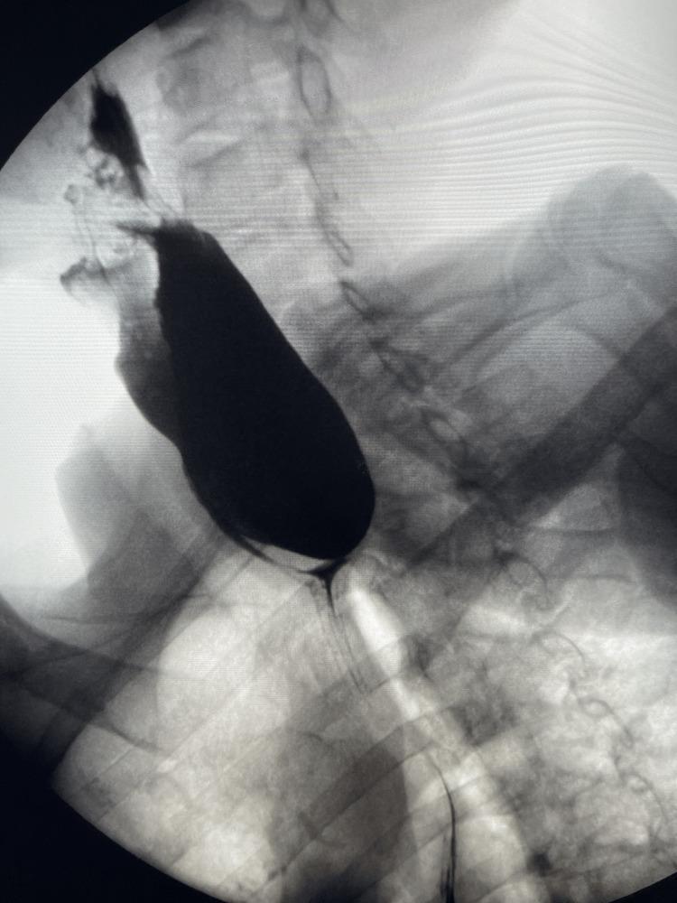
This diagnostic image is a lateral view barium swallow (fluoroscopy/X-ray radiography) of the neck and upper aerodigestive tract. The radiopaque barium contrast medium outlines the pharynx and proximal esophagus. Visible anatomical landmarks include the cervical vertebrae, the mandible, and the soft tissues of the throat. The key pathological finding is a small, posterior outpouching of barium at the level of the pharyngoesophageal junction, specifically at Killian's triangle. This pouch measures approximately 1.1 cm and is characteristic of a Zenker's diverticulum (ZD). The contrast is seen filling the diverticulum as it passes from the hypopharynx into the esophageal lumen. This imaging study illustrates a classic case of a pulsion diverticulum, highlighting its relationship to the posterior pharyngeal wall and its clinical relevance in patients presenting with dysphagia or regurgitation.
This anteroposterior (AP) barium swallow radiograph of the chest and upper abdomen demonstrates a prominent Zenker's diverticulum. The primary finding is a large, pouch-like radiopaque structure located in the superior mediastinum, superimposed on the upper thoracic vertebrae and positioned just above the level of the clavicles. The diverticulum exhibits a distinct air-fluid level, with the inferior portion filled with dense barium contrast and the superior portion containing air. The trachea remains midline, although the large size of the diverticulum may cause minor posterior or lateral displacement not fully appreciated in the AP view. The lung fields appear clear without evidence of active consolidation, and the skeletal structures, including the ribs and clavicles, are unremarkable. This diagnostic image is characteristic of a pharyngoesophageal diverticulum, typically occurring through Killian's dehiscence, and is a key educational example for gastrointestinal radiology and the evaluation of chronic dysphagia.
epiphrenic esophageal diverticulum barium swallow
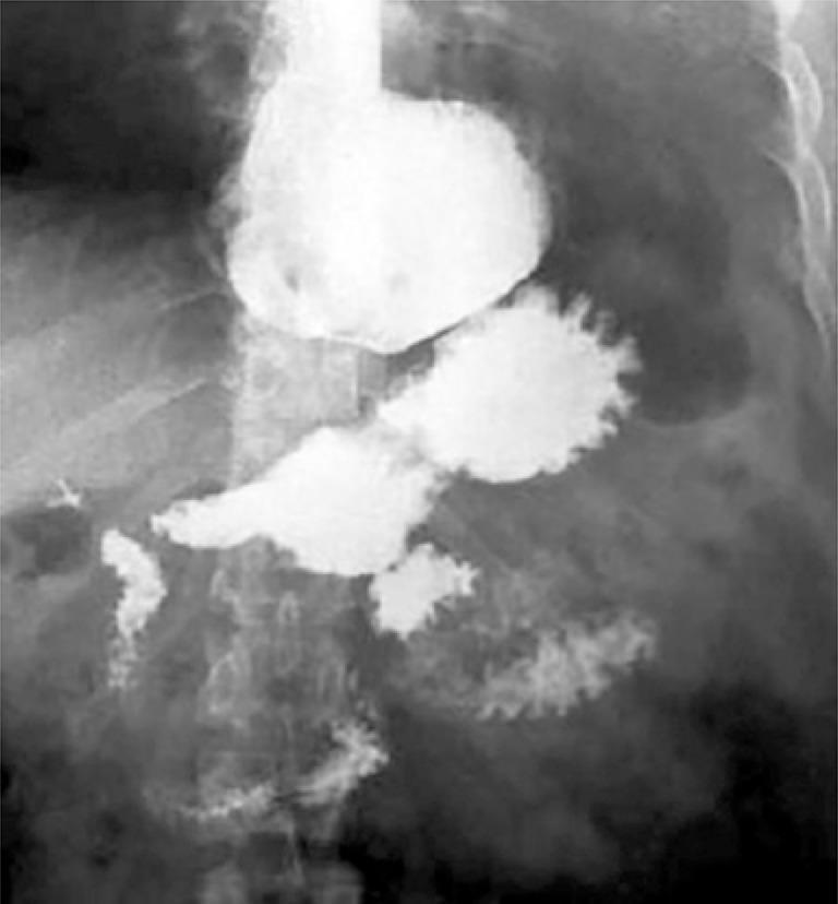
This diagnostic image is an upper gastrointestinal (UGI) seriography, or barium swallow study, utilizing positive contrast to visualize the esophagus, stomach, and duodenum. The primary finding is a large, contrast-filled epiphrenic diverticulum located in the distal esophagus just above the gastroesophageal junction. The diverticulum appears as a prominent, rounded, lateral outpouching of the esophageal lumen. Distal to this, the contrast outlines the gastric fundus, body, and antrum. The proximal duodenum is also visible with characteristic mucosal fold enhancement. The image demonstrates the hallmark radiographic presentation of a large esophageal diverticulum, which can lead to clinical symptoms such as dysphagia and regurgitation. The study is typical of gastroenterology and radiology educational materials focused on esophageal motility disorders and structural abnormalities.
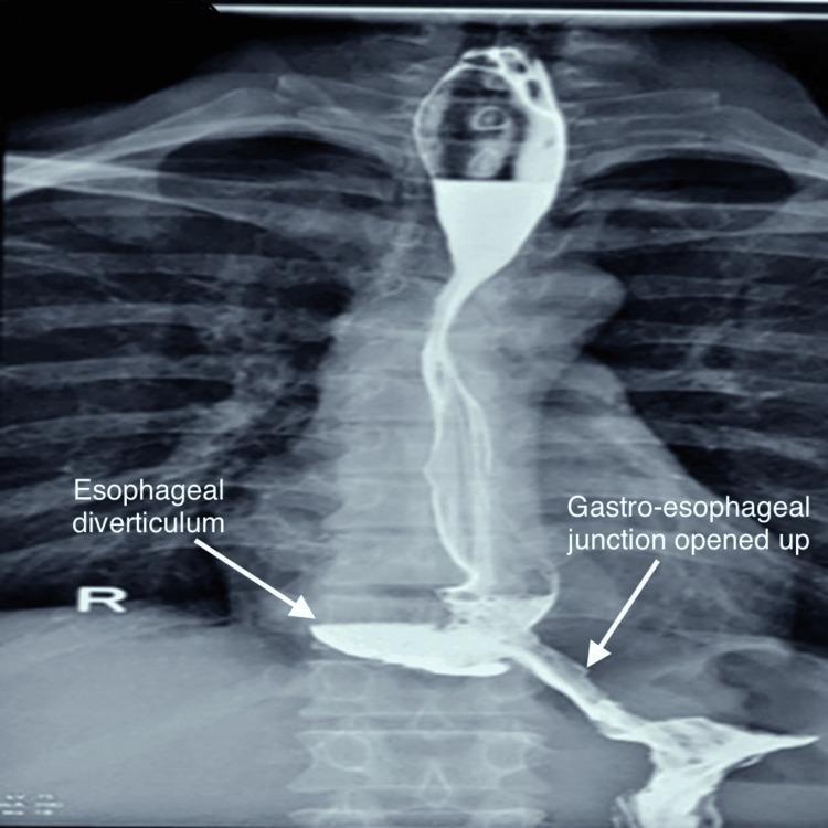
Postoperative barium swallow study (upper gastrointestinal contrast radiography) demonstrating the distal esophagus and gastro-esophageal junction (GEJ). The image shows the esophagus opacified with radiopaque contrast agent, revealing a significant out-pouching on the right posterior aspect labeled as an 'Esophageal diverticulum' (specifically an epiphrenic diverticulum). This diverticulum is well-defined and filled with contrast, communicating with the esophageal lumen. Distal to the diverticulum, the GEJ is clearly visualized and labeled as 'opened up', indicating surgical patency (post-Heller’s cardiomyotomy). Contrast is seen flowing smoothly through the junction into the stomach without evidence of luminal narrowing, stricture, stenosis, or extravasation. The contrast boundaries are sharp, suggesting preserved mucosal integrity and no postoperative leakage. This diagnostic image illustrates the successful management of esophageal motility disorders or structural abnormalities, confirming appropriate bolus transit into the gastric cardia.
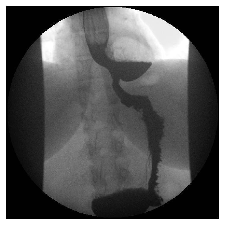
This diagnostic image is a barium swallow radiograph (fluoroscopy) of the upper gastrointestinal tract, focusing on the thoracic esophagus. The image shows a prominent, radiopaque curvilinear structure representing the esophagus opacified with barium contrast. A large, saccular outpouching or epiphrenic diverticulum is visible in the distal esophagus, extending laterally. A distinct horizontal air-fluid level is demonstrated within the diverticulum, characterized by a radiolucent (darker) gas-filled superior portion and a radiopaque (lighter) fluid/contrast-filled inferior portion. The distal esophageal lumen appears narrowed inferior to the diverticulum, and there is evidence of mucosal irregularity and proximal reflux of contrast material. The surrounding anatomical context includes visible vertebral bodies of the thoracic spine and the faint silhouette of the diaphragm. This image serves as a classic educational example of an esophageal diverticulum and associated stasis, useful for demonstrating diagnostic imaging findings in patients presenting with chronic dysphagia and regurgitation.
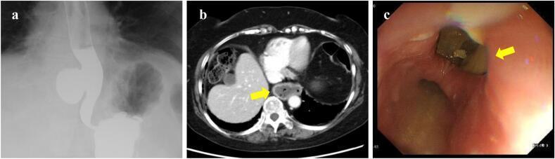
This composite figure displays diagnostic findings for a 50-mm epiphrenic diverticulum located in the lower thoracic esophagus. Panel (a) is a contrast esophagogram (barium swallow) showing a prominent, saccular outpouching on the right wall of the distal esophagus, approximately 5 cm above the esophagogastric junction. Panel (b) is an axial contrast-enhanced CT scan of the lower mediastinum identifying the diverticulum (indicated by a yellow arrow) as a distinct, air- and fluid-filled structure adjacent to the esophageal lumen. Panel (c) provides an endoscopic view showing the large diverticular orifice (yellow arrow) within the lower esophagus, containing visible food residue. The surrounding esophageal mucosa appears erythematous, consistent with mild reflux esophagitis. Together, these images illustrate the radiographic, tomographic, and endoscopic features of a pulsion-type esophageal diverticulum, which is frequently associated with esophageal motility disorders like EGJ outflow obstruction.
Types of Esophageal Diverticulum
Classification by Location
1. Zenker's Diverticulum (Pharyngoesophageal)
- Most common type
- Arises posteriorly through Killian's triangle/dehiscence - a natural area of weakness between the thyropharyngeus and cricopharyngeus muscles, just above the upper esophageal sphincter
- Technically located in the hypopharynx, proximal to the true esophagus, though it's conventionally discussed with esophageal diverticula (Cummings Otolaryngology, p. 160-169)
- Pulsion diverticulum - a false diverticulum (herniation of mucosa/submucosa only, without muscle layer), caused by increased intraluminal pressure against a poorly relaxing cricopharyngeus
- Presents with dysphagia, regurgitation of undigested food, halitosis, aspiration risk
2. Midesophageal (Traction) Diverticulum
- Also called parabronchial diverticulum
- The only true diverticula that form in the esophagus - the outpouching contains all layers of the esophageal wall (mucosa, submucosa, and muscle) (Cummings Otolaryngology, p. 166-169)
- Classic mechanism: traction from adjacent mediastinal inflammatory lymphadenopathy (historically tuberculosis, now also fungal infections or other granulomatous disease) that scars and pulls the esophageal wall outward
- Usually asymptomatic and found incidentally
- Located near the tracheal bifurcation/hilum, related anatomically to adjacent nodes
3. Epiphrenic (Supradiaphragmatic) Diverticulum
- Arises in the distal esophagus, just above the LES, typically within the last 10 cm
- A pulsion, false diverticulum, strongly associated with underlying esophageal motility disorders - especially achalasia, diffuse esophageal spasm, and EGJ outflow obstruction (Robbins Pathologic Basis of Disease, p. 1624-1627)
- Increased intraluminal pressure from dysmotility/functional obstruction pushes mucosa through the muscular wall
- Can develop complications: stasis esophagitis, and rarely squamous cell carcinoma has been reported within the pouch
4. Intramural Pseudodiverticulosis
- Multiple tiny outpouchings representing dilated submucosal glands/ducts, not true diverticula of the lumen
- Often associated with chronic esophagitis, candidiasis, diabetes, or alcohol use
- Gives a characteristic "thorny" or serrated appearance on barium study
Classification by Mechanism (Summary)
| Type | Mechanism | Wall layers involved | True or False |
|---|---|---|---|
| Zenker's | Pulsion (high pressure against poorly relaxing cricopharyngeus) | Mucosa + submucosa only | False |
| Midesophageal | Traction (external inflammatory scarring, e.g., TB nodes) | All layers | True |
| Epiphrenic | Pulsion (motility disorder, functional obstruction) | Mucosa + submucosa only | False |
| Intramural pseudodiverticulosis | Ductal dilation (chronic inflammation) | Submucosal gland ducts | Neither (pseudo) |
Quick Clinical Correlation
- Zenker's → cervical dysphagia, regurgitation of undigested food, aspiration, halitosis; managed with cricopharyngeal myotomy (endoscopic or open) or Z-POEM
- Midesophageal (traction) → usually asymptomatic, incidental finding; treat underlying granulomatous cause if active
- Epiphrenic → dysphagia/regurgitation; workup must include manometry since an underlying motility disorder (achalasia, spasm) is typically the driver and needs to be addressed alongside the diverticulum itself