
AXILLARY ARTERY AND ANASTOMOSIS AROUND SCAPULA
Reading File
The axillary artery is the continuation of the subclavian artery and supplies the shoulder and axilla, with the scapular anastomosis being a clinically important collateral network.
Searching sources...
Finding Sources
Finding Sources
Finding Sources
Reading File
Reading File
Finding Sources
Reading File
Reading File
Reading File
Loading Image
Loading Image
Loading Image
Here is a comprehensive account of the axillary artery and the anastomosis around the scapula, based on Gray's Anatomy for Students and the THIEME Atlas of General Anatomy.
Axillary Artery
Overview and Extent
The axillary artery is the direct continuation of the subclavian artery. It begins at the lateral margin of rib I and ends at the inferior margin of teres major, where it continues as the brachial artery. It passes through the axilla, accompanied medially/anteriorly by the axillary vein, and is surrounded by the cords of the brachial plexus.
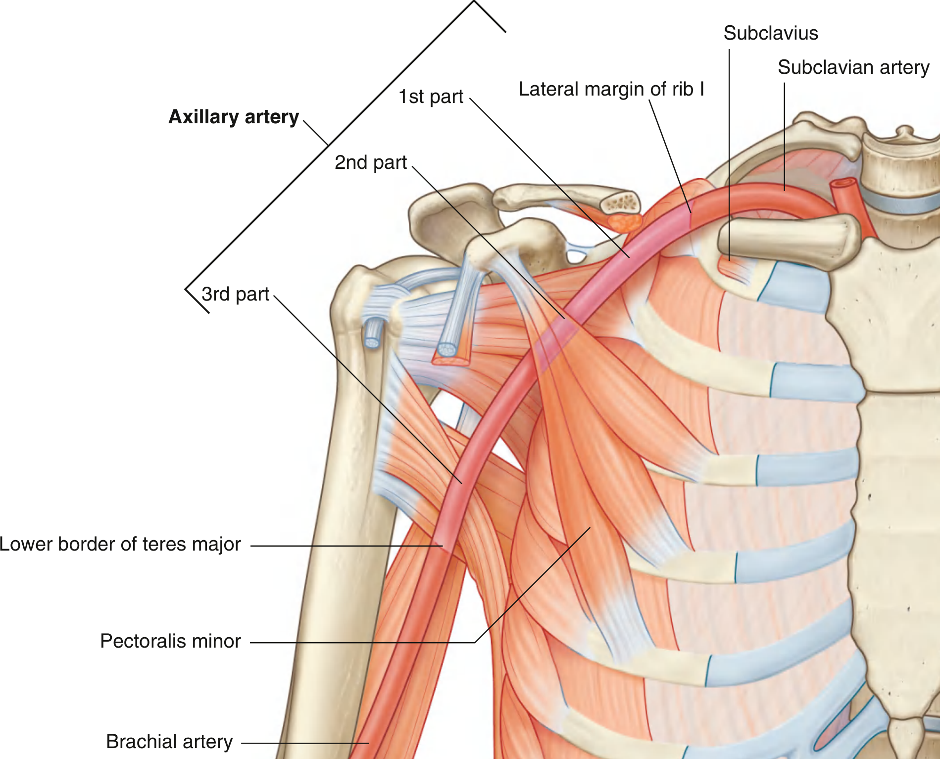
Three Parts (Divided by Pectoralis Minor)
The pectoralis minor muscle crosses anterior to the axillary artery and divides it into three parts:
| Part | Position relative to pectoralis minor | No. of branches |
|---|---|---|
| 1st part | Proximal (medial) to pectoralis minor | 1 branch |
| 2nd part | Posterior (behind) pectoralis minor | 2 branches |
| 3rd part | Distal (lateral) to pectoralis minor | 3 branches |
A useful mnemonic: 1-2-3 rule (1st part = 1 branch, 2nd part = 2 branches, 3rd part = 3 branches).
Branches
1st Part (1 Branch)
1. Superior thoracic artery
- Small vessel from the anterior surface
- Supplies the upper medial and anterior walls of the axilla
2nd Part (2 Branches)
2. Thoraco-acromial artery
- Originates just posterior to the medial (superior) margin of pectoralis minor
- Curves around the superior margin of the muscle, pierces the clavipectoral fascia
- Immediately divides into 4 branches: pectoral, deltoid, clavicular, and acromial
- Pectoral branch contributes to breast supply; deltoid branch accompanies the cephalic vein in the deltopectoral (clavipectoral) triangle
3. Lateral thoracic artery
- Arises from the anterior surface, posterior to the lateral (inferior) margin of pectoralis minor
- Follows the muscle to the thoracic wall
- Supplies medial and anterior walls of the axilla
- In women, contributes branches to the breast around the inferior margin of pectoralis major
3rd Part (3 Branches)
4. Subscapular artery - the largest branch of the axillary artery
- Arises from the posterior surface, follows the inferior margin of subscapularis
- Divides into two terminal branches:
- Circumflex scapular artery - passes through the triangular space (between subscapularis, teres major, and long head of triceps), enters the infraspinous fossa, and anastomoses with the suprascapular and dorsal scapular arteries
- Thoracodorsal artery - follows the lateral border of the scapula to the inferior angle; supplies the posterior and medial axillary walls (and latissimus dorsi)
5. Anterior circumflex humeral artery
- Small; from the lateral side of the 3rd part
- Passes anterior to the surgical neck of the humerus
- Anastomoses with the posterior circumflex humeral artery
- Supplies the glenohumeral joint and head of humerus
6. Posterior circumflex humeral artery
- Larger; from the lateral surface of the 3rd part, just posterior to the anterior circumflex humeral artery
- Passes through the quadrangular space (bounded by teres minor, teres major, long head of triceps, and surgical neck of humerus) along with the axillary nerve
- Curves around the surgical neck of the humerus
- Anastomoses with the anterior circumflex humeral artery, profunda brachii, suprascapular, and thoraco-acromial arteries
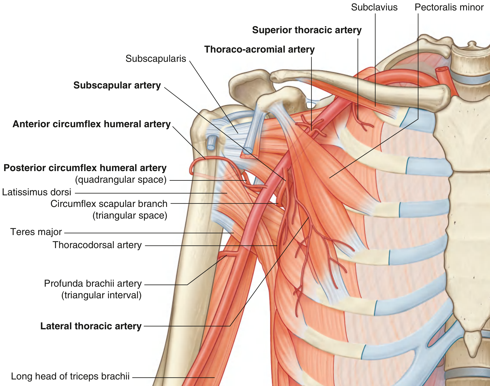
Anatomical Variants (Lippert & Pabst)
- Normal pattern (40% of cases): all 6 branches arise as described above
- Thoraco-acromial artery arises from the lateral thoracic artery (10%)
- Common origin of lateral thoracic artery and subscapular artery (10%)
- Posterior circumflex humeral artery arises from the subscapular artery (20%)
- Common origin of anterior and posterior circumflex humeral arteries - the "common circumflex humeral artery" (20%)
Anastomosis Around the Scapula (Scapular Arcade)
This is a clinically important collateral network on the posterior scapular surface, formed by the anastomosis of three vessels:
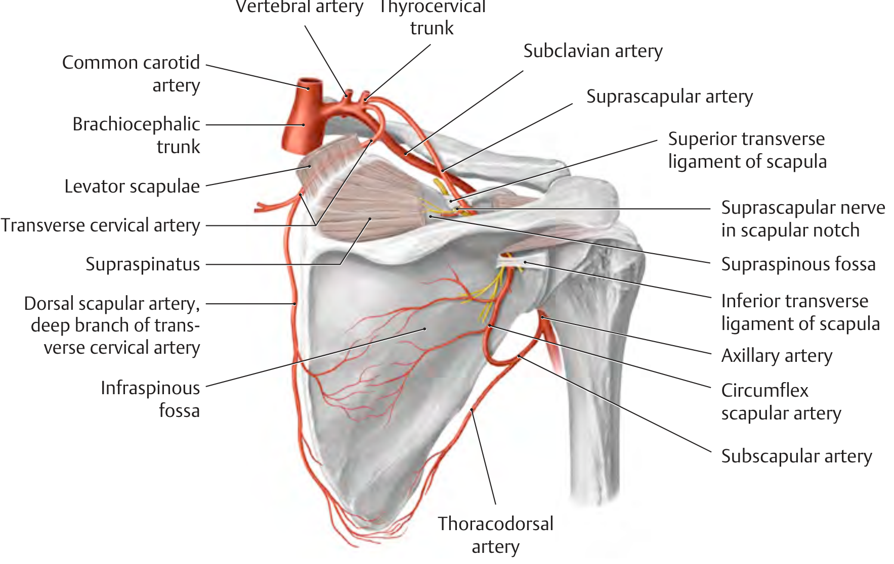
The Three Contributing Arteries
| Artery | Origin | Route |
|---|---|---|
| Suprascapular artery | Thyrocervical trunk (branch of subclavian) | Passes over the superior transverse scapular ligament (nerve passes under); enters supraspinous fossa, then passes under inferior transverse ligament (often absent) into the infraspinous fossa |
| Circumflex scapular artery | Subscapular artery (from 3rd part of axillary) | Passes through the triangular space (medial axillary foramen) to reach the infraspinous fossa |
| Dorsal scapular artery (deep branch of transverse cervical artery) | Transverse cervical artery (from thyrocervical trunk) | Runs along the medial border of the scapula |
Anastomotic Network
All three vessels converge and anastomose in the infraspinous fossa, forming a rich anastomotic arcade around the posterior scapula.
Clinical Significance
This anastomosis is critically important because:
- If the axillary artery is ligated or occluded (e.g., trauma, surgery), the scapular arcade provides a collateral pathway to reconstitute blood flow to the distal upper limb
- The collateral blood can flow: subclavian artery → suprascapular artery / dorsal scapular artery → scapular arcade → circumflex scapular artery → subscapular artery → axillary artery (distal to occlusion)
- This is why ligation of the axillary artery proximal to the subscapular artery origin is generally safe and compatible with limb viability
- The anastomosis also explains why the suprascapular nerve passes under the superior transverse scapular ligament (while the artery passes over it) - this spatial separation is a classic exam point
Neurovascular Passageways Around the Scapula
| Space | Boundaries | Structures transmitted |
|---|---|---|
| Triangular space (medial axillary foramen) | Teres minor (above), teres major (below), long head of triceps (lateral) | Circumflex scapular artery |
| Quadrangular space (lateral axillary foramen) | Teres minor (above), teres major (below), long head of triceps (medial), humerus (lateral) | Posterior circumflex humeral artery + axillary nerve |
| Triceps hiatus (triangular interval) | Teres major (above), long head of triceps (medial), humerus (lateral) | Deep artery of arm (profunda brachii) + radial nerve |
Sources:
- Gray's Anatomy for Students, pp. 853-855
- General Anatomy and Musculoskeletal System, THIEME Atlas of Anatomy, pp. 375, 404, 410
This is a shared conversation. Sign in to Orris to start your own chat.