
Tell me about the shop part. I mean the, um... The olive tarsic. The olive thoracic.
You're likely thinking of the optic tarsus - but let me check the medical library to give you accurate information.
Searching sources...
Reading File
Tell me about the wall of thoracic
The thoracic wall (chest wall) is made up of bones, muscles, and soft tissues that enclose and protect the thoracic cavity. Let me pull detailed information from the medical library.
Searching sources...
Finding Sources
Finding Sources
Good results from Gray's Anatomy for Students. Let me read the key sections now.
Reading File
Reading File
Now let me read the muscles, vessels, and nerves sections:
Reading File
Reading File
Excellent - I now have comprehensive content. Let me fetch the key diagram images to include:
Loading Image
Loading Image
Here is a thorough overview of the thoracic wall based on Gray's Anatomy for Students:
The Thoracic Wall
The thoracic wall is segmental in design and made up of skeletal elements and muscles. It extends between:
- Superiorly - the superior thoracic aperture (bordered by vertebra T1, rib I, and the manubrium of the sternum)
- Inferiorly - the inferior thoracic aperture (bordered by vertebra T12, rib XII, rib XI, the costal margin, and the xiphoid process)
1. Skeletal Framework
The skeleton of the thoracic wall has three regions:
Posteriorly:
- 12 thoracic vertebrae with their intervertebral discs
- Each vertebra is characterized by articulations with ribs
- A typical thoracic vertebra has a heart-shaped body, a long spinous process, and three sites on each side for rib articulation: two demifacets on the body (for the rib head) and one transverse costal facet (for the rib tubercle)
Laterally:
- 12 pairs of ribs, forming the lateral walls along with three layers of intercostal muscles
- True ribs (I-VII): costal cartilages articulate directly with the sternum
- False ribs (VIII-X): costal cartilages articulate with the cartilage above them
- Floating ribs (XI-XII): do not articulate with the sternum or other ribs at all - their costal cartilages only cover their tips
Anteriorly:
- The sternum, made up of three parts:
- Manubrium - the uppermost part
- Body - the main part
- Xiphoid process - the inferior tip
- The manubrium and body meet at the sternal angle (angle of Louis), a key clinical landmark used in physical examination of the thorax
2. Muscles of the Thoracic Wall
The thoracic wall muscles alter rib/sternum position to change thoracic volume during breathing, and also reinforce the wall.
Intercostal Muscles (3 layers)
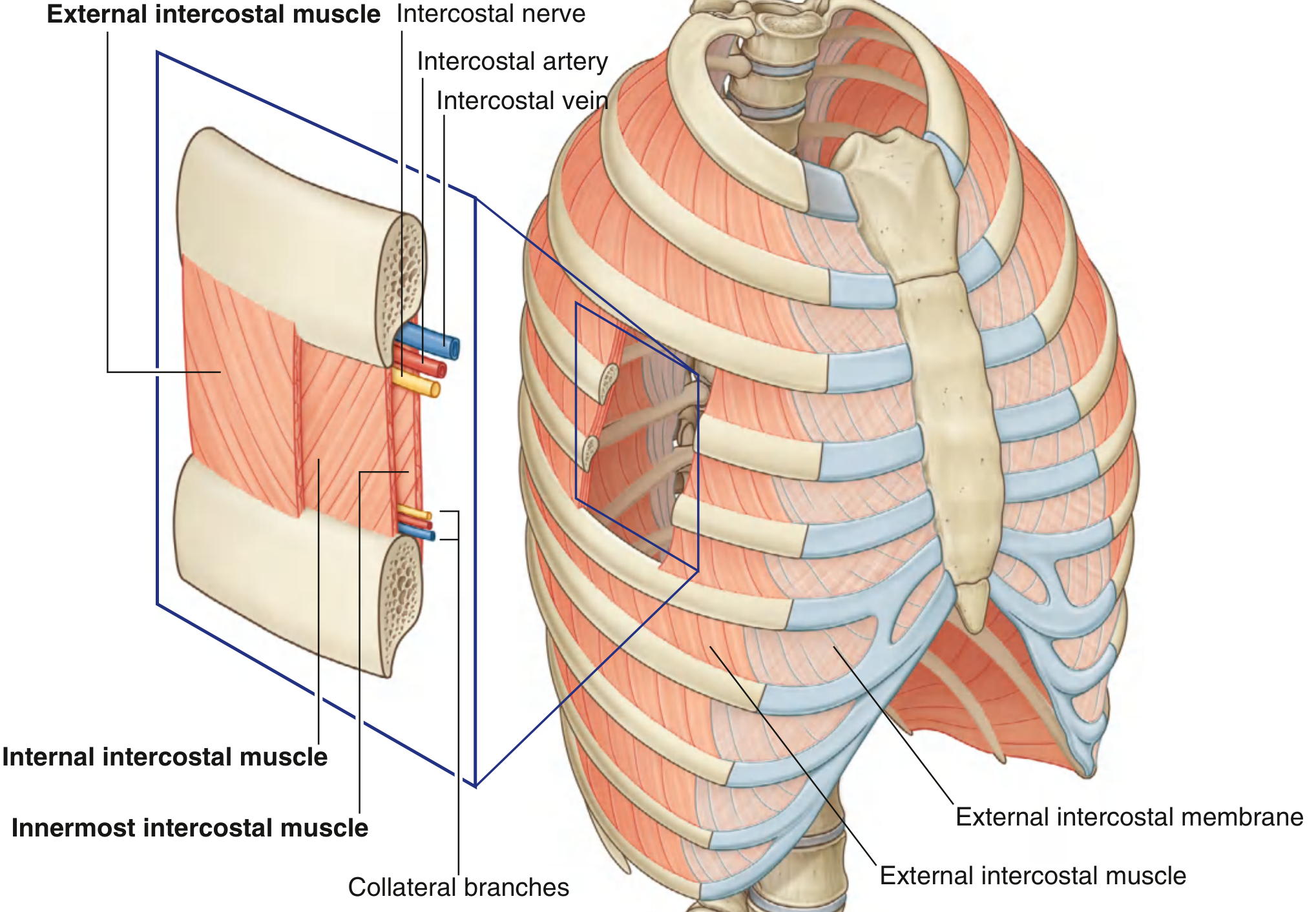
Three flat muscles fill each intercostal space between adjacent ribs:
| Muscle | Attachment | Innervation | Function |
|---|---|---|---|
| External intercostal | Inferior margin of rib above → superior margin of rib below | Intercostal nerves T1-T11 | Most active during inspiration; elevates ribs |
| Internal intercostal | Costal groove of rib above → superior margin of rib below | Intercostal nerves T1-T11 | Most active during expiration; depresses ribs |
| Innermost intercostal | Medial edge of costal groove → internal aspect of rib below | Related intercostal nerves | Acts with internal intercostals |
| Subcostales | Internal surface of lower ribs | Related intercostal nerves | May depress ribs |
| Transverse thoracis | Costal cartilages of ribs II-VI | Related intercostal nerves | Depresses costal cartilages |
Additional muscles include the levatores costarum and serratus posterior superior/inferior, which also move the ribs during breathing.
3. Neurovascular Supply
The neurovascular supply is segmental, mirroring the wall's segmental structure.
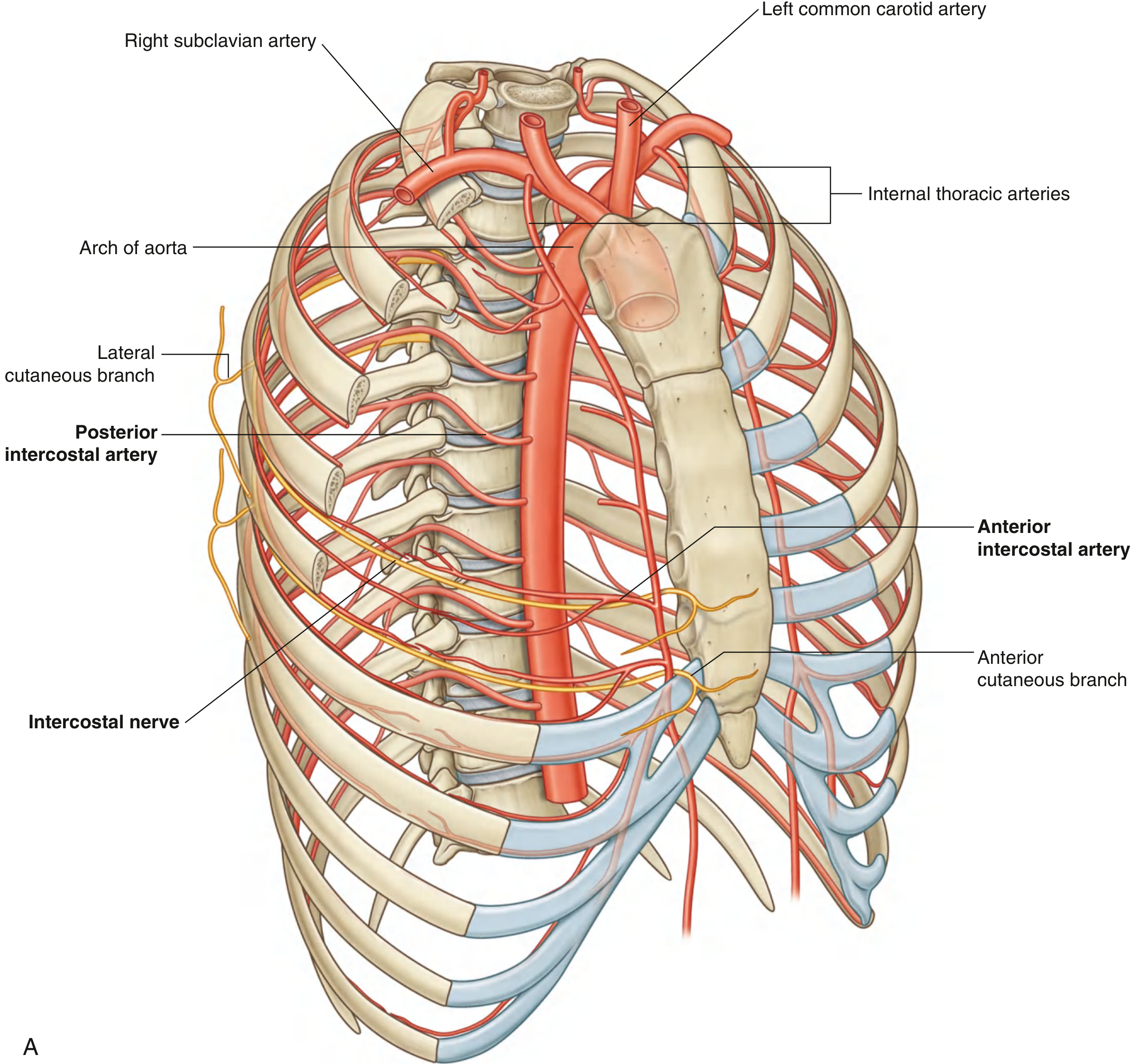
Arteries
Blood supply comes from two main sources:
- Thoracic aorta (posterior mediastinum) - gives off posterior intercostal arteries that pass laterally around the wall along the inferior margin of each rib
- Internal thoracic arteries - run along the deep aspect of the anterior thoracic wall on either side of the sternum; give off anterior intercostal arteries
Nerves
- Intercostal nerves (anterior rami of spinal nerves T1-T11) run with the vessels and provide:
- Somatic motor supply to the muscles of the wall
- Somatic sensory supply from the skin and parietal pleura
- Postganglionic sympathetic fibers
Dermatomes
- T2 is the highest dermatome on the anterior chest wall (T1 is mostly in the upper limb)
- Skin over the xiphoid process = T6
- T7-T12 dermatomes curve down onto the anterior abdominal wall
Clinical note: The intercostal nerve and vessels run along the inferior margin of each rib in the costal groove. When inserting chest tubes or needles through the thoracic wall, the instrument should pass along the superior margin of the rib below to avoid these structures.
4. Apertures
- Superior thoracic aperture - a narrow inlet bounded by T1, the medial border of rib I, and the manubrium. Structures passing between the thorax and neck travel through here (trachea, esophagus, great vessels, nerves).
- Inferior thoracic aperture - a wide outlet closed by the diaphragm. It is bordered by T12, rib XII, rib XI, the costal margin, and the xiphoid.
Source: Gray's Anatomy for Students
This is a shared conversation. Sign in to Orris to start your own chat.