
I feel tent on cervical and bent on lumbar
cervical lordosis tent shape lumbar kyphosis bent spine clinical examination sign
Spinal Curvatures: "Tent on Cervical, Bent on Lumbar"
- "Tent" on cervical = Cervical Lordosis - the cervical spine curves inward (anteriorly), making a tent-like or forward arch shape (concave posteriorly). This is the normal "C" curve of the neck.
- "Bent" on lumbar = Lumbar Lordosis - the lumbar spine also curves inward (anteriorly), creating a "bent-back" inward arch (concave posteriorly). This gives the lower back its characteristic inward curve.
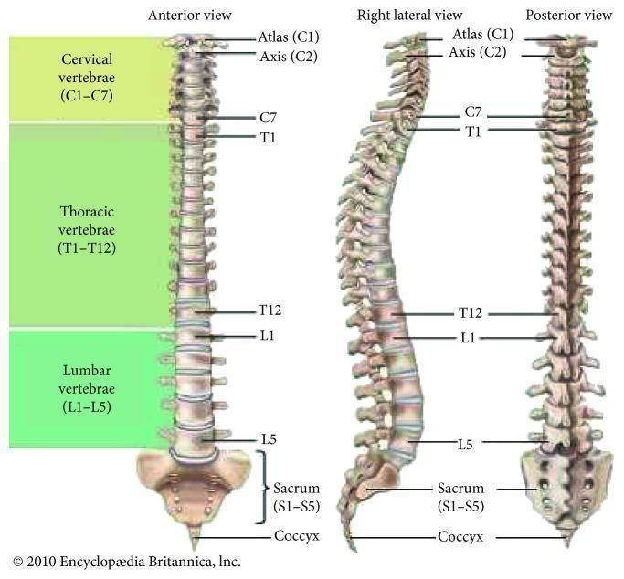
The Four Normal Spinal Curvatures
| Region | Normal Curve | Direction | Shape |
|---|---|---|---|
| Cervical (C1-C7) | Lordosis | Concave posteriorly (curves forward) | Like a tent / forward arch |
| Thoracic (T1-T12) | Kyphosis | Concave anteriorly (curves backward) | Rounded outward hump |
| Lumbar (L1-L5) | Lordosis | Concave posteriorly (curves forward) | Inward arch / "bent inward" |
| Sacral | Kyphosis | Concave anteriorly | Fixed posterior curve |
Why These Curves Matter Clinically
- Causes: trauma, disc disease, ankylosing spondylitis, post-surgical
- Symptoms: neck pain, reduced range of motion, potential cord compression
- Clinically normal cervical lordosis is 31-40 degrees on lateral X-ray
- Causes: ankylosing spondylitis (classic), disc prolapse (muscle spasm flattens the curve), fracture, TB spine
- In ankylosing spondylitis: progressive loss of lumbar lordosis + increasing thoracic kyphosis = the stooped "question mark" posture; knees bent to maintain balance - S Das Manual on Clinical Surgery
- In acute disc prolapse: patient stands with characteristic lumbar scoliosis, loss of lordosis, slight hip/knee flexion
- Causes: hip flexion contractures, pregnancy, obesity, congenital hip dislocation
- Signs: stomach appears thrust forward, buttocks more pronounced, visible gap under lower back when lying flat
Quick Memory Aid
Cervical and Lumbar = Lordosis (both curve the same way - inward/anteriorly)
Thoracic and Sacral = Kyphosis (both curve outward/posteriorly)
I have neck hump
neck hump causes treatment buffalo hump dorsocervical fat pad dowager's hump
buffalo hump dorsocervical fat pad neck back
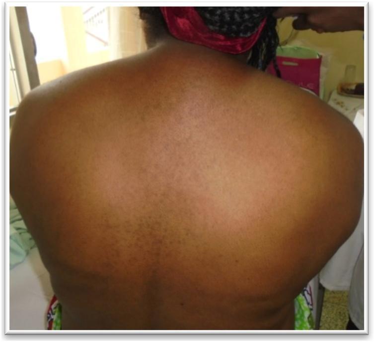
A clinical photograph of the posterior aspect of a patient's upper back and neck region, demonstrating a 'buffalo hump' (dorsocervical fat pad). The image shows a prominent, localized accumulation of subcutaneous adipose tissue in the upper thoracic and lower cervical region, creating a rounded, convex deformity between the shoulder blades and at the base of the neck. The skin overlying the fat pad appears stretched but otherwise intact, with subtle hyperpigmentation and scattered small macules. This physical finding is a classic clinical manifestation of hypercortisolism (Cushing's syndrome), characterized by centripetal fat redistribution. The photograph is taken from a posterior-superior perspective, highlighting the anatomical prominence relative to the scapulae and spine, which is useful for medical students and clinicians in identifying physical signs of endocrine dysfunction.
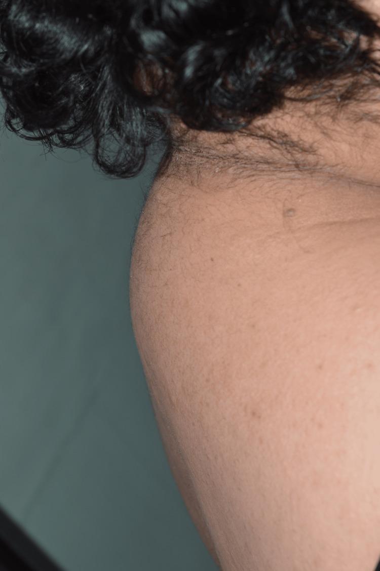
This clinical photograph shows a lateral view of the posterior neck and upper back of a patient, demonstrating a prominent dorsocervical fat pad, commonly referred to as a 'buffalo hump.' The anatomical region involved is the posterior cervicothoracic junction, specifically overlying the C7-T1 vertebrae. The growth is characterized by a rounded, localized accumulation of adipose tissue that protrudes posteriorly, altering the natural contour of the neck and upper spine. The overlying skin appears intact with visible fine terminal hairs and follicular orifices, typical of the nuchal region. No acute inflammatory signs, ulceration, or primary skin lesions are visible on the surface of the fat pad. This clinical finding is frequently associated with metabolic disturbances, hypercortisolism (Cushing's syndrome), or certain pharmacological side effects, such as those seen in antiretroviral therapy for HIV. The image serves as an educational example of abnormal fat distribution (lipodystrophy) in a clinical diagnostic context.
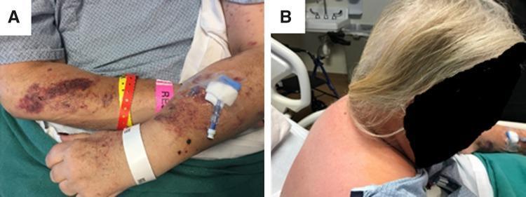
Clinical photograph presenting characteristic physical stigmata of Cushing's syndrome in a patient. Panel A displays a top-down view of the patient's forearm and antecubital fossa, demonstrating significant skin fragility and easy bruisability. Multiple irregular, confluent, purplish-red ecchymotic patches and smaller petechiae are visible across the skin surface. Clinical equipment, including multiple hospital identification bands and an indwelling intravenous catheter with a blue-and-white luer-lock hub, is present on the arm. Panel B illustrates the posterior neck and upper back of the same patient, highlighting a dorsocervical fat pad, commonly known as a 'buffalo hump.' This finding appears as a distinct, rounded, soft-tissue prominence at the level of the cervicothoracic junction, creating an abnormal posterior contour. These visual findings are hallmark cutaneous and musculoskeletal manifestations of hypercortisolism, useful for medical students and clinicians in identifying metabolic or endocrine dysfunction during physical examination.
Neck Hump - What It Is and What Causes It
Type 1: Buffalo Hump (Dorsocervical Fat Pad)
| Cause | Details |
|---|---|
| Cushing's syndrome | Excess cortisol (from pituitary tumor, adrenal tumor, or ectopic ACTH) causes fat redistribution to the back of neck, face (moon face), and abdomen. Classic triad: buffalo hump + moon face + truncal obesity |
| Long-term steroid use | Prednisone, dexamethasone, or other corticosteroids taken for months/years mimic Cushing's syndrome |
| HIV antiretroviral therapy | Protease inhibitors and thymidine analogues (stavudine, zidovudine) cause lipodystrophy with fat redistribution - Harrison's Internal Medicine 22E |
| Obesity | General weight gain with preferential fat deposition at the back of the neck |
| Madelung's disease | Rare condition causing diffuse fatty deposits, often in the neck region |
Type 2: Dowager's Hump (Kyphosis)
- Osteoporosis - vertebral compression fractures cause wedging of vertebral bodies, most common in post-menopausal women and elderly
- Ankylosing spondylitis - progressive spinal stiffening and kyphosis (as discussed earlier)
- Poor posture / forward head posture - chronic rounding of the upper back over years
- Scheuermann's disease - osteochondritis of the vertebral end plates in teenagers
- Fracture - trauma causing vertebral wedge fracture
How to Tell Them Apart
| Feature | Buffalo Hump | Dowager's Hump |
|---|---|---|
| Feel on touch | Soft, doughy | Firm, bony |
| Location | Fat pad at C7/T1 level | Curved upper spine |
| Spine shape | Normal curve | Exaggerated outward curve |
| Associated symptoms | Moon face, weight gain, stretch marks | Back pain, height loss, posture change |
| Main cause | Hormonal/metabolic | Bone/posture |
What You Should Do
- Feel the hump - soft (fat) vs. firm (bone)
- Check for other Cushing's signs - moon face, purple stretch marks, easy bruising, high blood pressure, unexplained weight gain
- Review your medications - are you on steroids?
- Order tests - morning cortisol, 24-hour urine cortisol, or dexamethasone suppression test if Cushing's is suspected; DEXA scan and X-rays if osteoporosis/kyphosis is suspected