
Defecation reflex
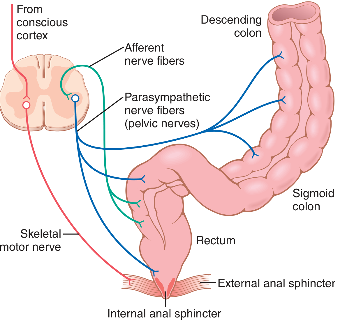
defecation reflex neural pathway diagram
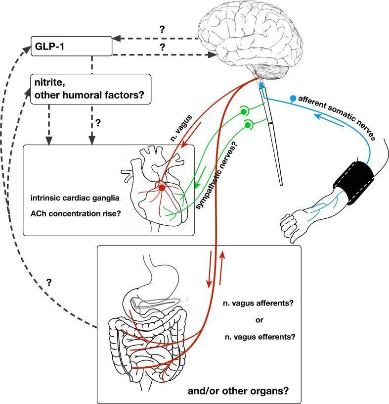
This pathophysiology diagram illustrates the 'remote preconditioning reflex arc,' a complex neural and humoral pathway involved in remote ischemic preconditioning (RIPre). The diagram depicts a stimulus applied to an arm via a blood pressure cuff, which triggers afferent somatic nerves that project to the brain. Efferent signals are shown descending from the central nervous system via the vagus nerve (n. vagus) and sympathetic nerves to the heart and other visceral organs, specifically the intestine. Key cardiovascular features include the involvement of intrinsic cardiac ganglia and a potential rise in acetylcholine (ACh) concentration in the myocardium. The diagram highlights several scientific uncertainties using question marks, specifically regarding the role of sympathetic nerves, whether abdominal vagal fibers are afferent or efferent, and the origin of humoral factors like GLP-1 and nitrite. Humoral pathways are shown as dashed lines connecting systemic factors to the brain and heart, indicating a parallel mechanism of cardioprotection alongside the primary neural reflex arc.
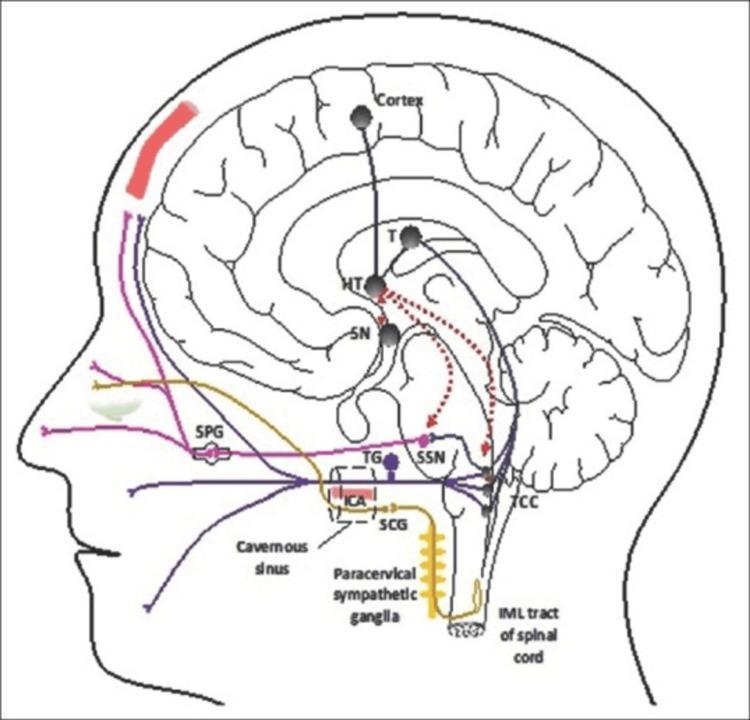
A pathophysiology diagram illustrating the neural pathways involved in cluster headaches within a sagittal view of the human head and brain. The diagram highlights the trigeminal-autonomic reflex arc. Key neural structures depicted include the hypothalamus (HT), which serves as a central hub with connections to the thalamus (T), suprachiasmatic nucleus (SN), superior salivatory nucleus (SSN), and the trigeminocervical complex (TCC). Sensory pathways are shown in purple and blue, originating from the dural structures and trigeminal nerve, synapsing in the TCC and projecting to the cortex. The parasympathetic pathway (pink) involves the SSN and sphenopalatine ganglion (SPG), leading to autonomic symptoms. The sympathetic pathway (yellow) is traced from the intermediolateral (IML) tract of the spinal cord through the superior cervical ganglion (SCG) and the plexus surrounding the internal carotid artery (ICA) in the cavernous sinus. Red dashed lines indicate the hypothalamic modulation of the SSN and TCC, which are critical in the pathogenesis of trigeminal autonomic cephalgias.
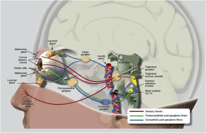
This medical pathophysiology diagram illustrates the neuroanatomical pathway of the nasolacrimal reflex and the lacrimal functional unit (LFU). The illustration overlays neural pathways onto a sagittal view of the human head, focusing on the eye, brainstem, and nasal region. Sensory nerves (red lines) are shown originating from the anterior ethmoidal nerve in the nasal cavity and the corneal sensory nerves, converging at the trigeminal ganglion before reaching the trigeminal nucleus caudalis in the brainstem. Parasympathetic post-ganglionic fibers (green lines) originate from the superior salivatory nucleus and pass through the geniculate and pterygopalatine ganglia to innervate the lacrimal glands, meibomian glands, and goblet cells. Sympathetic post-ganglionic fibers (blue lines) are shown following the carotid artery and passing through the ciliary ganglion. Key anatomical labels include the lacrimal gland, meibomian glands, trigeminal ganglion, and the motor nucleus of CN VII. This diagram serves as an educational tool for understanding dry eye disease pathophysiology and the mechanisms of neurostimulation for tear production.
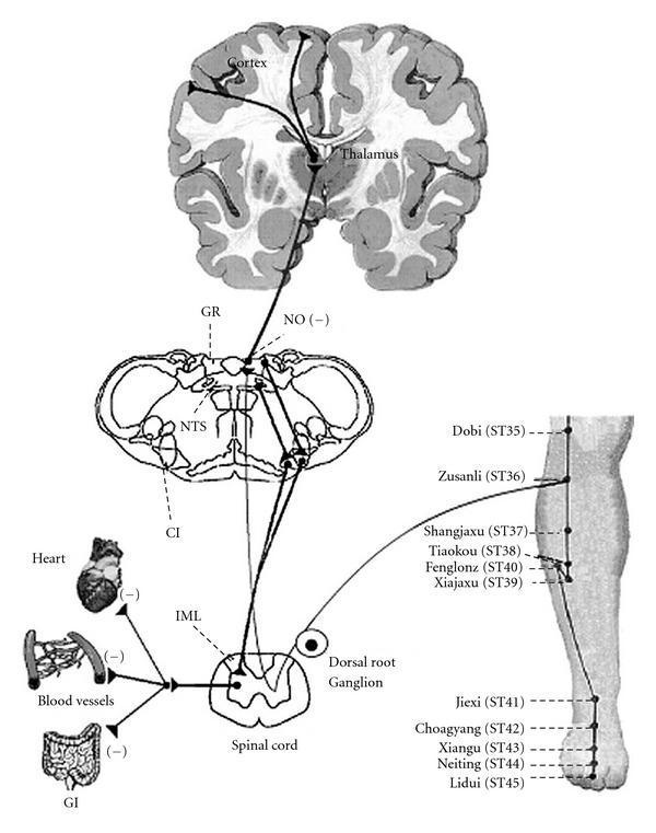
This pathophysiology diagram illustrates the neural circuits of the somatosympathetic reflex and the gracile-thalamic-cortex pathway, specifically in the context of acupuncture stimulation. The visual flow begins at acupuncture points on the lower leg (notably ST36 Zusanli), where afferent signals travel through the dorsal root ganglion to the spinal cord. In the spinal cord, a branch connects to the intermediolateral nucleus (IML), showing inhibitory output (-) to effector organs: the heart, blood vessels, and gastrointestinal (GI) tract. The ascending pathway continues through the medulla oblongata, highlighting the nucleus of the solitary tract (NTS) and the gracile nucleus (GR). Signals then ascend to the thalamus and ultimately to the cerebral cortex. The diagram also identifies key neurochemical modulators such as nitric oxide (NO) with inhibitory signs, and labels secondary structures like the cardioinhibitory center (CI). This illustration is designed for medical education on autonomic nervous system regulation and the neuroanatomical basis of peripheral nerve stimulation effects on internal organs.
Defecation Reflex
Overview
Anatomy of Continence
- Internal anal sphincter (IAS) - a thickening of circular smooth muscle, tonically contracted under autonomic control. Sympathetic innervation is excitatory (maintains tone); parasympathetic is inhibitory (relaxes it during defecation).
- External anal sphincter (EAS) - striated voluntary muscle, innervated by the pudendal nerve (somatic nervous system, S2-S4). It is kept in tonic contraction and is under conscious/voluntary control.
Triggering Event
The Two Defecation Reflexes
1. Intrinsic Myenteric Reflex (Local / Enteric)
- Arc: Rectal wall stretch → afferent signals in the myenteric plexus → peristaltic waves through the descending colon, sigmoid colon, and rectum → inhibitory signals relax the internal anal sphincter
- Nature: This reflex operates entirely within the enteric nervous system of the gut wall
- Limitation: By itself it is relatively weak - capable of initiating peristalsis but not powerful enough to produce effective defecation alone
2. Parasympathetic Defecation Reflex (Spinal / Extrinsic)
- Arc: Rectal distension → afferent signals travel via pelvic nerves → sacral spinal cord (S2-S4, conus medullaris) → efferent parasympathetic signals return via pelvic nerves → descending colon, sigmoid, rectum, and anus
- Effect: Greatly intensifies peristaltic waves and produces powerful relaxation of the internal anal sphincter
- Extent: Can empty the bowel all the way from the splenic flexure to the anus
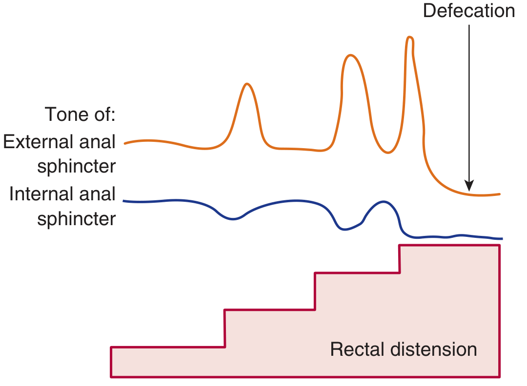
Sphincter Responses to Rectal Distension
Voluntary Control and Facilitation
| Component | Mechanism |
|---|---|
| Voluntary deferral | Conscious contraction of the EAS via pudendal nerve; signal ascends to cortex; conscious decision overrides the reflex |
| Voluntary facilitation | Deep breath → diaphragm descends; Valsalva maneuver → abdominal muscles contract; intra-abdominal pressure rises; pelvic floor lowers 1-3 cm; puborectalis relaxes; anorectal angle straightens (from ~90-100° to >15° wider) |
| Result of straining | EAS voluntarily relaxed + straightened anorectal angle = defecation occurs before the 55 mmHg threshold is reached |
Associated Reflexes
- Deep inspiration (diaphragm descends)
- Glottis closure (enables Valsalva)
- Abdominal wall contraction (increases intra-abdominal pressure)
- Pelvic floor relaxation (pulls anal ring outward, evaginates feces)
Effect of Spinal Cord Injury
- Cord transection above sacral segments: The intrinsic myenteric reflex and the spinal sacral arc remain intact, so automatic reflex defecation still occurs - but there is no voluntary control. Defecation happens at inconvenient, unpredictable times.
- Low spinal cord / cauda equina lesion: The sacral defecation reflex itself is disrupted. Digital stimulation may be ineffective. Management relies on antidiarrheal agents to prevent continuous soiling.
- This also explains why newborns defecate automatically - the cortical inhibitory pathway is not yet fully developed. (Guyton & Hall, p. 796; Sleisenger & Fordtran's, p. 3524)
Clinical Relevance
| Condition | Mechanism |
|---|---|
| Constipation from habit | Chronically inhibiting the natural defecation reflex blunts its sensitivity over time |
| Hirschsprung's disease | Absent myenteric ganglia in rectosigmoid → no intrinsic reflex → functional obstruction |
| Neurogenic bowel | Spinal cord injury disrupts voluntary EAS control or the sacral arc |
| Fecal incontinence | Pudendal nerve damage → EAS weakness → loss of voluntary continence |
| Parkinson's disease | Impaired CNS dopaminergic modulation of the sacral defecation reflex contributes to constipation |
- Guyton and Hall Textbook of Medical Physiology, Chapter 64 (pp. 795-796)
- Ganong's Review of Medical Physiology, 26th ed., Chapter 27 (p. 505)
- Sleisenger and Fordtran's Gastrointestinal and Liver Disease (spinal cord injury section)
Frey syndrome
Frey syndrome Minor starch iodine test gustatory sweating parotid
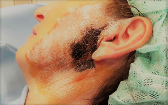
This clinical photograph displays a patient's left profile, focusing on the preauricular and parotid region during a Minor starch-iodine test. The skin is coated with a white, powdery layer of starch. A prominent, irregularly shaped black-to-dark-blue patch is visible anterior to the tragus of the ear and extending onto the cheek, representing a positive result where gustatory sweating has occurred. This chemical reaction occurs when moisture triggers iodine to form a dark complex with the amylose in starch, mapping the territory of Frey's syndrome (auriculotemporal syndrome). Faint reddish and brownish vertical lines on the lower cheek and neck indicate the application of iodine solution or anatomical markings for subsequent treatment. The image illustrates the diagnostic localization of aberrant gustatory sweating prior to therapeutic interventions like botulinum toxin-A injections. Target audience includes medical students and practitioners in neurology, ENT, or maxillofacial surgery.
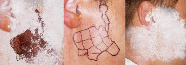
This sequence of clinical photographs illustrates the diagnostic and therapeutic management of Frey syndrome (gustatory sweating) following a parotidectomy. The left image demonstrates a positive Minor’s starch-iodine test; the presence of sweat on the preauricular and facial skin has reacted with the starch and iodine to create a focal, deep blue-black discoloration, identifying the pathological sweating area. The middle image shows the clinical mapping phase where the identified sweating region is outlined and subdivided into a grid of 'boxes' using a waterproof marker. This grid serves as a guide for localized intracutaneous Botulinum Toxin A (BTA) injections to ensure precise coverage of the affected territory. The right image shows a follow-up Minor’s test after BTA treatment; the skin remains covered only by the white starch powder with no blue-black reaction visible during gustatory stimulation, indicating successful chemical denervation of the eccrine sweat glands and resolution of symptoms. This series serves as an educational resource for dermatological and ENT surgery follow-up, emphasizing clinical testing and injection mapping for localized hyperhidrosis.
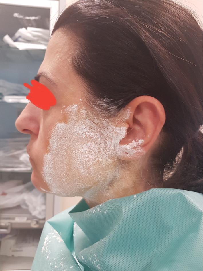
Clinical photograph of a patient undergoing the Minor starch-iodine test, a diagnostic tool for evaluating localized hyperhidrosis or Frey syndrome. The image shows the lateral profile of a patient's face with a thick, white layer of starch powder applied over a dried iodine solution on the right cheek, preauricular region, and extending toward the mandibular angle. The starch appears as a matte, powdery coating with variations in density at the peripheral margins near the ear and temple. This setup is used to visualize abnormal gustatory sweating; when sweat is produced, the chemical reaction between iodine and starch in the presence of moisture results in a characteristic blue-black or violet discoloration. The patient is draped in a medical-grade blue protective gown. Anonymization is maintained via a digital red marking over the eye. The clinical significance of this procedure is to map the specific area of autonomic dysfunction for targeted treatment, such as botulinum toxin injections.
Frey Syndrome (Auriculotemporal Syndrome / Gustatory Sweating)
Definition
Anatomy of Normal Parotid Innervation
- Preganglionic parasympathetic fibers arise from the inferior salivatory nucleus and travel via the glossopharyngeal nerve (CN IX)
- The Jacobson's nerve (branch of CN IX) enters the middle ear via the inferior tympanic canaliculus, forms the tympanic plexus, then exits as the lesser petrosal nerve through the foramen ovale
- The lesser petrosal nerve synapses in the otic ganglion
- Postganglionic secretomotor fibers join the auriculotemporal nerve (branch of CN V3) and travel to the parotid gland to stimulate salivation
- Separately, postganglionic sympathetic fibers innervate the sweat glands of the overlying skin
Pathophysiology - Misdirected Nerve Regeneration
- The postganglionic parasympathetic fibers of the auriculotemporal nerve (to parotid)
- The postganglionic sympathetic fibers to local sweat glands
Causes / Triggers
| Category | Examples |
|---|---|
| Most common | Parotidectomy (superficial or total) |
| Trauma | Temporomandibular joint trauma, condylar fracture |
| Surgery | Carotid endarterectomy, TMJ surgery |
| Childhood | Forceps delivery injury to parotid region (often misdiagnosed as food allergy) |
| Infection | Herpes zoster, parotitis |
| Other glands | Submandibular gland surgery/trauma (without auriculotemporal nerve involvement) |
Incidence
Diagnosis
Clinical
- Sweating and flushing over the preauricular/parotid region during eating
- Ipsilateral facial skin involvement
- History of prior parotid surgery or trauma
Minor's Starch-Iodine Test (Gold Standard)
- Paint the affected facial skin with iodine solution and allow to dry
- Dust with starch powder
- Patient chews a sialagogue (e.g., lemon wedge) for several minutes
- Positive result: Dark blue-black spots appear where sweat dissolves the starch-iodine complex - visually mapping the affected territory
Treatment
| Option | Details |
|---|---|
| Observation | Many patients tolerate mild symptoms and require no treatment |
| Topical antiperspirants | Aluminium chlorohydrate-containing agents applied to affected skin; simple first-line measure |
| Glycopyrrolate 1% roll-on | Topical anticholinergic lotion; effective in controlling symptoms |
| Botulinum toxin A (intracutaneous) | Most effective treatment; injected into the mapped affected area; benefit lasts ~1.5 years in 60% of patients; the Minor's test guides the injection grid |
| Tympanic neurectomy | Surgical interruption of the Jacobson's nerve (preganglionic parasympathetic supply); reserved for cases not responding to simpler measures |
Prevention
- Thick skin flap during surgery
- Partial superficial parotidectomy (less disruption)
- Sternocleidomastoid muscle flap - commonly used but systematic reviews suggest it is unlikely to achieve complete prevention
- AlloDerm or other interposition materials - evidence remains inconclusive
Key Summary Points
- Frey syndrome = gustatory sweating + flushing over auriculotemporal nerve territory after parotid injury
- Mechanism: aberrant parasympathetic reinnervation of sympathetically-innervated sweat glands; ACh stimulates both
- Latent period of weeks to months (rarely years) before onset
- Diagnosis confirmed by Minor's starch-iodine test
- Most effective treatment: botulinum toxin A injections
- Subclinical incidence is very high (~96%) even when patients don't complain
- Scott-Brown's Otorhinolaryngology Head & Neck Surgery (pp. 6776-6818)
- Cummings Otolaryngology Head and Neck Surgery (pp. 2032-2038)
- Bailey and Love's Short Practice of Surgery, 28th ed. (pp. 854, 4334)