
Lateral reticulospinal tract originates from medulla oblongata or is it originate from all the brain stem?
lateral reticulospinal tract origin medulla or brainstem anatomy
Lateral Reticulospinal Tract - Origin
The Two Reticulospinal Tracts
| Tract | Also Called | Origin | Location in Spinal Cord |
|---|---|---|---|
| Pontine reticulospinal tract | Medial reticulospinal tract | Pontine reticular formation (ventral pons) | Anterior/ventral funiculus |
| Medullary reticulospinal tract | Lateral reticulospinal tract | Medullary reticular formation (nucleus reticularis gigantocellularis, rostral medulla) | Lateral funiculus |
Key Points
-
Origin is the medulla, not the whole brainstem. The lateral (medullary) reticulospinal tract arises specifically from the nucleus reticularis gigantocellularis in the medullary reticular formation. The Kenhub anatomy reference states: "The medullary reticulospinal tract arises from the medullary nuclei of the reticular formation, located in the rostral medulla."
-
Why the confusion? The reticular formation itself runs the entire length of the brainstem (midbrain through medulla). However, each reticulospinal tract has a specific region of the reticular formation as its primary origin:
- Pons → pontine (medial) reticulospinal tract
- Medulla → medullary (lateral) reticulospinal tract
-
Function of the lateral reticulospinal tract: It inhibits antigravity (extensor) muscles and liberates them from reflex control. This is the opposite of the pontine reticulospinal tract, which facilitates extensor stretch reflexes. - Neuroscience: Exploring the Brain, 5th Ed., p. 1331
-
Cortical input: Both tracts receive descending signals from the motor cortex (via corticoreticular fibers from the premotor cortex and supplementary motor area), which synapse bilaterally onto the reticular formation nuclei before the fibers descend as the reticulospinal tracts.
Clinical Relevance
- Damage to the lateral reticulospinal tract (medullary origin) contributes to spasticity of extensor muscles, since the inhibitory influence on extensors is lost.
- As noted in StatPearls on Reticular Formation, "The lateral medullary reticulospinal tract functions to inhibit excitatory axial extensor muscles as well as control autonomic functions of breathing."
Bottom Line
Vestibular nucleus are present in quantomedullary junction. So, lateral vestibulospinal tract and medial vestibulospinal tract both arise from lateral vestibular nucleus or from different nucleus like lateral and medial vestibulolar nucleus.
lateral vestibulospinal tract medial vestibulospinal tract origin vestibular nucleus anatomy
Vestibulospinal Tracts - Origins from Different Nuclei
The Four Vestibular Nuclei (Quick Map)
| Nucleus | Also Known As | Key Projections |
|---|---|---|
| Lateral | Deiters' nucleus (giant cells) | Lateral vestibulospinal tract |
| Medial | Largest of the four | Medial vestibulospinal tract (primary), MLF |
| Inferior | Descending/spinal | Contributes to both tracts |
| Superior | Mainly ascending via MLF to eye muscle nuclei |
Lateral Vestibulospinal Tract (LVST)
- Primary origin: Lateral vestibular nucleus (Deiters' nucleus) - this is considered the sole or predominant source
- Also receives contribution from the inferior vestibular nucleus
- The lateral vestibular nucleus contains characteristic giant Deiters cells that are somatotopically organized:
- Rostral + ventral portion → fibers to cervical cord
- Dorsal + caudal portion → fibers to lumbosacral cord
- Course: Descends ipsilaterally (uncrossed) throughout the entire length of the spinal cord
- Function: Facilitates extensor/antigravity muscle tone; inhibits flexors - helps maintain balance and upright posture by responding to utricular "gravity detector" input
- - Neuroanatomy through Clinical Cases, 3rd Ed., p. 551-552; Localization in Clinical Neurology, 8e, p. 824
Medial Vestibulospinal Tract (MVST)
- Primary origin: Medial vestibular nucleus (the largest of the four nuclei)
- Additional contributions from the inferior vestibular nucleus and, to a lesser degree, the lateral vestibular nucleus
- Course: Descends bilaterally (both crossed and uncrossed fibers) - travels in the medial longitudinal fasciculus (MLF)
- Reach: Only extends to cervical and upper thoracic spinal cord (does NOT reach lumbosacral levels)
- Function: Controls neck and head position; exerts both excitatory and inhibitory effects on contralateral cervical motor neurons (vestibulocolic reflex)
- - Localization in Clinical Neurology, 8e, p. 824; Gray's Anatomy for Students, p. 1332
Summary Comparison Table
| Feature | Lateral VST | Medial VST |
|---|---|---|
| Main nucleus of origin | Lateral (Deiters') | Medial |
| Additional contribution | Inferior nucleus | Inferior + lateral nuclei |
| Decussation | Uncrossed (ipsilateral) | Bilateral (crossed + uncrossed) |
| Spinal cord extent | Entire cord (cervical to sacral) | Cervical + upper thoracic only |
| Location in cord | Anterior funiculus | Medial longitudinal fasciculus |
| Main function | Extensor tone, antigravity balance | Neck/head position control |
| Reflex mediated | Vestibulospinal reflex | Vestibulocolic reflex |
One Important Nuance
Posterior spinocerebellar tract and anterior spinocerebellar tract. Posterior spinocerebellar tract first order neuron release in Clarks column. А-а-а, and in anterior spinocerebellar tract, does it release in Clarks column or in lamina 5-8 of lumbosacral segment of spinal cord, like Redix lamina?
Posterior vs. Anterior Spinocerebellar Tracts - Second-Order Neuron Origins
Posterior (Dorsal) Spinocerebellar Tract - Clarke's Column
- 1st order neuron: Dorsal root ganglion (DRG) cells - large Ia/Ib afferent fibers from muscle spindles, Golgi tendon organs, touch and pressure receptors of the lower limb and trunk
- 1st order axons enter the spinal cord and ascend briefly in the gracile fasciculus (posterior column)
- 2nd order neuron (relay station): Nucleus dorsalis of Clarke (Clarke's column)
- Rexed lamina VII (intermediate zone)
- Spans T1 to L2 levels of the spinal cord
- Receives input from lower thoracic, lumbar, and sacral levels (below L2, 1st order fibers first ascend in posterior column to reach Clarke's column at L2)
- Axons from Clarke's column ascend ipsilaterally in the dorsolateral funiculus
- Enters cerebellum via the inferior cerebellar peduncle
- Function: Carries afferent (sensory) proprioceptive feedback - actual information about limb position/movement
Anterior (Ventral) Spinocerebellar Tract - Laminae V-VIII / Spinal Border Cells
- 1st order neuron: Same DRG cells from lower limb
- 2nd order neuron (relay station): NOT Clarke's column
- Arises from "spinal border cells" - neurons located along the outer edge of the central gray matter at the border of laminae V-VIII (the intermediate zone and base of the dorsal horn/ventral horn region)
- These are scattered neurons in the intermediate zone of the lumbosacral spinal cord
- Corresponds to Rexed laminae V, VI, VII, and VIII
- Key difference in crossing: Axons cross in the ventral commissure to ascend contralaterally in the anterolateral funiculus
- Then at the brainstem level they cross AGAIN to enter the ipsilateral cerebellum via the superior cerebellar peduncle
- This double-crossing means the information ultimately reaches the same-side cerebellum (ipsilateral) as where the movement occurred
- Function: Carries information about spinal cord interneuron activity - reflecting the activity of descending motor commands (efference copy/internal state of spinal circuits), NOT just raw sensory input
Comparison Table
| Feature | Posterior (Dorsal) SCT | Anterior (Ventral) SCT |
|---|---|---|
| 2nd order neuron location | Clarke's column (nucleus dorsalis) | Spinal border cells / laminae V-VIII |
| Rexed lamina | Lamina VII (intermediate zone) | Laminae V-VIII (intermediate zone + borders) |
| Spinal cord levels | T1-L2 (Clarke's column) | Lumbosacral enlargement |
| Crossing | Does NOT cross - ipsilateral | Crosses TWICE (double-crossed - net ipsilateral) |
| Cerebellar peduncle | Inferior cerebellar peduncle | Superior cerebellar peduncle |
| Information carried | Proprioceptive afferent input | Interneuron activity (efferent copy) |
| Upper limb equivalent | Cuneocerebellar tract | Rostral spinocerebellar tract |
The Diagram (from Neuroanatomy through Clinical Cases, 3rd Ed.)
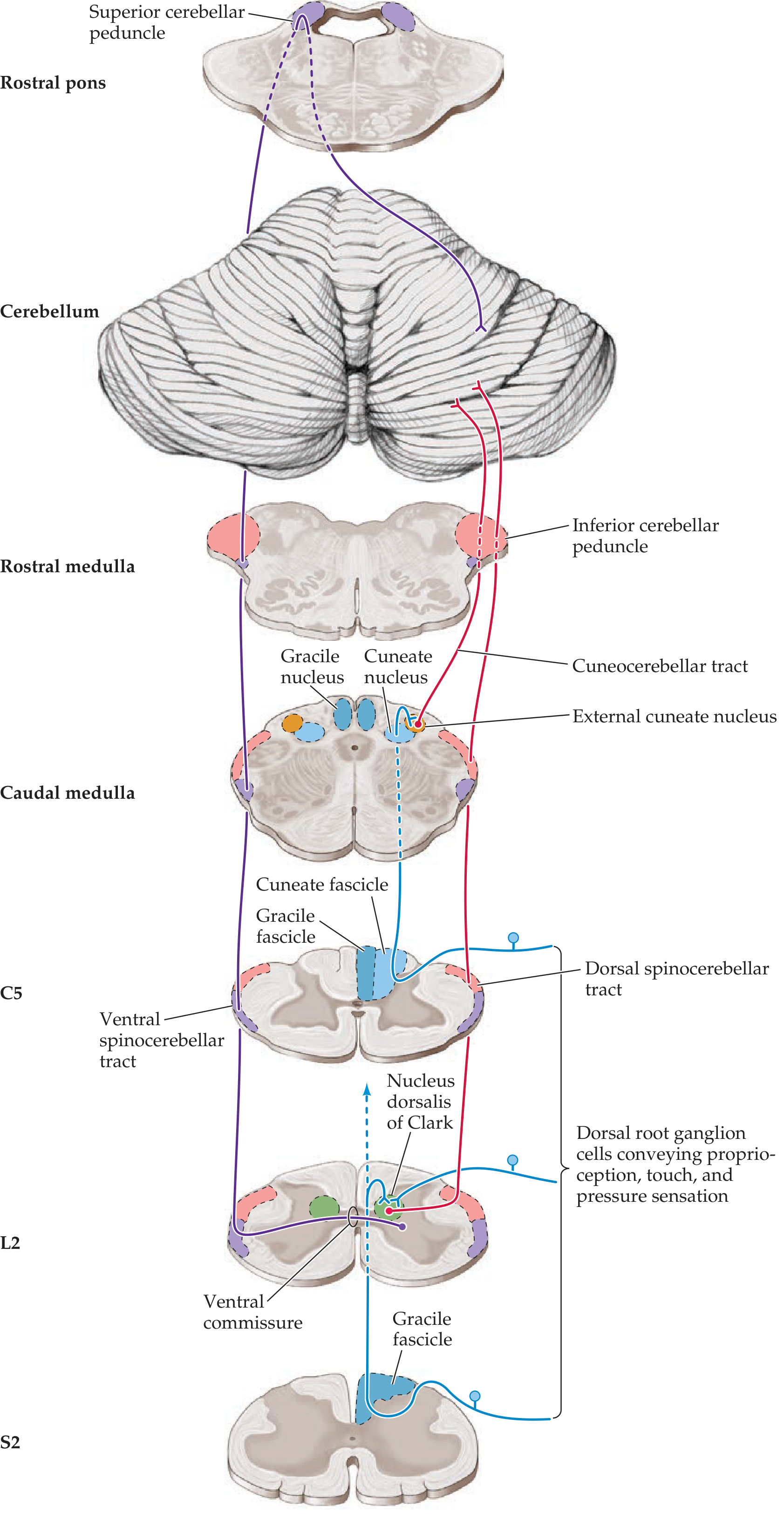
- At L2: the Nucleus dorsalis of Clark is clearly shown (green) as the relay for the dorsal spinocerebellar tract (red/blue)
- At S2: the ventral spinocerebellar tract (purple) originates from scattered cells at the periphery of the gray matter, crosses via the ventral commissure, and ascends to the superior cerebellar peduncle (top left, purple)
To Directly Answer Your Question
Right crura arises from right side of anterior surface of bodies of L1 to L3 vertebrae and corresponding IV disc or IV disc. What does it mean IV disc?
- The anterior surface of the bodies of L1, L2, and L3 vertebrae
- AND the intervertebral discs between those vertebrae (i.e., the L1-L2 disc and the L2-L3 disc)