
Diabetes for endocrine complete detail
Reading File
Finding Sources
Finding Sources
Finding Sources
Finding Sources
Finding Sources
Finding Sources
Finding Sources
Now I have the key sections. Let me read the main Harrison's diabetes block (block45) which has the most comprehensive coverage, and also the Goldman-Cecil comprehensive DM section.
Reading File
The file starts with obesity. The diabetes section starts around line 497 (GDM) and 1047 (Type 1). Let me read from the diabetes-specific sections:
Reading File
Excellent — I now have extremely rich content. Let me read more of this section (the treatment, complications portions) and the pathology section for histology:
Reading File
Searching Images
diabetes mellitus islets of Langerhans pancreas histology type 1 type 2
Reading File
Searching Images
diabetic retinopathy nephropathy neuropathy complications
I now have very comprehensive material. Let me read the earlier sections covering definition, epidemiology, pathophysiology, and diagnosis more thoroughly:
Reading File
I have very comprehensive data from Harrison's. Let me now also look at block44 for the definition/intro section and block46 for more clinical data:
Reading File
That was a different section. I already have extremely comprehensive content from the block45 read. Let me now compile all the information into a complete, well-structured answer.# Diabetes Mellitus — Complete Endocrine Review
Source: Harrison's Principles of Internal Medicine 22E (2025), Goldman-Cecil Medicine, Robbins Pathology
1. DEFINITION & OVERVIEW
Diabetes mellitus (DM) is a group of metabolic diseases characterized by hyperglycemia resulting from defects in insulin secretion, insulin action, or both. Chronic hyperglycemia causes long-term damage, dysfunction, and failure of various organs — especially the eyes, kidneys, nerves, heart, and blood vessels.
The global burden is staggering: in 2021, the IDF estimated ~537 million adults worldwide had DM, with ~90% having Type 2. DM accounts for roughly 1.5 million deaths/year directly and is the leading cause of new-onset blindness (ages 20–74), end-stage renal disease, and non-traumatic lower-limb amputations.
2. CLASSIFICATION
| Type | Key Feature |
|---|---|
| Type 1 DM (T1DM) | Autoimmune β-cell destruction → absolute insulin deficiency |
| Type 2 DM (T2DM) | Range from predominant insulin resistance + relative deficiency to secretory defect |
| Other specific types | Monogenic (MODY), endocrinopathies, drug-induced, infections, etc. |
| Gestational DM (GDM) | Glucose intolerance first diagnosed in 2nd/3rd trimester of pregnancy |
Other Specific Causes (Type III)
- Genetic defects of β-cell function (MODY): Mutations in HNF-4α, glucokinase (GCK), HNF-1α, insulin promoter factor-1, HNF-1β, NeuroD1, KLF11, PAX4, GATA4/6, GLIS3, mitochondrial DNA
- Neonatal DM: Mutations in ATP-sensitive K⁺ channel subunits (KATP), RFX6, insulin gene (onset <6 months)
- Pancreatic disease: Pancreatitis, pancreatectomy, cystic fibrosis, hemochromatosis, fibrocalculous pancreatopathy
- Endocrinopathies: Acromegaly (GH excess), Cushing's syndrome (glucocorticoid excess), glucagonoma, pheochromocytoma, hyperthyroidism, somatostatinoma, aldosteronoma
- Drug/chemical-induced: Glucocorticoids, calcineurin & mTOR inhibitors, pentamidine, nicotinic acid, statins, thiazides, antipsychotics (second-generation), PCSK9 inhibitors, antiretrovirals
- Infections: Congenital rubella, CMV, coxsackievirus
- Immune-mediated: Stiff-person syndrome, anti-insulin receptor antibodies, checkpoint inhibitor therapy
- Genetic syndromes: Down syndrome, Turner syndrome, Klinefelter syndrome, Wolfram syndrome, Prader-Willi syndrome, Friedreich's ataxia
3. NORMAL PANCREATIC ENDOCRINOLOGY
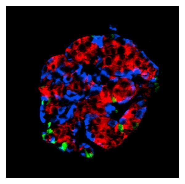
The islets of Langerhans (~1 million in the human pancreas) contain:
- β-cells (~65–80%): secrete insulin and C-peptide and amylin (IAPP)
- α-cells (~15–20%): secrete glucagon
- δ-cells (~5–10%): secrete somatostatin
- PP cells: secrete pancreatic polypeptide
Insulin Secretion
Glucose enters β-cells via GLUT2, is metabolized → ATP rises → KATP channels close → membrane depolarization → voltage-gated Ca²⁺ channels open → Ca²⁺ influx → exocytosis of insulin granules. First-phase insulin release (rapid, within 2 min) is lost early in T2DM.
4. TYPE 1 DIABETES MELLITUS
Pathophysiology
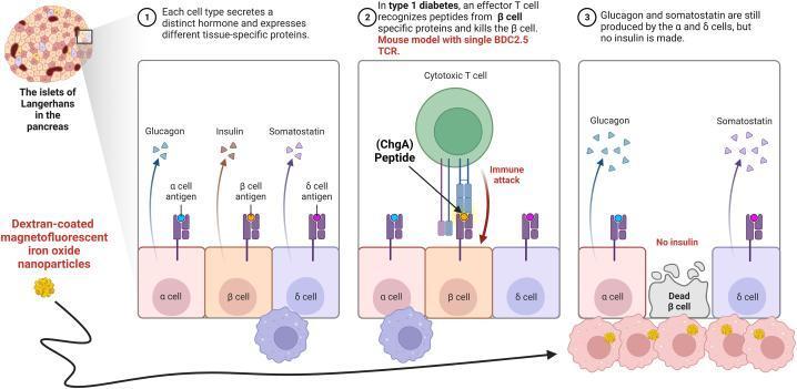
T1DM results from autoimmune destruction of pancreatic β-cells. Key features:
- Selective β-cell destruction by autoreactive CD8⁺ cytotoxic T cells (insulitis); α and δ cells remain intact
- HLA associations: DR3-DQ2 and DR4-DQ8 haplotypes confer highest risk; HLA accounts for ~50% of genetic risk
- Non-HLA genes: INS (insulin gene), PTPN22, CTLA4, IL2RA, IFIH1, etc.
- Concordance in identical twins: ~50% → environmental triggers required
- Autoantibodies (appear years before clinical onset): anti-GAD65, anti-IA-2, anti-ZnT8, anti-insulin (IAA), ICA (islet cell antibodies)
- Absolute insulin deficiency → tendency toward diabetic ketoacidosis (DKA)
Staging (ADA 2024)
- Stage 1: ≥2 autoantibodies, normoglycemia, presymptomatic
- Stage 2: ≥2 autoantibodies + dysglycemia (IFG and/or IGT), presymptomatic
- Stage 3: Clinical hyperglycemia, symptomatic
Clinical Presentation
- Peak onset: childhood/adolescence, but can occur at any age (incl. LADA in adults)
- Classic triad: polyuria, polydipsia, weight loss
- Often acute/abrupt onset, may present with DKA
- DKA precipitants: infection, missed insulin, new-onset T1DM
5. TYPE 2 DIABETES MELLITUS
Pathophysiology
T2DM results from a combination of:
- Insulin resistance (peripheral tissues — muscle, liver, adipose)
- Relative insulin deficiency (β-cell secretory failure)
- Incretin defect: Reduced GLP-1 and GIP response to meals
- Increased glucagon secretion from α-cells
- Increased renal glucose reabsorption (upregulated SGLT2)
- Central appetite dysregulation
- Gut microbiome alterations
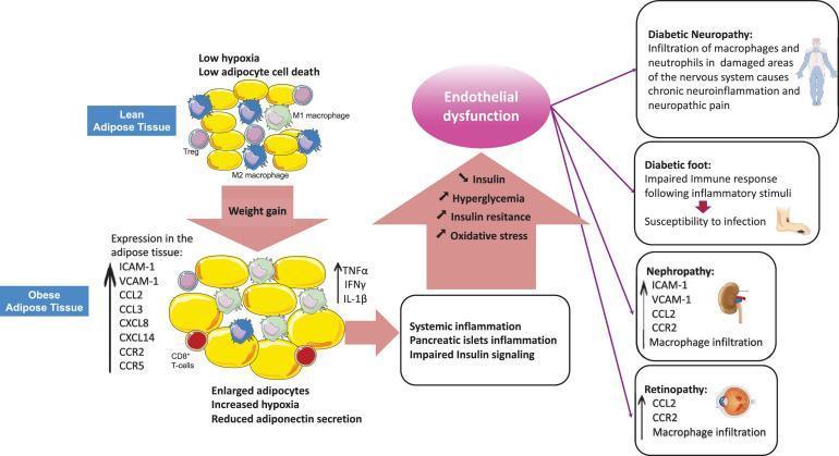
Insulin Resistance Mechanism
- Obesity → hypertrophied adipocytes → hypoxia → M1 macrophage infiltration → pro-inflammatory cytokines (TNF-α, IL-1β, IL-6, IFN-γ) → impaired insulin receptor signaling (IRS-1 phosphorylation at serine instead of tyrosine)
- Ectopic fat in liver (hepatic steatosis) and muscle → diacylglycerol and ceramide accumulation → PKC activation → impaired insulin signaling
β-cell Failure
- Progressive loss of β-cell mass over time
- Deposition of amyloid (IAPP/amylin) in islets — characteristic histology of T2DM
- Glucotoxicity and lipotoxicity worsen β-cell function ("exhaustion")
Risk Factors
- Obesity (BMI >25), especially central/visceral adiposity
- Physical inactivity
- Family history (genetic predisposition is polygenic)
- Age >45
- Prior GDM or prediabetes (IFG/IGT)
- Polycystic ovary syndrome (PCOS)
- Ethnic background (Asian, South Asian, Hispanic, Black populations at higher risk)
6. GESTATIONAL DIABETES (GDM)
- Glucose intolerance in the 2nd or 3rd trimester due to pregnancy-related insulin resistance (placental hormones: HPL, progesterone, cortisol)
- IDF 2021: affects ~16% of all pregnancies worldwide
- Risk factors: obesity, prior GDM, family history, PCOS
- Complications: macrosomia, shoulder dystocia, neonatal hypoglycemia, pre-eclampsia
- Mother and child have ↑ lifetime risk of T2DM
- Screening: 75g OGTT at 24–28 weeks
- Diabetes diagnosed in 1st trimester = preexisting (pregestational) DM
7. DIAGNOSIS
| Test | Normal | Prediabetes | Diabetes |
|---|---|---|---|
| Fasting plasma glucose (FPG) | <5.6 mmol/L (<100 mg/dL) | 5.6–6.9 mmol/L (IFG) | ≥7.0 mmol/L (≥126 mg/dL) |
| 2-hr plasma glucose (OGTT 75g) | <7.8 mmol/L (<140 mg/dL) | 7.8–11.0 mmol/L (IGT) | ≥11.1 mmol/L (≥200 mg/dL) |
| HbA1c | <5.7% | 5.7–6.4% | ≥6.5% |
| Random plasma glucose + symptoms | — | — | ≥11.1 mmol/L + classic symptoms |
In the absence of classic hyperglycemic symptoms, diagnosis requires two abnormal tests (same or different assays), or confirmation on a repeat test.
HbA1c
- Reflects mean glucose over preceding 8–12 weeks (RBC lifespan ~120 days)
- Each 1% rise in HbA1c ≈ 1.6 mmol/L (29 mg/dL) rise in mean plasma glucose
- Affected by conditions altering RBC turnover: hemolytic anemia, sickle cell disease, pregnancy → use FPG or OGTT instead
8. PATHOLOGY (Histology)
Type 1 DM
- Insulitis: Lymphocytic infiltration (CD4⁺ and CD8⁺ T cells) of islets
- Selective β-cell destruction; α, δ, PP cells preserved
- In established T1DM: islets appear small and atrophic; β-cells absent
Type 2 DM
- Amyloid deposition in islets (composed of IAPP/amylin) — appears as waxy eosinophilic material on H&E; Congo red positive, birefringent under polarized light
- Mild reduction in β-cell mass (~50% at diagnosis)
- No significant insulitis
9. PATHOPHYSIOLOGY OF COMPLICATIONS
Central mechanism: Chronic hyperglycemia → cellular damage via multiple pathways:
- Advanced Glycation End-Products (AGEs): Non-enzymatic glycosylation of intra- and extracellular proteins → cross-linking → glomerular dysfunction, endothelial dysfunction, atherosclerosis; AGE examples: pentosidine, glucosepane, carboxymethyllysine
- Polyol pathway: Aldose reductase converts glucose → sorbitol (osmotic damage to nerves, lens)
- PKC activation: Diacylglycerol accumulation → PKC → altered gene transcription, VEGF upregulation
- Hexosamine pathway: Excess glucose → glucosamine → altered protein function via O-GlcNAcylation
- Oxidative stress: Mitochondrial superoxide production → activates all above pathways (unifying mechanism)
Epigenetic changes: Hyperglycemia induces epigenetic modifications affecting gene expression in target cells — may explain "metabolic memory."
10. CHRONIC COMPLICATIONS
A. Microvascular Complications
1. Diabetic Retinopathy
Leading cause of new blindness in adults aged 20–74 in the US.
| Stage | Features |
|---|---|
| Non-proliferative (NPDR) | Microaneurysms, blot hemorrhages, cotton-wool spots; altered venous caliber; intraretinal microvascular abnormalities (IRMA) |
| Proliferative (PDR) | Neovascularization at optic nerve/macula; vitreous hemorrhage; fibrosis; retinal detachment |
| Macular edema | Can occur at any stage; leading cause of moderate visual loss; detectable by OCT/FA |

- Pathomechanism: Loss of retinal pericytes → increased vascular permeability → retinal ischemia → VEGF-A upregulation → neovascularization
- Management: Tight glycemic control, BP control; laser photocoagulation (NPDR → PDR); intravitreal anti-VEGF (ranibizumab, bevacizumab, aflibercept) for PDR and macular edema; vitrectomy for vitreous hemorrhage/tractional detachment
- Screening: Annual dilated fundoscopy (T1DM: start 5 years after diagnosis; T2DM: at diagnosis)
2. Diabetic Nephropathy (Diabetic Kidney Disease — DKD)
Leading cause of end-stage renal disease (ESRD) in the US.
Stages (Mogensen classification):
- Hyperfiltration (↑GFR)
- Silent (normal albumin)
- Incipient nephropathy: microalbuminuria (30–300 mg/day)
- Overt nephropathy: macroalbuminuria (>300 mg/day), declining GFR
- ESRD: GFR <15 mL/min
Histology: Glomerular basement membrane (GBM) thickening, mesangial expansion, Kimmelstiel-Wilson nodules (nodular glomerulosclerosis — pathognomonic), afferent and efferent arteriolar hyalinosis
Management: ACE inhibitors/ARBs (first-line for proteinuria/HTN), SGLT2 inhibitors (finerenone/empagliflozin — renoprotective effects), tight glycemic control, BP target <130/80 mmHg
3. Diabetic Neuropathy
Most common complication; affects ~50% of patients with long-standing DM.
| Type | Features |
|---|---|
| Distal symmetric sensorimotor polyneuropathy | Most common; "stocking-glove" loss of vibration, proprioception, pain, temperature → foot ulcers/Charcot joint |
| Autonomic neuropathy | Gastroparesis, orthostatic hypotension, erectile dysfunction, neurogenic bladder, sudomotor dysfunction, cardiac (↓HRV) |
| Painful diabetic neuropathy | Burning, shooting, stabbing pain — predominantly nocturnal |
| Mononeuropathy | Cranial nerve palsies (CN III most common), carpal tunnel syndrome, mononeuritis multiplex |
| Radiculopathy/plexopathy | Thoracic radiculopathy, lumbosacral plexopathy (diabetic amyotrophy) |
Management of peripheral neuropathy: Pregabalin, duloxetine, gabapentin, TCAs; capsaicin; foot care, regular podiatric exams
Management of gastroparesis: Metoclopramide, domperidone, erythromycin; dietary modifications
B. Macrovascular Complications
Diabetes doubles the risk of cardiovascular disease; CVD accounts for ~50% of deaths in T2DM.
- Mechanisms: Hyperglycemia + insulin resistance → dyslipidemia (↑TG, ↓HDL, small dense LDL) + hypertension + chronic inflammation + endothelial dysfunction → accelerated atherosclerosis
- Adipose lipolysis → excess fatty acid delivery → ectopic triglyceride/diacylglycerol/ceramide accumulation in liver, muscle, heart → lipotoxicity
| Complication | Features |
|---|---|
| Coronary artery disease | Leading cause of death; often silent (impaired pain sensation); more diffuse/multivessel disease |
| Cerebrovascular disease | 2–4× ↑ stroke risk |
| Peripheral arterial disease (PAD) | Claudication → critical limb ischemia → amputation |
| Diabetic cardiomyopathy | Cardiac dysfunction independent of CAD/HTN |
CV risk reduction: Statins (all DM patients >40 yr), aspirin (secondary prevention), ACEI/ARB, BP <130/80, smoking cessation, GLP-1 RA (cardiovascular benefit proven: liraglutide, semaglutide), SGLT2 inhibitors (empagliflozin, canagliflozin — reduce MACE, heart failure hospitalizations, cardiovascular death)
11. ACUTE COMPLICATIONS
A. Diabetic Ketoacidosis (DKA)
Primarily T1DM (rare in T2DM); life-threatening.
Precipitants: Infection (most common), missed insulin, new-onset T1DM, MI, surgery, trauma, drugs (glucocorticoids, SGLT2i in T1DM)
Pathophysiology: Absolute/relative insulin deficiency + glucagon excess → unchecked lipolysis → excess FFA → hepatic ketogenesis (acetoacetate, β-hydroxybutyrate, acetone) → high anion-gap metabolic acidosis
Diagnostic criteria:
- Blood glucose usually >250 mg/dL (though "euglycemic DKA" possible with SGLT2i use)
- pH <7.3 (or bicarbonate <18 mEq/L)
- Ketonemia/ketonuria (serum β-hydroxybutyrate ≥3 mmol/L)
- Anion gap >12
Clinical features: Nausea/vomiting, abdominal pain, Kussmaul respiration, fruity/acetone breath, altered consciousness (severe), dehydration
Management:
- IV fluids (0.9% NaCl initially — correct dehydration)
- Insulin infusion (regular insulin 0.1 U/kg/h; do NOT give before K⁺ ≥3.5 mEq/L)
- Potassium replacement (always needed — insulin drives K⁺ into cells)
- Bicarbonate only if pH <6.9 (controversial)
- Monitor: glucose hourly, BMP q2–4h, AG, urine output
- Treat precipitant
- Transition to SC insulin when AG normalized, patient tolerating PO
B. Hyperglycemic Hyperosmolar State (HHS)
Primarily T2DM; very high mortality (~15%).
- Blood glucose markedly elevated (typically >600 mg/dL)
- Serum osmolality >320 mOsm/kg (often >350)
- Minimal or no ketoacidosis (enough endogenous insulin to prevent ketosis)
- Profound dehydration, neurologic changes (focal deficits, seizures, coma)
- Management: aggressive fluid replacement (often 8–10 L deficit), slower insulin infusion, electrolyte monitoring
C. Hypoglycemia
The major limiting factor in achieving tight glycemic control.
- Plasma glucose <70 mg/dL (threshold for symptoms); severe <54 mg/dL
- Causes in DM: Excess insulin/secretagogues, skipped meals, alcohol, exercise, renal failure (↓ insulin clearance)
- Symptoms: Adrenergic (diaphoresis, tremor, palpitations, anxiety — at ~60 mg/dL) → neuroglycopenic (confusion, seizure, coma — at ~50 mg/dL)
- Hypoglycemia unawareness: Loss of adrenergic warning symptoms after recurrent hypoglycemia (autonomic failure)
- Management: 15g oral fast-acting glucose (glucose tablets/juice) → recheck in 15 min ("15-15 rule"); glucagon IM/SC or IV dextrose if unconscious
12. GLYCEMIC TARGETS
| Parameter | ADA Target (Most Adults) |
|---|---|
| HbA1c | <7% (more stringent <6.5% if safely achievable; less stringent <8% for elderly/comorbid) |
| Fasting/preprandial glucose | 80–130 mg/dL (4.4–7.2 mmol/L) |
| Peak postprandial glucose | <180 mg/dL (<10 mmol/L) |
| Time in Range (TIR 70–180) | >70% |
13. MANAGEMENT
A. Non-Pharmacologic
- Lifestyle modification: Cornerstone of T2DM prevention and treatment
- Medical nutrition therapy: Low glycemic index foods, reduced saturated fat, adequate fiber
- Physical activity: ≥150 min/week moderate aerobic exercise + resistance training
- Weight loss: Even 5–10% weight loss significantly improves glycemic control
- Patient education and self-monitoring of blood glucose (SMBG) / CGM
- Bariatric surgery: For BMI >35 with T2DM; 68.2% achieve complete remission within 5 years; Swedish Obese Subjects study showed 78% reduction in T2DM incidence
B. Insulin Therapy
| Type | Onset | Peak | Duration | Examples |
|---|---|---|---|---|
| Rapid-acting | 10–30 min | 0.5–3 hr | 3–5 hr | Lispro, Aspart, Glulisine |
| Short-acting (Regular) | 30–60 min | 2–4 hr | 5–8 hr | Regular insulin (Humulin R) |
| Intermediate-acting (NPH) | 1–2 hr | 4–6 hr | 10–16 hr | NPH (Humulin N) |
| Long-acting (Basal) | 1–2 hr | Flat/minimal | 20–24+ hr | Glargine, Detemir, Degludec |
| Ultra-long-acting | 0.5–1.5 hr | Flat | >42 hr | Degludec (iDegLira) |
| Premixed | Biphasic | — | — | 70/30, 75/25, 50/50 |
Intensive insulin regimens: Basal-bolus (1 long-acting + 3 rapid-acting pre-meal) = most physiologic; Insulin pump (CSII) = continuous subcutaneous infusion of rapid-acting insulin
Complications of insulin therapy: Hypoglycemia, weight gain (~2–4 kg), lipohypertrophy (rotate injection sites), rarely lipodystrophy or local allergy
C. Non-Insulin Pharmacotherapy (T2DM)
| Drug Class | Mechanism | HbA1c ↓ | Key Benefits | Key Risks |
|---|---|---|---|---|
| Metformin | ↓Hepatic glucose production (↓gluconeogenesis via AMPK/Complex I mitochondrial inhibition) | 1–2% | First-line; weight neutral/↓; cheap; CV neutral; no hypoglycemia; may ↓ cancer risk | GI side effects; lactic acidosis (rare; hold for contrast/surgery/eGFR<30); B12 deficiency |
| SGLT2 inhibitors (empagliflozin, dapagliflozin, canagliflozin) | ↓Renal glucose reabsorption (glycosuria) | 0.5–1% | ↓CV events (MACE), ↓HF hospitalization, ↓CKD progression, weight loss, ↓BP | UTI, genital mycotic infections, DKA (rare), Fournier's gangrene (rare), volume depletion, ↑ amputations (canagliflozin) |
| GLP-1 Receptor Agonists (liraglutide, semaglutide, dulaglutide, exenatide) | Glucose-dependent insulin secretion, ↓glucagon, slows gastric emptying, central satiety | 0.8–1.5% | ↓CV events (MACE), weight loss, ↓BP; weekly dosing available (semaglutide) | Nausea/vomiting (GI side effects), pancreatitis (rare), C-cell hyperplasia (rodent), injection site reactions |
| DPP-4 inhibitors (sitagliptin, saxagliptin, linagliptin) | ↑Endogenous GLP-1 and GIP by inhibiting DPP-4 enzyme | 0.5–0.8% | Weight neutral; well tolerated; CV neutral | Nasopharyngitis, UTI; HF risk (saxagliptin); joint pain |
| Sulfonylureas (glipizide, glyburide, glimepiride) | Close KATP channels on β-cells → ↑insulin secretion (glucose-independent) | 1–2% | Cheap; rapid action | Hypoglycemia (especially glyburide); weight gain; ↓effect over time |
| Thiazolidinediones (pioglitazone, rosiglitazone) | PPAR-γ agonist → ↑insulin sensitivity, ↓hepatic fat | 0.5–1.4% | Pioglitazone: ↓TG, ↑HDL; ↓recurrent stroke (IRIS trial) | Weight gain (2–3 kg), edema, CHF (CI in class III/IV), fractures (postmenopausal women), bladder cancer (pioglitazone), macular edema |
| Meglitinides (repaglinide, nateglinide) | Close KATP channels (short-acting → taken with meals) | 0.5–1.5% | Flexible dosing; useful in irregular meal patterns | Hypoglycemia; weight gain; frequent dosing |
| α-Glucosidase inhibitors (acarbose, miglitol) | ↓Intestinal glucose absorption (delay oligosaccharide breakdown) | 0.5–0.8% | ↓Postprandial glucose spike; weight neutral | GI (flatulence, diarrhea, bloating); CI if IBD, gastroparesis, Cr >2 |
| Pramlintide | Synthetic amylin analog → ↓glucagon, slows gastric emptying | 0.5–0.7% | Used with insulin; weight neutral/↓ | Nausea; hypoglycemia with insulin dose reduction needed |
| Colesevelam | Bile acid sequestrant → ↓hepatic glucose production | 0.5% | Also ↓LDL | GI; drug interactions |
| Bromocriptine | Dopamine agonist → ↑insulin sensitivity | 0.5% | Uncertain role | Nausea, dizziness, orthostatic hypotension |
SGLT2 Inhibitor Key Trials
- EMPA-REG OUTCOME (empagliflozin): ↓MACE 14%, ↓CV death 38%, ↓HF hospitalization 35%
- CANVAS (canagliflozin): ↓MACE, renal outcomes; ↑amputations
- DECLARE-TIMI 58 (dapagliflozin): ↓HF hospitalization, ↓CKD progression
- CREDENCE/DAPA-CKD: Significant renoprotection even without CV disease
GLP-1 RA Key Trials
- LEADER (liraglutide): ↓MACE 13%, ↓CV death, ↓renal outcomes
- SUSTAIN-6 (semaglutide SC): ↓MACE 26%
- REWIND (dulaglutide): ↓MACE, primary prevention population
D. Type 1 DM Immunotherapy
- Teplizumab (anti-CD3 monoclonal antibody): Approved 2022 by FDA to delay Stage 3 T1DM in high-risk Stage 2 patients — delays onset by ~2 years. Mechanism: tolerizes autoreactive T cells
14. MONITORING
| Parameter | Method | Frequency |
|---|---|---|
| Short-term glucose | SMBG (glucometer) / CGM (continuous glucose monitor) | As needed; CGM preferred in T1DM |
| Long-term glycemic control | HbA1c | Every 3 months (if not at goal) or every 6 months |
| Renal function | Urine albumin-to-creatinine ratio (ACR) + serum creatinine/eGFR | Annually |
| Retinal screening | Dilated fundoscopy / retinal photography | Annually (T1DM: 5 yr after dx; T2DM: at dx) |
| Foot exam | Monofilament + vibration | Annually |
| Lipids | Fasting lipid panel | Annually |
| Blood pressure | At every visit | Goal: <130/80 mmHg |
| Thyroid (T1DM) | TSH | At dx, then periodically (associated autoimmune disease) |
15. SPECIAL SITUATIONS
Diabetes in Pregnancy (Pregestational DM)
- Congenital anomalies (cardiac, neural tube) linked to periconceptional hyperglycemia — preconception HbA1c <7% target
- Pre-eclampsia, macrosomia, stillbirth risk ↑
- Only insulin approved during pregnancy (metformin/glyburide used off-label in GDM)
- Target: HbA1c <6–6.5% during pregnancy (if achievable without hypoglycemia)
Sick-Day Rules
- Never stop insulin in T1DM during illness
- Monitor glucose every 2–4 hours; check ketones if >250 mg/dL
- "Hold" metformin and SGLT2i during acute illness (dehydration/contrast risk)
Perioperative Diabetes Management
- Target glucose 140–180 mg/dL intraoperatively
- Hold oral hypoglycemics on day of surgery; insulin drip for major procedures
16. PREDIABETES AND PREVENTION
- Prediabetes (IFG or IGT or HbA1c 5.7–6.4%): affects ~374 million adults globally
- Diabetes Prevention Program (DPP): Intensive lifestyle modification reduced T2DM incidence by 58% (vs. 31% with metformin) over 3 years
- Metformin: indicated for prevention in prediabetes especially with BMI >35, age <60, prior GDM
- GLP-1 RAs (semaglutide) and SGLT2i also being studied for prevention
17. SUMMARY — KEY DISTINGUISHING FEATURES
| Feature | T1DM | T2DM |
|---|---|---|
| Age of onset | Usually <30 yr (but any age) | Usually >40 yr (but rising in youth) |
| Onset | Acute | Gradual |
| Body habitus | Normal/thin | Obese (usually) |
| Insulin levels | Absent/very low | Normal/high early → low late |
| C-peptide | Low/absent | Present |
| Autoantibodies | Positive (GAD65, IA-2, ZnT8, IAA) | Negative |
| HLA association | Strong (DR3/DR4) | Weak |
| DKA | Common | Rare (HHS more common) |
| Treatment | Insulin always required | Lifestyle → oral agents → insulin |
| Islet histology | Insulitis → atrophic islets | Amyloid (IAPP) deposition |
*Sources: Harrison's Principles of Internal Medicine 22E (2025), pp. 3227–3292; Goldman-Cecil Medicine; Robbins & Kumar Basic Pathology; Goodman & Gilman's Pharmacological Basis of Therapeutics.*This is a comprehensive endocrine review of Diabetes Mellitus covering all major domains. Here is a quick orientation to the 17 sections above:
What's covered:
| Section | Topic |
|---|---|
| 1 | Definition & global burden |
| 2 | Full classification (T1, T2, MODY, other specific types, GDM) |
| 3 | Normal pancreatic endocrinology (β-cell insulin secretion mechanism) |
| 4 | T1DM — autoimmune pathophysiology, HLA genetics, staging, autoantibodies |
| 5 | T2DM — insulin resistance, β-cell failure, "Ominous Octet," risk factors |
| 6 | Gestational DM — screening, complications, management |
| 7 | Diagnostic criteria — FPG, OGTT, HbA1c thresholds (ADA standards) |
| 8 | Pathology/histology — insulitis (T1DM), amyloid/IAPP deposition (T2DM) |
| 9 | Mechanisms of chronic complications — AGEs, polyol pathway, PKC, oxidative stress |
| 10 | Chronic complications — retinopathy, nephropathy (Kimmelstiel-Wilson), neuropathy, macrovascular disease |
| 11 | Acute complications — DKA (management protocol), HHS, hypoglycemia |
| 12 | Glycemic targets (HbA1c <7%, TIR >70%) |
| 13 | Management — lifestyle, insulin types, all oral/injectable drug classes with mechanisms, key CV outcomes trials |
| 14 | Monitoring schedule |
| 15 | Special situations — pregnancy, sick-day rules, perioperative care |
| 16 | Prediabetes & prevention (DPP trial — 58% risk reduction) |
| 17 | T1DM vs T2DM distinguishing features (comparison table) |
This is a shared conversation. Sign in to Orris to start your own chat.