
Bed side pulmonary function tests full details for md exams 10 marks answer
Reading File
Now let me search the medical library and web simultaneously for comprehensive information on bedside pulmonary function tests.
Finding Sources
Finding Sources
Searching the Web
bedside pulmonary function tests spirometry peak flow MD exam detailed notes
Now let me search for more specific content from the medical library on spirometry and bedside tests, and extract the slideshare content:
Finding Sources
Finding Sources
Reading Web Page
https://www.slideshare.net/slideshow/bedside-pulmonary-funct…
Now let me read the relevant textbook sections for detailed content:
Reading File
Reading File
Reading File
Reading File
Now let me fetch a key spirometry diagram image and search for more on Snider's test, FET, and other bedside tests:
Loading Image
Finding Sources
Finding Sources
I now have comprehensive information to write a complete MD exam answer. Let me compile everything:
Bedside Pulmonary Function Tests - Full Details (MD Exam 10 Marks Answer)
Definition and Introduction
Pulmonary Function Tests (PFTs) are objective measurements used to assess the functional capacity of the lungs, characterize pulmonary dysfunction, determine severity of lung disease, monitor treatment response, and evaluate patients before surgery. Bedside PFTs are those that can be performed at the patient's bedside without specialized laboratory equipment.
(Bailey & Love's Short Practice of Surgery, 28th Ed., p. 998)
Classification of Bedside PFTs
Bedside pulmonary function tests include the following:
- Snider's Match Blowing Test
- Forced Expiratory Time (FET)
- Seberese's Single Breath Count
- Seberese's Breath Holding Test
- Cough Test
- DeBono's Whistle Blowing Test
- Wright's Peak Flow Meter (PEFR measurement)
- Spirometry with Pocket-Sized Spirometer
1. Snider's Match Blowing Test
Principle: Tests the ability to blow out a lighted match held at a distance from the mouth, correlating to forced expiratory flow.
Method:
- Patient takes a maximal inspiration
- A lighted match is held 15 cm (6 inches) in front of the open mouth
- Patient blows as hard as possible (mouth wide open, not pursed lips)
- Observation: whether the flame is extinguished
Interpretation:
- Positive (Able to blow out): FEV1 > 1.6 L (adequate respiratory reserve)
- Negative (Unable to blow out): FEV1 < 1.6 L - suggests significant airflow obstruction
- If patient cannot blow it out at even 6 cm, FEV1 is likely < 1 L - high operative risk
- The test is effort dependent
Clinical significance: Used in preoperative assessment. If patient cannot blow out the match at 15 cm, they are at increased risk for postoperative pulmonary complications.
2. Forced Expiratory Time (FET)
Principle: The time taken to forcibly exhale the lungs from full inspiration to full expiration (equivalent to the time to exhale all of FVC). Normally corresponds to the spirometric forced expiration.
Method:
- Patient takes a maximal deep inspiration
- Exhales as hard and fast as possible
- Examiner auscultates over the trachea with a stethoscope and times the duration of audible expiratory airflow
Interpretation:
- Normal: < 3 seconds (some say < 4 seconds)
- 3-6 seconds: Borderline / mild obstruction (FEV1/FVC ~ 70%)
- > 6 seconds: Significant obstructive pattern - correlates with FEV1/FVC < 50%
Significance: Prolonged FET is a reliable bedside indicator of obstructive airway disease (asthma, COPD, emphysema).
3. Seberese's Single Breath Count (SBC)
Principle: Measures the maximum number of digits a patient can count aloud in one breath after maximal inspiration. This correlates with vital capacity.
Method:
- Patient takes a maximal deep breath
- Counts aloud in a normal conversational tone (1, 2, 3, 4...) until they run out of breath
Interpretation:
- Normal: > 30 (correlates with VC > 2 L)
- 15-30: Reduced VC (~1-2 L)
- < 15: Severely reduced VC (< 1 L) - high surgical risk
- Each count approximately corresponds to ~100 mL of vital capacity
Significance: Quick, non-invasive proxy for vital capacity. Used preoperatively to stratify pulmonary risk.
4. Seberese's Breath Holding Test
Principle: Tests respiratory reserve and cardiovascular reserve by measuring how long a patient can voluntarily hold their breath after maximal inspiration (Valsalva maneuver).
Method:
- Patient breathes normally for a few cycles
- Takes a maximal deep breath and holds it as long as possible
- Time from full inspiration to next breath is recorded
Interpretation:
- Normal: > 25-30 seconds
- 15-25 seconds: Fair reserve (moderate risk)
- < 15 seconds: Poor reserve - high operative risk, likely significant cardiorespiratory compromise
Significance: Breath hold time is affected by both respiratory and cardiovascular reserve, making it a quick combined screening tool.
5. Cough Test
Principle: Evaluates whether a patient can generate an effective cough, which is important for postoperative airway clearance.
Normal effective cough requirements:
- FVC > 20 mL/kg
- FEV1 > 15 mL/kg
- PEFR > 200 L/min
- Vital capacity should be approximately 3x the tidal volume for effective cough generation
Interpretation:
- If patient can produce a strong, sharp cough: adequate cough mechanism
- Wet productive cough / self-propagating paroxysms of coughing: suggests susceptibility to pulmonary complications
- Inadequate cough = PEFR < 200 L/min - high risk of postoperative atelectasis and pneumonia
Significance: Poor cough efficiency predicts inability to clear secretions postoperatively.
6. DeBono's Whistle Blowing Test
Principle: Measures PEFR indirectly using a variable-resistance whistle device.
Method:
- Patient blows down a wide-bore tube at the end of which is a whistle
- On the side is a hole with an adjustable knob (leak port)
- As the patient blows, the whistle sounds; the leak hole is gradually increased until the whistle sound disappears
- The last position at which the whistle can still be blown is read off a scale
Interpretation: Provides a semi-quantitative estimate of PEFR.
Significance: Simple alternative to the Wright Peak Flow Meter for measuring PEFR at the bedside.
7. Wright's Peak Flow Meter (PEFR Measurement)
Principle: Measures Peak Expiratory Flow Rate (PEFR) - the maximum airflow velocity achievable during a forced expiratory maneuver from total lung capacity.
(Bailey & Love's, p. 998; Roberts & Hedges' Clinical Procedures in Emergency Medicine)
Equipment: The "mini-Wright" peak flow meter - a handheld, portable device.
Method:
- Patient stands or sits upright
- Takes a maximal deep inspiration
- Seals lips around the mouthpiece
- Exhales as hard and fast as possible
- Best of 3 attempts is recorded
Normal Values:
- Males: 450-700 L/min
- Females: 300-500 L/min
Interpretation (Traffic Light System - used in Asthma):
| Zone | PEFR (% predicted) | Action |
|---|---|---|
| Green | > 80% | Good control |
| Yellow | 50-80% | Caution required |
| Red | < 50% | Emergency |
Severity classification for surgical risk:
- PEFR < 200 L/min: impaired cough efficiency - significant risk
Clinical uses:
- Monitoring asthma control and response to bronchodilators
- Diagnosing airflow obstruction
- Preoperative pulmonary assessment
- Differentiating COPD from CHF (limited utility)
Limitations:
- Effort dependent
- Affected by thoracic/abdominal wound pain
- Only tests large airway patency (does not detect small airway disease)
- Does not measure FVC or provide flow-volume loop data
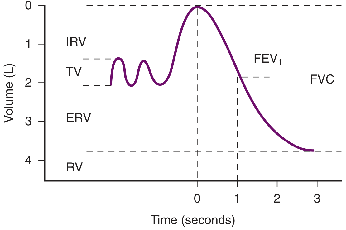
8. Spirometry with Pocket-Sized (Handheld) Spirometer
Principle: Measures flow and volume of air during forced expiratory maneuver. Gives the most complete picture of lung mechanics among bedside tests.
(Roberts & Hedges' Clinical Procedures in Emergency Medicine, p. 61)
Key Parameters Measured:
| Parameter | Definition | Normal |
|---|---|---|
| FVC (Forced Vital Capacity) | Total volume exhaled during forced expiration from full inspiration to full expiration | 80-120% predicted |
| FEV1 | Volume exhaled in first 1 second of forced expiration | > 75% predicted |
| FEV1/FVC ratio | Ratio of FEV1 to FVC | ≥ 0.70 (70%) |
| FEF 25-75% | Mean flow during middle 50% of FVC (sensitive for small airway disease) | > 79% predicted |
| PEFR | Maximum flow during forced expiration | 450-700 L/min (M) |
Procedure:
- Calibrate device per manufacturer's instructions
- Patient stands or sits upright, chin slightly extended
- Normal breathing for a few cycles
- Maximal inspiration
- Seal lips firmly around mouthpiece
- Exhale as hard and as fast as possible until lungs are completely empty
- Best of 3 technically acceptable efforts recorded
Contraindications:
- Severe respiratory compromise requiring immediate treatment
- Pneumothorax / pneumomediastinum (forced expiration increases intrathoracic pressure)
- Suspected aortic aneurysm or cerebral aneurysm
- Unable to cooperate (unconscious, heavily sedated, children < 4-5 years)
Interpretation of Spirometry Results
(Bailey & Love's, p. 998; Murray & Nadel's Textbook of Respiratory Medicine)
Pattern Recognition:
| Parameter | Obstructive Pattern | Restrictive Pattern | Normal |
|---|---|---|---|
| PEFR | ↓↓ | Normal or ↓ | Normal |
| FEV1 | ↓↓ | Normal or ↓ | Normal |
| FVC | Normal or ↓ | ↓↓ | Normal |
| FEV1/FVC ratio | < 70% | > 80% | 70-80% |
| TLC | Increased (air trapping) | Decreased | Normal |
Obstructive diseases: Asthma, COPD, emphysema, bronchiectasis
Restrictive diseases: Pulmonary fibrosis, pleural effusion, chest wall deformities, neuromuscular disease
Severity of Obstruction (based on FEV1 % predicted):
| Grade | FEV1 % Predicted |
|---|---|
| Mild | > 70% |
| Moderate | 60-69% |
| Moderately severe | 50-59% |
| Severe | 35-49% |
| Very severe | < 35% |
Bronchodilator Reversibility:
- Repeat spirometry 15-20 min after bronchodilator (salbutamol 400 mcg)
- Significant reversibility: FEV1 increase of ≥ 12% AND ≥ 200 mL from baseline
- Positive test suggests asthma (reversible obstruction) vs. fixed obstruction (COPD, emphysema)
Indications for Bedside PFTs
- Preoperative assessment - especially for thoracic, abdominal, upper GI surgeries
- Acute exacerbations of asthma - to quantify severity and monitor response to treatment
- Acute COPD exacerbations - though not routinely recommended in guidelines
- Differentiating dyspnea causes
- Neuromuscular disease evaluation (e.g., Guillain-Barre, MG)
- Monitoring chronic lung disease
Preoperative Assessment Criteria (Surgical Risk Stratification)
Using bedside PFTs before lung resection or major surgery:
| Test | Low Risk | Moderate Risk | High Risk |
|---|---|---|---|
| SBC (Single Breath Count) | > 30 | 15-30 | < 15 |
| Breath Holding Time | > 30 sec | 15-25 sec | < 15 sec |
| PEFR | > 300 L/min | 200-300 | < 200 L/min |
| FEV1 | > 2 L | 1-2 L | < 1 L |
| FEV1/FVC | > 70% | 50-70% | < 50% |
Summary Table - Bedside PFTs at a Glance
| Test | What it Measures | Normal | Significance |
|---|---|---|---|
| Snider's Match Test | Approx. FEV1 | Blows out at 15 cm | FEV1 < 1.6 L if fails |
| Forced Expiratory Time | Airway obstruction | < 3 sec | > 6 sec = severe obstruction |
| Single Breath Count | Vital capacity | > 30 | < 15 = VC < 1 L |
| Breath Holding Test | Combined cardiorespiratory reserve | > 30 sec | < 15 sec = high risk |
| Cough Test | Cough effectiveness | Strong, sharp cough | PEFR < 200 = inadequate cough |
| DeBono's Whistle | PEFR (semi-quantitative) | Blows whistle at max opening | Semiquantitative |
| Wright's Peak Flow | PEFR | Males 450-700; Females 300-500 L/min | < 200 = impaired cough |
| Pocket Spirometry | FVC, FEV1, FEV1/FVC, PEFR | FEV1/FVC > 70% | Complete pattern diagnosis |
Key Points for MD Exam
- Forced expiratory time > 6 seconds is the simplest bedside indicator of significant obstructive lung disease
- PEFR < 200 L/min indicates inadequate cough efficiency and high postoperative pulmonary risk
- FEV1/FVC < 70% defines obstructive pattern; FVC reduced with normal FEV1/FVC defines restrictive pattern
- Spirometry with a handheld device provides the most complete bedside assessment - diagnoses type, severity, and bronchodilator reversibility
- All bedside PFTs (except breath holding) are effort dependent and require patient cooperation
- Bedside PFTs are suitable for age > 4-5 years, cooperative patients only
- Bronchodilator reversibility testing distinguishes asthma (reversible) from COPD (partially reversible or fixed)
Sources: Bailey & Love's Short Practice of Surgery 28th Ed. | Roberts & Hedges' Clinical Procedures in Emergency Medicine | Murray & Nadel's Textbook of Respiratory Medicine | Goldman-Cecil Medicine
This is a shared conversation. Sign in to Orris to start your own chat.