
Ameoba
Amoeba biology medical importance overview
Entamoeba histolytica life cycle trophozoite cyst
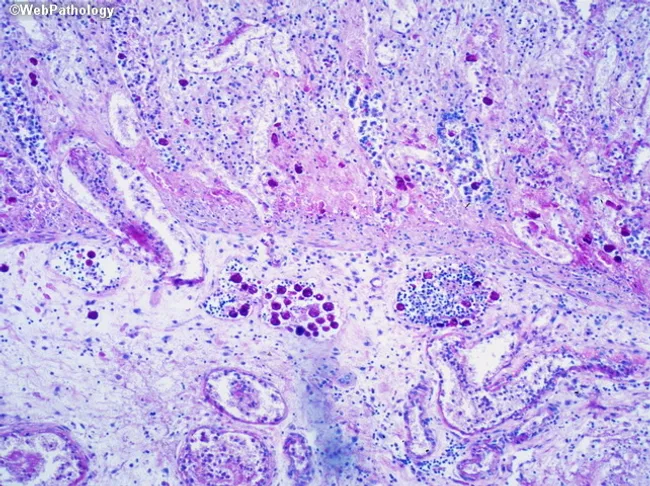
This image is a brightfield light microscopy slide of colonic mucosa stained with Periodic Acid–Schiff (PAS), highlighting Entamoeba histolytica trophozoites within inflamed tissue. The specimen type is gastrointestinal mucosa biopsy. The primary subject includes magenta PAS-positive amoebic trophozoites, visible as irregular, vacuolated cytoplasm containing ingested material, often adjacent to neutrophilic and mononuclear inflammatory cells. The tissue architecture shows disrupted mucosal glands, ulceration, and lamina propria infiltration, typical of amoebic colitis. The magenta organisms stand out against a pale pink to purple backdrop; chromotrope-rich cytoplasm and endosome-like vacuoles are evident; occasional red blood cells may be within trophozoite cytoplasm if present. This staining pattern (PAS) enhances detection of the cytoplasmic glycogen-rich trophozoites, aiding differentiation from host cells and other pathogens. Clinically, histopathology confirmation supports a diagnosis of Entamoeba histolytica infection and guides anti-amoebic therapy. The image demonstrates classic features of tissue-invasive amebiasis in the colon, with potential for flask-shaped ulcers, necrosis, and focal microabscess formation. This content is relevant for gastroenterology, pathology education, infectious disease diagnosis, and histology training, illustrating the diagnostic significance of PAS-positive trophozoites. Potential differential diagnoses include nonpathogenic Entamoeba dispar and PAS-positive fungal organisms; but cooperation of clinical data is essential.
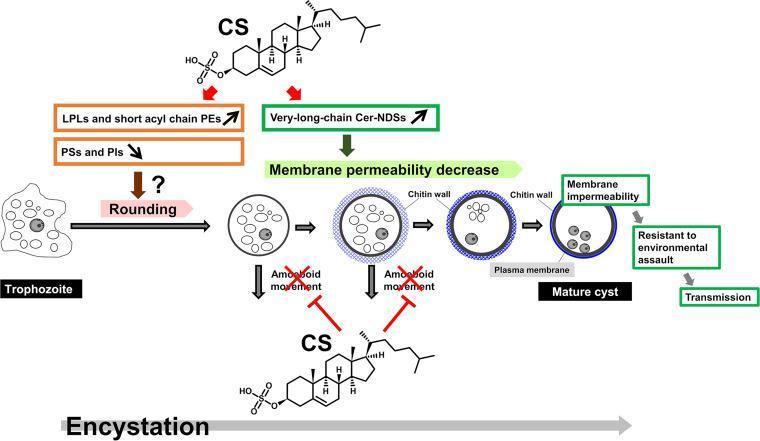
This pathophysiology diagram illustrates the molecular and morphological stages of Entamoeba encystation, highlighting the pleiotropic roles of Cholesterol Sulfate (CS). The process begins with an irregular trophozoite transitioning into a spherical shape (rounding). This morphological change is associated with an increase in lysophospholipids (LPLs) and short acyl chain phosphatidylethanolamines (PEs), and a decrease in phosphatidylserines (PSs) and phosphatidylinositols (PIs). Simultaneously, CS triggers an increase in very-long-chain Ceramide non-hydroxy-fatty acid dihydrosphingosines (Cer-NDSs), which leads to decreased membrane permeability. Structurally, the cell develops a chitin wall around the plasma membrane, eventually forming a mature cyst characterized by membrane impermeability. The diagram shows that CS also functions to inhibit amoeboid movement during this transition, facilitating the formation of a rigid, spherical cyst. The clinical significance of this process is the acquisition of resistance to environmental assault, which is essential for successful parasite transmission. Key terms depicted include trophozoite, mature cyst, chitin wall, and various lipid signaling molecules involved in protozoan development.
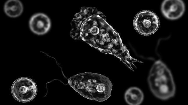
This medical illustration depicts the three morphological life-cycle stages of Naegleria fowleri, a thermophilic excavate amoeba responsible for Primary Amebic Meningoencephalitis (PAM). The visual showcases the following forms against a high-contrast dark background: 1) Cyst: Small, spherical structures (approx. 10–15 µm) with a distinct, smooth double cell wall and internal granular content, representing the dormant, resistant stage. 2) Trophozoite: The larger, pathogenic amoeboid form (approx. 10–35 µm) characterized by an irregular shape and active pseudopodia (lobopodia). Internal features include a prominent nucleus with a dense central nucleolus and multiple contractile vacuoles. 3) Flagellate: A pear-shaped transitional stage featuring two long, anterior flagella used for rapid locomotion in unfavorable environmental conditions. The illustration utilizes high-resolution 3D-style rendering to emphasize cellular morphology, membrane integrity, and specialized locomotory organelles, serving as an educational tool for parasitology and infectious disease diagnostics.
Amoeba - Overview
What is an Amoeba?
- Outer plasma membrane surrounded by a stiff, clear ectoplasm
- Central granular endoplasm containing food vacuoles, nucleus, and a contractile vacuole
- No fixed mouth or anus - food is taken in and expelled at any point on the surface
- Reproduce mainly by binary fission
Medically Important Amoeba Species
1. Entamoeba histolytica (Most Important)
- Worldwide distribution, major health problem in South/Southeast Asia, China, West Asia, Latin America, and Africa
- Approximately 50 million people carry E. histolytica globally; ~500,000 cases of invasive disease per year; ~50,000 deaths annually
- In India, approximately 15% of the population is affected
- In high-endemic areas, stool prevalence averages 10%; amoebic liver abscess incidence can reach 21 per 100,000 population
- Transmitted via the faecal-oral route - contaminated food or water
| Form | Location | Role |
|---|---|---|
| Trophozoite | Large intestine | Invasive, causes disease |
| Cyst (infective) | Excreted in stool | Survives in environment, infects new hosts |
- Ingested cysts hatch in the small bowel, releasing trophozoites that colonize the colon
- Trophozoites form characteristic flask-shaped ulcers in the submucosa
- Some trophozoites enter the portal circulation and become trapped in the liver's interlobular veins, causing focal hepatocyte infarction and liquefactive necrosis via proteolytic enzymes
- Coalescing necrosis forms an amoebic liver abscess - classic "anchovy sauce" (chocolate-coloured, odourless) pus
- Most infections (>90%) are asymptomatic
- Symptomatic range: mild abdominal discomfort and loose stools to acute fulminating amoebic dysentery (bloody diarrhoea, tenesmus, cramping)
- Liver - amoebic liver abscess (most common extraintestinal site; right lobe in 80% of cases, left in 10%)
- Lungs, brain, spleen, skin (rarer)
- E. histolytica can be differentiated into at least 18 zymodemes (populations differing by electrophoretic enzyme mobilities)
- 7 zymodemes are pathogenic, 11 are non-pathogenic
- Note: the morphologically identical but non-pathogenic Entamoeba dispar was previously misclassified as E. histolytica
2. Naegleria fowleri ("Brain-eating amoeba")
- Free-living thermophilic amoeba found in warm freshwater (lakes, hot springs, poorly chlorinated pools)
- Causes Primary Amoebic Meningoencephalitis (PAM) - rare but nearly always fatal
- Enters through the nasal mucosa during water activities, migrates along olfactory nerves to the brain
- Three life-cycle forms: trophozoite (amoeboid), flagellate (transitional), and cyst
- Presents like bacterial meningitis: severe headache, fever, neck stiffness, progressing rapidly to coma and death
3. Acanthamoeba spp.
- Free-living amoeba found in soil, water, and air
- Causes:
- Acanthamoeba keratitis - painful, vision-threatening corneal infection, especially in contact lens wearers
- Granulomatous Amoebic Encephalitis (GAE) - in immunocompromised individuals (HIV, transplant patients)
Amoeba in Biology Research
Histopathology Image
Quick Summary Table
| Species | Habitat | Disease | Key Feature |
|---|---|---|---|
| Entamoeba histolytica | Human gut | Amoebiasis, liver abscess | Flask-shaped ulcers; anchovy-sauce pus |
| Naegleria fowleri | Warm freshwater | PAM (fatal meningitis) | Enters via nose; has flagellate form |
| Acanthamoeba | Soil, water | Keratitis, GAE | Contact lens risk; immunocompromised |
| Amoeba proteus | Freshwater | Non-pathogenic | Model organism in research |